Natural product based apoptosis inducers
- Summary
- Abstract
- Description
- Claims
- Application Information
AI Technical Summary
Benefits of technology
Problems solved by technology
Method used
Image
Examples
example 1
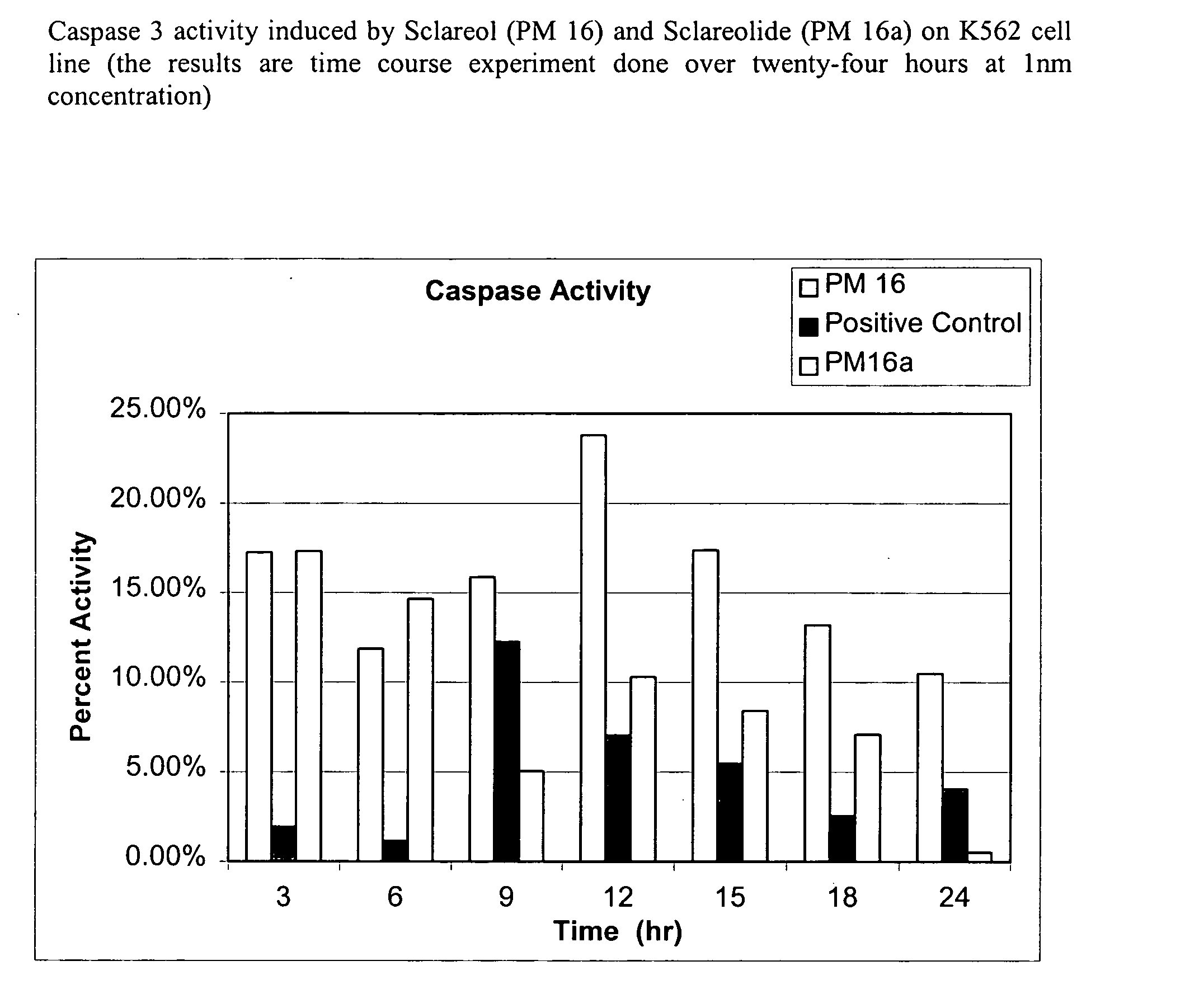
Screening for Caspase Activity in Compositions Comprising Plant Extract.
[0174] 1. Thaw the (100×) substrate Z-DEVD-R110 and Apo-ONE™ Homogeneous Caspase-3 / 7 Buffer (available from Promega Corporation) to room temperature. Avoid multiple freeze-thaw cycles of the Substrate and Buffer. [0175] 2. Mix each component by inversion or vortexing. [0176] 3. Dilute the Substrate (1:100) with Buffer to make the desired amount of the Homogeneous Caspase-3 / 7 Reagent. Store the Reagent, protected from light, at room temperature until use. The Reagent may be stored at 4° C. for 24 hours. [0177] 4. Set up assay, blank, and positive or negative control reactions as appropriate. [0178] 5. Add Homogeneous Caspase-3 / 7 Reagent to each well of a black or white 96 well plate, maintaining a 1:1 ratio of Reagent to sample. [0179] 6. Gently mix contents by shaking at 300-500 rpm on a plate shaker from 30 seconds up to read time. Incubate the reactions for 30 minutes to 18 hours. [01...
example 2
DNA Fragmentation Assays for Apoptosis Protocol
Protocol I: Triton X-100 Lysis Buffer
[0186] In 96 flat-wells plate, incubate 4×10⁶ target cells (40 wells of 105 per well) with desired concentration of effectors (105 target cells per well). After incubation, collect the cell sample in 1.5 ml eppendorf tube, spin down, resuspend with 0.5 ml PBS in 1.5 ml eppendorf tubes, and add 55 ul of lysis buffer for 20 min on ice (4° C.). Centrifuge the eppendorf tubes in cold at 12,000 g for 30 minutes. Transfer the samples to new 1.5 ml eppendorf tubes and then extract the supernatant with 1:1 mixture of phenol:chloroform (gentle agitation for 5 min followed by centrifugation) and precipitate in two equivalence of cold ethanol and one-tenth equivalence of sodium acetate. Spin down, decant, and resuspend the precipitates in 30 ul of deionized water-RNase solution (0.4 ml water+5 ul of RNase) and 5 ul of loading buffer for 30 minutes at 37° C. Also insert 2 ul of Hindi III marker (12 ul of Sto...
example 3
Acridine Orange / Ethidium Bromide Staining for Apoptosis Cells (AO Staining)
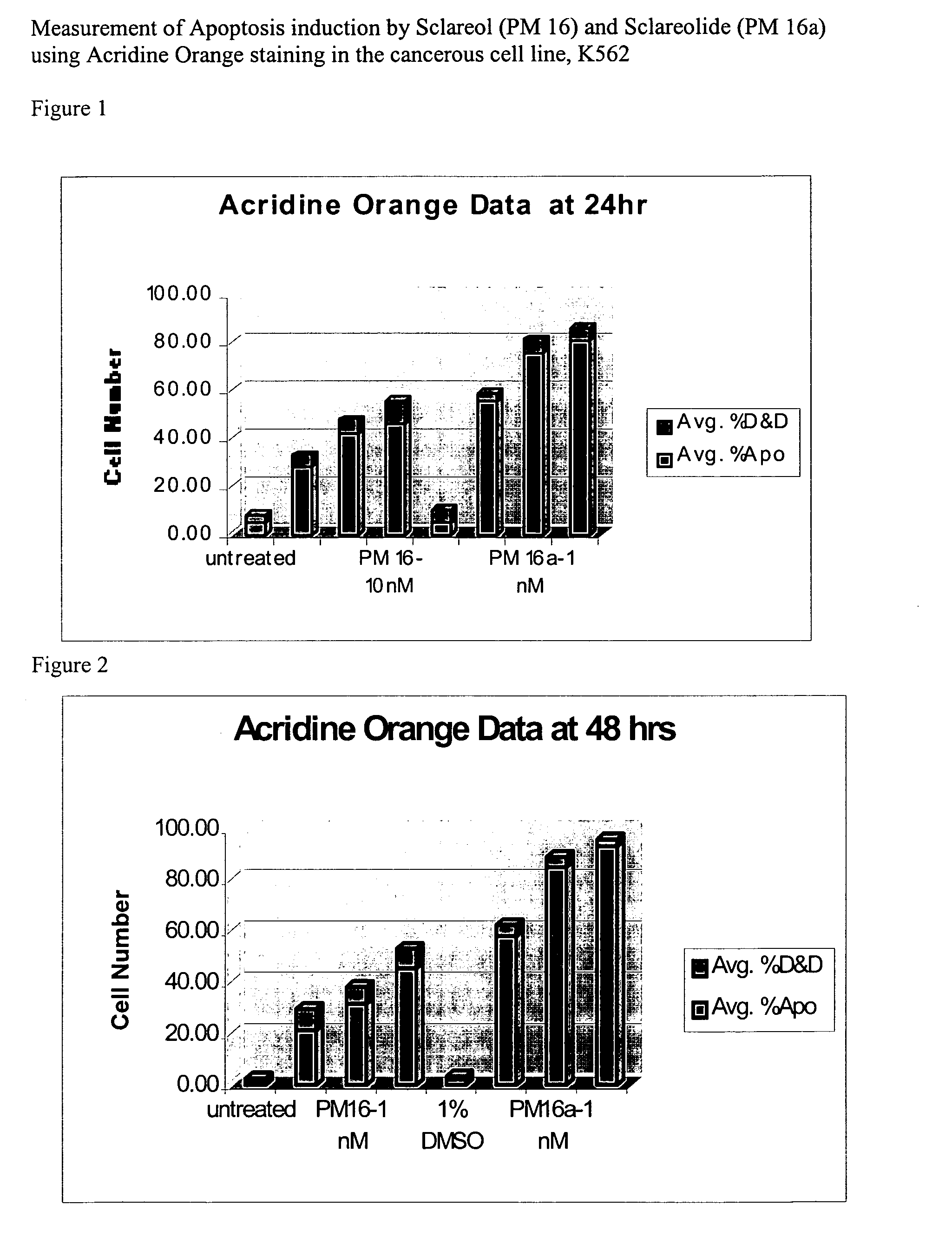
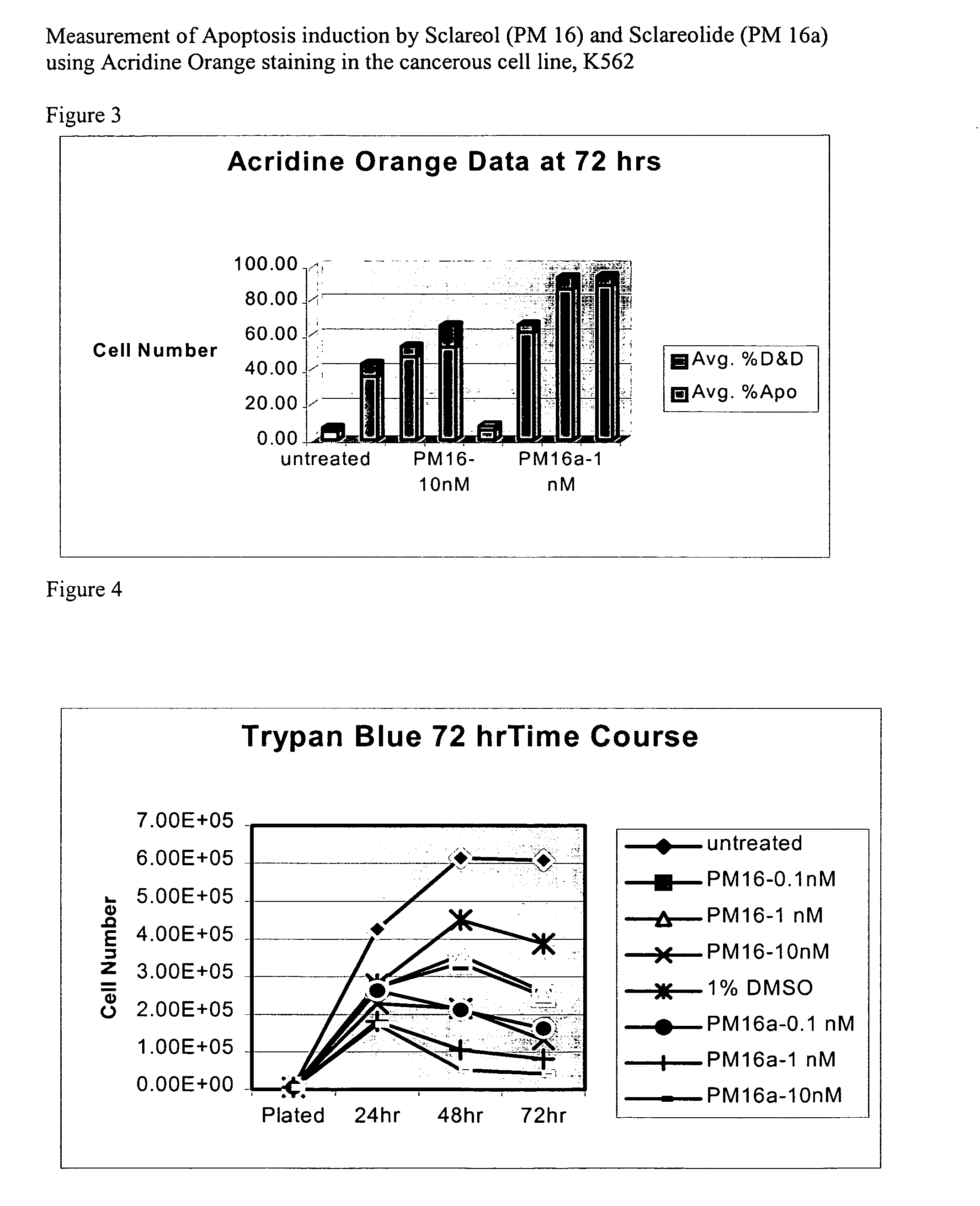
[0201] Acridine Orange (AO) is an intercalating fluorescence dye that can enter the nucleus of a cell to stain DNA. This AO-staining method has an advantage of high staining-specificity, but with the disadvantage that samples can only be observed for a short period of time, usually within 24 hours. The AO stain can be used to test cell viabilities in a cell sample in conjunction with propidium iodide (PI). AO / PI fluoresce green under dark field fluorescence microscopy, while nonviable cells fluoresce orange.
[0202] Acridine orange (AO) / Ethidium bromide (EtBr) staining for Apoptosis cells (AO staining) Solutions: [0203] (i) AO stock solution: 1 mg / ml as 0.001 g AO+1 ml PBS [0204] (ii) EtBr stock solution: 1 mg / ml: 0.001 g+1 ml PBS [0205] (iii) Working dye solution: 0.1 mg / ml AO stock solution, 0.1 mg / ml EtBr stock solution is prepared by mixing 100 μl of each stock solution plus (+) 800 μl of PBS [0206] Make...
PUM
 Abstract
Description
Claims
Application Information
 - R&D
- Intellectual Property
- Life Sciences
- Materials
- Tech Scout
- Unparalleled Data Quality
- Higher Quality Content
- 60% Fewer Hallucinations
Browse by: Latest US Patents, China's latest patents, Technical Efficacy Thesaurus, Application Domain, Technology Topic, Popular Technical Reports.
© 2025 PatSnap. All rights reserved.Legal|Privacy policy|Modern Slavery Act Transparency Statement|Sitemap|About US| Contact US: help@patsnap.com


