Enzyme-catalyzed metal deposition for the enhanced detection of analytes of interest

a metal deposition and enzyme-catalyzed technology, applied in the field of immunochemistry, can solve the problems of inability to detect small peptides, most drugs, and low analyte concentration, and achieve the effect of high assay sensitivity, accurate determination and rapid

Active Publication Date: 2005-05-12
VENTANA MEDICAL SYST INC
View PDF31 Cites 150 Cited by
  • Summary
  • Abstract
  • Description
  • Claims
  • Application Information

AI Technical Summary

Benefits of technology

[0026] In the present invention, regardless of assay employed, the detection and quantification of the biological analyte of interest is based on a metal precipitate forming system that is catalyzed by an enzyme, such as, e.g., alkaline phosphatase. The enzyme catalyzes the dephosphorylation of a reducing agent precursor into an active reducing agent, which in turn reduces a metal ion into a detectable and quantifiable metal precipitate. By using the methods of the invention, low concentrations of analyte in assays such as in situ histochemical assays, immunoassays such as ELISA, or hybridization to DNA microarrays on glass slides, can be determined accurately, rapidly, and with high assay sensitivity.

Problems solved by technology

A disadvantage of the existing ELISA techniques, however, is that the adsorption and detection phases, to secure assay sensitivity, can be very time consuming for samples having low analyte concentrations.
In such samples, the rate of adsorption of analyte to the surface is proportional to the concentration of analyte in the solution, and thus will also become very low, even in well-stirred systems.
Accordingly, such sandwich assays cannot be used for the detection of small peptides, most drugs, or synthetic drug candidates.

Method used


Image

  • Enzyme-catalyzed metal deposition for the enhanced detection of analytes of interest
  • Enzyme-catalyzed metal deposition for the enhanced detection of analytes of interest
  • Enzyme-catalyzed metal deposition for the enhanced detection of analytes of interest

Examples


example 1

[0126] Phosphates for Alkaline-Phosphatase Mediated Reduction of Silver (I)

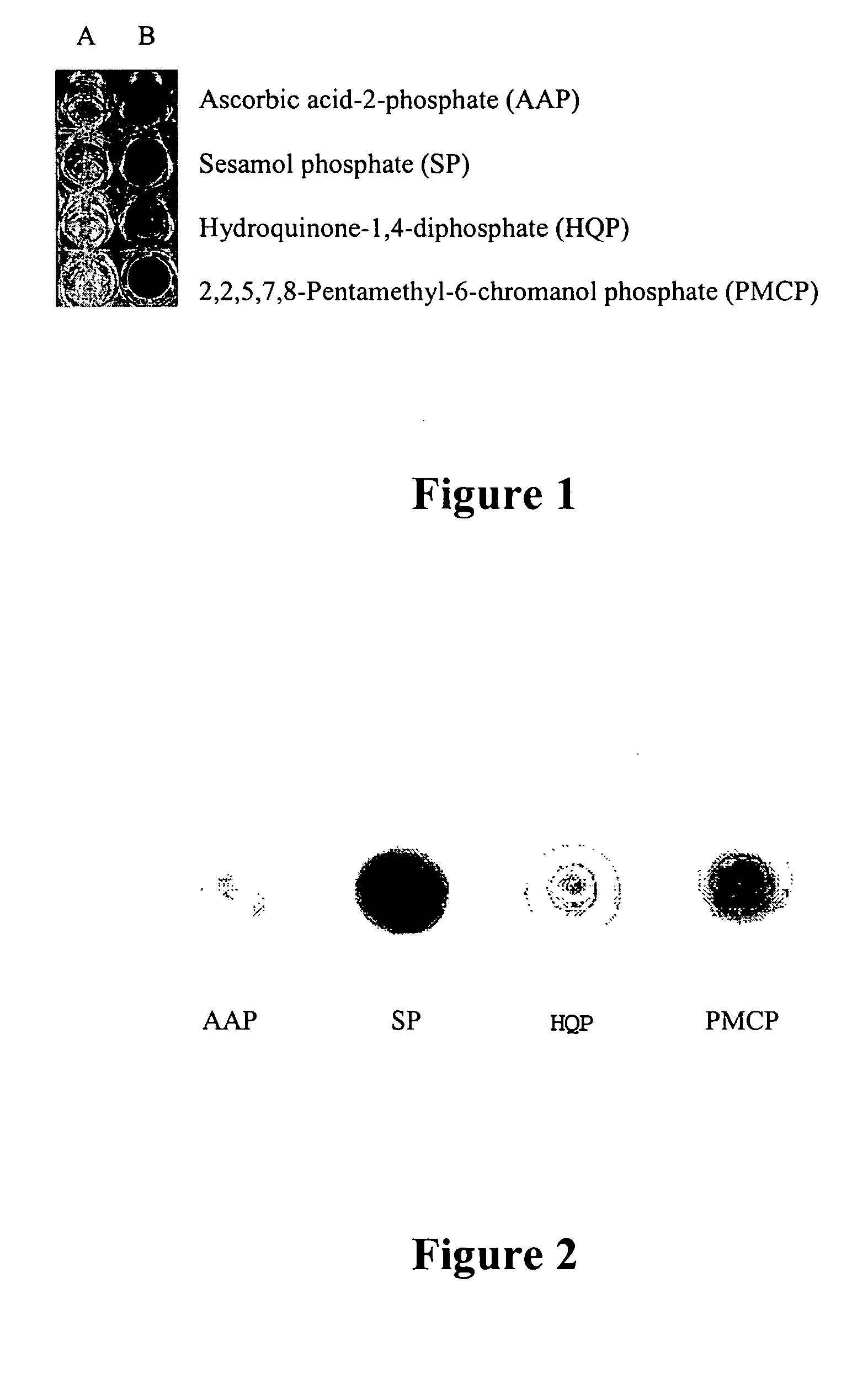
[0127] Silver nitrate (100 μL of 50 mM AgNO3) was aliquoted into the wells of a microtiter plate (see FIG. 1). Addition of 100 μL of a 50 mM solution of either ascorbic acid-2-phosphate (wells A1, B1), sesamol phosphate (wells A2, B2), hydroquinone-1,4-diphosphate (wells A3, B3), or 2,2,5,7,8-pentamethyl-6-chromanol phosphate (wells A4, B4) to the silver solution did not elicit any reaction. If calf-intestinal alkaline phosphatase (5 μL of 0.2 mg / mL) was added to the Column B wells, each solution formed metallic silver (0) as either a fine, black precipitate or silvering of the container walls.

[0128] Column A: 100 μL of 50 mM AgNO3 and 100 μL of 50 mM substrate phosphate.

[0129] Column B: 100 μL of 50 mM AgNO3, 100 μL of 50 mM substrate phosphate, and 5 μL of 0.2 mg / mL calf-intestinal alkaline phosphatase (Pierce Chemical, Rockford, Ill.) in 100 mM Tris, pH 7.0.

example 2

Alkaline Phosphatase, Silver Nitrate, And Substrate Phosphates On Nitrocellulose

[0130] Alkaline phosphatase (5 μL of 0.2 mg / mL) was added to nitrocellulose paper and dried (see FIG. 2). Each of the phosphates were spotted on the nitrocellulose as 5 μL of 0.05M solution in 0.1M Tris, pH 9. When 5 μL of 0.05M AgNO3 was added to each dried spot, a black precipitate was observed only at the spot where alkaline phosphatase was applied.

example 3

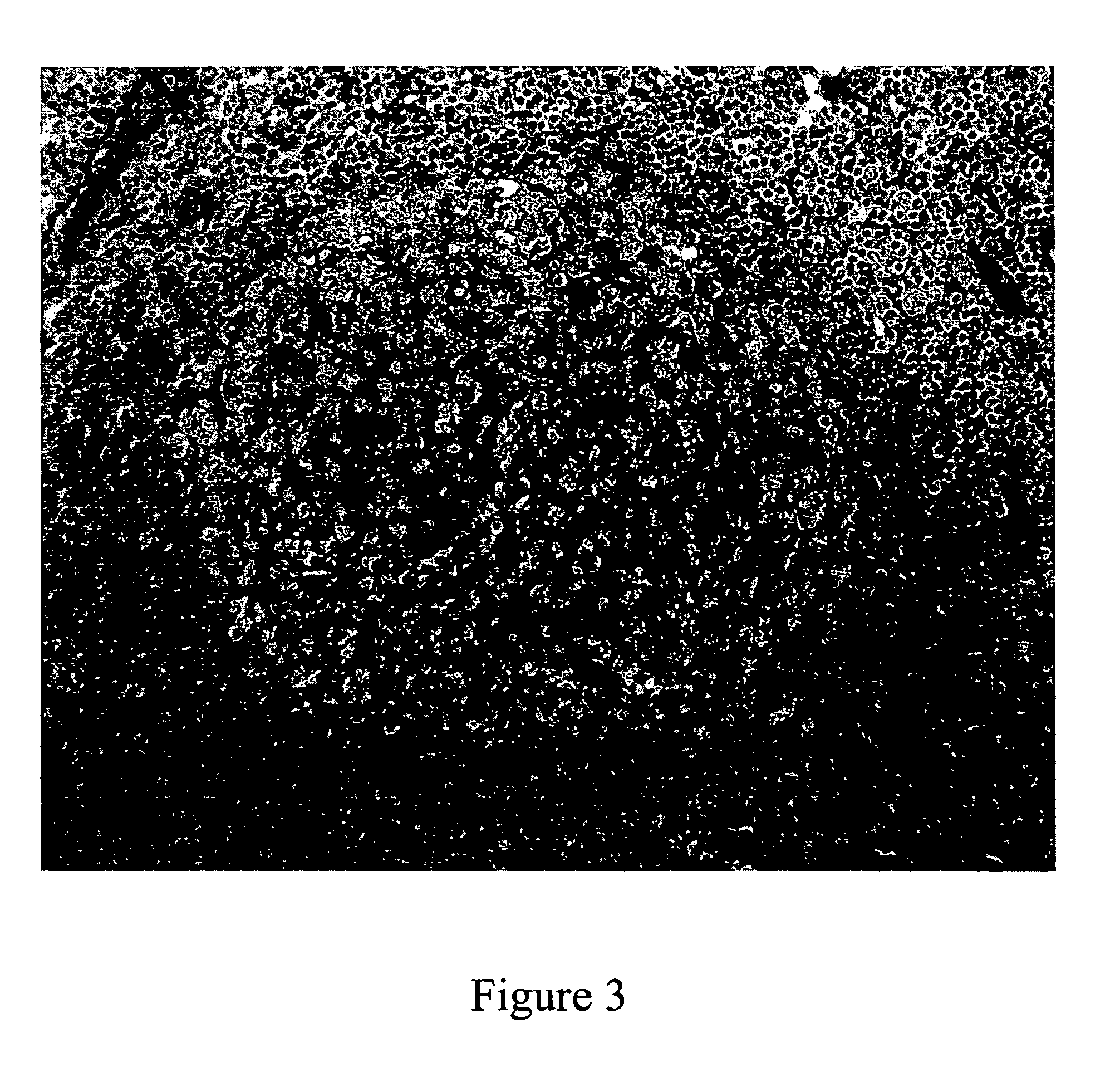
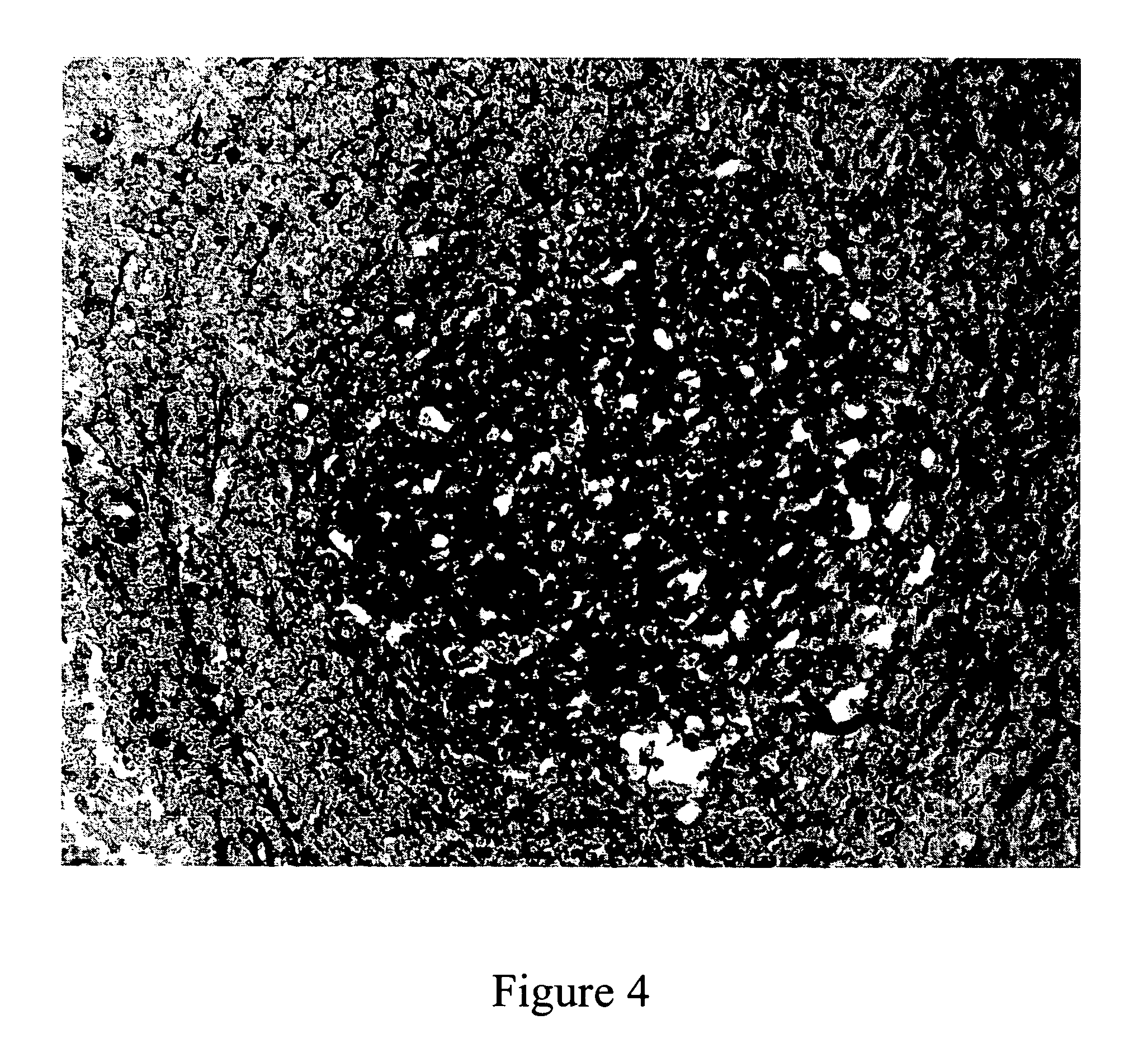
Offline Development of Ag Nanoparticles on Tonsil Using the AP-SA Conjugate at High pH

[0131] The preparation of the slides for analysis was done on a Ventana Benchmark® automated staining instrument (Ventana Medical Systems, Inc., Tucson, Ariz.). The slides were removed for manual development of signal. The use of the term “buffer” within this Example refers to a 0.1 M Tris buffer (Trizma base (Sigma-Aldrich) in dI H2O pH to 9 using glacial acetic acid (Sigma-Aldrich). The following is the adapted procedure from the instrument: the paraffin-coated tissue on the slide was heated to 75° C. for 4 minutes and treated twice with EZPrep™ (Ventana, Tucson, Ariz.) volume adjusted at 75° C. before application of the liquid cover slip with EZPrep volume adjust. After 4 minutes at 75° C., the slide was rinsed and automated deparaffinization volume adjust was added along with liquid cover slip to deparaffinize the tissue at 76° C. for 4 minutes. The slide was cooled to 40° C. and rinsed three ...


PUM

PropertyMeasurementUnit
pHaaaaaaaaaa
pHaaaaaaaaaa
pHaaaaaaaaaa

Abstract

The invention is directed to enhanced methods for detecting an analyte of interest in situ, by immunoassay, or by hybridization comprising binding an enzyme-labeled conjugate molecule to an analyte of interest in the presence of a redox-inactive reductive species and a soluble metal ion. The enzyme catalyzes the conversion of the inactive reductive species to an active reducing agent, which in turn reduces the metal ion to a metal atom thereby providing an enhanced means of detecting the analyte via metal deposition.

Description

CROSS REFERENCE TO RELATED APPLICATIONS [0001] This application is a continuation-in-part of U.S. application Ser. No. 10 / 877,919, filed Jun. 24, 2004, which claims the benefit of U.S. Provisional Appl. No. 60 / 482,596, filed Jun. 24, 2003, the contents of which are hereby incorporated by reference in their entireties.BACKGROUND OF THE INVENTION [0002] 1. Field of the Invention [0003] The present invention relates generally to the field of chemistry, and in particular, immunochemistry. More specifically, the present invention relates to a method for the enhanced detection of an analyte by using an enzyme-catalyzed reaction for the localized deposition of metal atoms. The present method of detection can be employed in various types of assays, such as, e.g., in-situ histochemical, immunoassays such as ELISA, and hybridization to nucleic acid microarrays. [0004] 2. Related Art [0005] Tissue staining is an ancient art by modem standards that goes back over one hundred years. Recently, ef...

Claims


Application Information

Patent Timeline
Patent Type & Authority Applications(United States)
IPC IPC(8): C07F9/09C07F9/655C07H15/20C07H15/24C07H15/26G01N33/58
CPCC07F9/093C07F9/65517C07F9/65522G01N33/581C07H15/24C07H15/26C07H15/20
Inventor BIENIARZ, CHRISTOPHERFARRELL, MICHAEL
Owner VENTANA MEDICAL SYST INC
Why Eureka
Try Eureka