Method of isolating cells and uses thereof

a cell and cell technology, applied in the field of non-invasive methods of retrieving and identifying cells, can solve the problems of high risk of congenital heart disease and leukaemia, high risk of miscarriage (1-2%), and samples obtained from other sources such as blood cannot be guaranteed, so as to facilitate cell identification and improve the yield of suspended single cells

Inactive Publication Date: 2005-06-09
MONASH UNIV +1
View PDF0 Cites 128 Cited by
  • Summary
  • Abstract
  • Description
  • Claims
  • Application Information

AI Technical Summary

Benefits of technology

[0006] Therefore a rapid, non-invasive diagnostic technique and preferably one which ensures the testing of a current fetus would significantly benefit all pregnant women of high or low genetic risk. A diagnosis within 24 hours would give them piece of mind and the opportunity to make an earlier decision regarding therapeutic abortion in the first trimester of their pregnancy.
[0013] It is preferred that the present method retrieves substantially intact cells that have substantially maintained their cell membrane integrity which allows for reliable identification such as through antibody testing.
[0014] In a preferred embodiment, the cervical mucus sample is further treated with a mucolytic agent prior to being treated with a collagenase and a protease to disassociate the cells. It has been found that by treating in this combined manner, there is a better yield of suspended single cells from the cervical mucus sample.

Problems solved by technology

Down syndrome is the most important genetic cause of mental retardation in humans and is also associated with a high risk of congenital heart disease and leukaemia.
Both these procedures are associated with a risk of miscarriage (1-2%).
Samples obtained from other sources such as the blood cannot ensure that the fetal cell so identified, may be derived from the current fetus or of a recently miscarried fetus because such cells can persist in the circulation for several years.
Such procedures are, lengthy and require a high level of technical expertise.
Further, results generally are not available to the patient for up to three weeks.

Method used


Image

  • Method of isolating cells and uses thereof

Examples


example 1

Diagnosis of Down Syndrome

[0174] (a) Collection and Preparation of Transcervical Cells

[0175] Collection of the transcervical cells from pregnant women during the first or second trimester is a similar procedure to a pap smear and involves direct aspiration of the cervical mucus, using a thin catheder (Aspiracath, Cook IVF), from the endocervical canal and the lower uterine pole. The tip of the catheder that contains the maternal mucus and transcervical cells is cut off into an eppendorf tube containing 0.5 ml of RPMI culture media and placed at 37° C. Gently the contents are mechanically removed from the inner tip of the catheder and suspended in culture media at 37° C.

[0176] (b) Identification and Isolation of Fetal Cells from Transcervical Samples

[0177] After collection, samples are treated with 0.5 ml of 20 mg / ml of N-acetyl-L-cysteine and further incubated with gentle shaking at 37° C. for 45 minutes. The entire sample is then washed twice in PBS before an incubation with 0....

example 2

Diagnosis of Down Syndrome II

[0185] (a ) The collection and preparation of transcervical cells is followed as described above in example 1.

[0186] (b) The Identification and Isolation of Fetal Cells

[0187] After collection, samples are spread onto slides and fixed in 100% ethanol. Immunohistochemistry is performed using first trimester fetal specific antibodies to identify the fetal cells. The slides are then dehydrated. Laser capture microdissection technology is used to remove positively stained cells from the slide onto membranes that can be directly transferred into PCR tubes.

[0188] (c, d and e) FL-PCR DNA fingerprinting for trisomy 21 and the analysis of FL-PCR products and diagnosis of aneuploidy is followed as described above in example 1

example 3

Simultaneous Diagnosis of Down Syndrome and Cystic Fibrosis DeltaF508 Mutation

[0189] (a & b) The collection and preparation of transcervical cells, and the identification and isolation of fetal cells is followed as described above in example 1.

[0190] (c) FL-PCR DNA Fingerprinting for Trisomy 21 and Cystic Fibrosis deltaF508 Diagnosis

[0191] The FL-PCR reaction is as described above with the following changes: the primer mix contains four informative chromosome 21 microsatellite markers along with the primer pair for deltaF508 mutation detection. Microsatellite markers outlined in Tables 2 & 3 are genotyped on parental genomic DNA to identify the heterozygous loci for incorporation into the DNA fingerprinting system. Final optimized primer pair concentrations are reaction and primer specific.

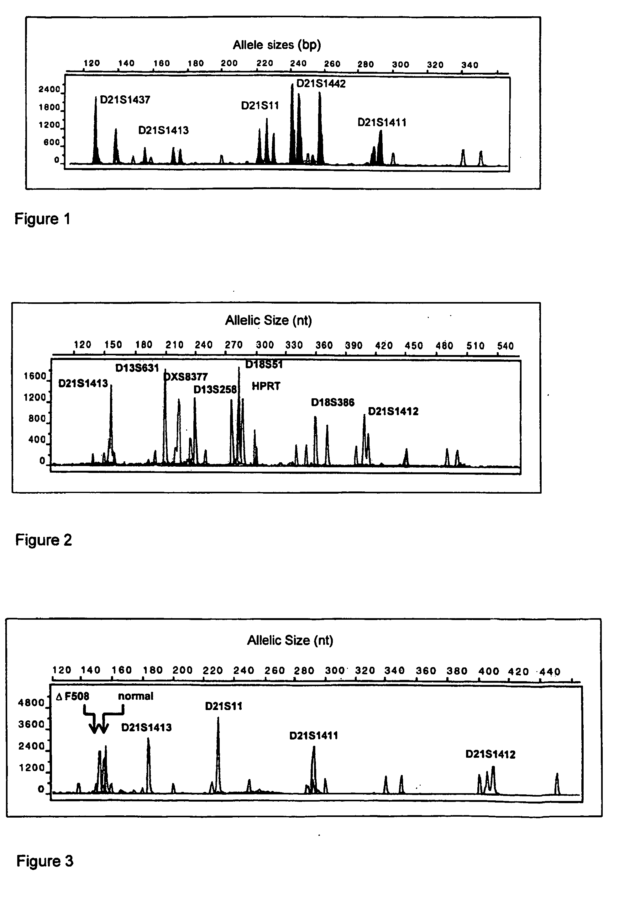
[0192] (d & e) Analysis of FL-PCR products and diagnosis of aneuploidy is as described above (see FIG. 3).

[0193] Finally it is to be understood that various other modifications and / or alterat...


PUM

PropertyMeasurementUnit
concentrationaaaaaaaaaa
volumeaaaaaaaaaa
volumeaaaaaaaaaa

Abstract

The present invention relates to a non-invasive method of retrieving and identifying cells particularly fetal cells and trophoblastic cells. The invention includes methods for use of the cells for identifying chromosomal abnormalities and mutations particularly for prenatal diagnosis by performing genetic diagnosis for chromosomal and single gene disorders. The invention also includes methods of confirming cells of fetal origin.

Description

[0001] The present invention relates to a non-invasive method of retrieving and identifying cells particularly fetal cells and trophoblastic cells. The invention includes methods for use of the cells for identifying chromosomal abnormalities and mutations particularly for prenatal diagnosis by performing genetic diagnosis for chromosomal and single gene disorders. The invention also includes methods of confirming cells of fetal origin. Introduction [0002] Approximately 0.5% of couples are at high risk of conceiving a child with a genetic disorder. Such genetic disorders include Cystic Fibrosis, Huntington's Disease, Beta Thalassaemia and Myotonic Dystrophy. For example, in Australia, 1 in 25 of the population is a carrier of a Cystic Fibrosis mutation and thus newborn screening for Cystic Fibrosis has recently been implemented to monitor all births. [0003] In addition to single gene disorders chromosomal abnormalities are the most common genetic disorders seen in spontaneous miscarr...

Claims


Application Information

Patent Timeline
Patent Type & Authority Applications(United States)
IPC IPC(8): G01N33/53C12N5/07C12N5/073C12N15/09C12Q1/68C12Q1/6881C12Q1/6883C12S3/24G01N33/50G01N33/569
CPCC12Q1/6881C12Q1/6883C12Q2600/16C12Q2600/156C12Q2600/158G01N33/56966
Inventor KATZ, MANDYCRAM, DAVID
Owner MONASH UNIV
Why Eureka
Try Eureka