Analysis of circulating tumor cells, fragments, and debris

a technology fragments, applied in the field of analysis fragments and debris, can solve the problems of spurious increase in the number of detected circulating cancer cells, increased cellular debris that can interfere, and increased the amount of circulating tumor cells. to achieve the effect of inhibiting further damage of

Inactive Publication Date: 2005-08-18
MENARINI SILICON BIOSYSTEMS SPA
View PDF28 Cites 173 Cited by
  • Summary
  • Abstract
  • Description
  • Claims
  • Application Information

AI Technical Summary

Benefits of technology

[0034] The methods and reagents described in this invention are used to analyze circulating tumor cells, fragments, and debris. Analysis is performed with a number of platforms, including multiparameter flow cytometry and the CellSpotter® fluorescent microscopy imaging system. It is possible to mimi...

Problems solved by technology

However, it appears that this notion is incorrect, and cancer is often a systemic disease by the time it is first detected using methods currently available.
Besides these external destructive factors, tumor cells damaged in a hostile environment may undergo programmed death or apoptosis.
Such tumor cell debris may still bear epitopes or determinants characteristic of intact cells and can lead to spurious increases in the number of detected circulating cancer cells.
Such changes increase the amount of cellular debris that can interfere with the isolation and detection of rare target cells such as CTC.
Detection of circulating tumor cells by microscopic imaging is similarly adversely affected by spurious decreases in classifiable tumor cells and a corresponding increase in interfering stainable debris.
Such delays are to be expected, since the techniques and equipment used in processing blood for this assay may not be readily available in every laboratory.
However, as the analysis of rare blood cells is more critical, the time window in which a blood sample can be analyzed is shorter.
In a cancer cell assay, larger volumes of blood have to be processed, and degradation of the blood sample can become more problematic as materials released by disintegrating cells, both from CTC and from hematopoietic cells, can increase the background and therefore decrease the ability to detect tumor cells.
All or some of these destructive processes may lead to formation of debris and/or aggregates including stainable DNA, DNA fragments and “DNA ladder” structures from disintegrating CTC as well as from inadvertent destruction of normal hematopoietic cells during drug therapy, since ...

Method used


Image

  • Analysis of circulating tumor cells, fragments, and debris
  • Analysis of circulating tumor cells, fragments, and debris
  • Analysis of circulating tumor cells, fragments, and debris

Examples


example 1

Sample Analysis Via Flow Cytometry

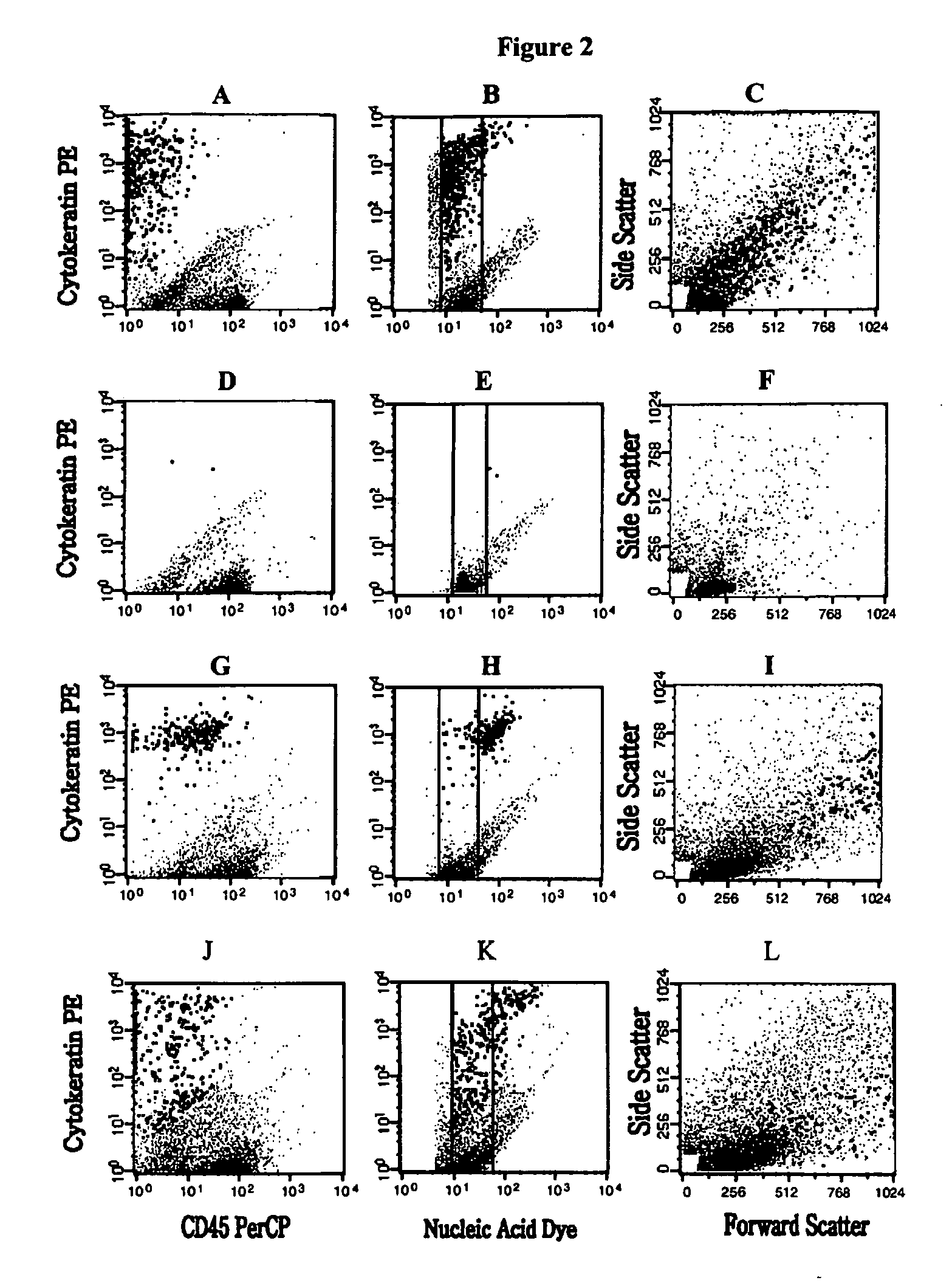
[0108] Samples were analyzed on a FACSCalibur flow cytometer equipped with a 488 nm argon ion laser (BDIS, San Jose, Calif.). Data acquisition was performed with CellQuest (BDIS, San Jose, Calif.) using a threshold on the fluorescence of the nucleic acid dye. The acquisition was halted after 8000 beads or 80% of the sample was analyzed. Multiparameter data analysis was performed on the listmode data (Paint-A-GatePRO, BDIS, San Jose, Calif.). Analysis criteria included size defined by forward light scatter, granularity defined by orthogonal light scatter, positive staining with the PE-labeled anti-cytokeratin MAb and no staining with the PerCP-labeled anti-CD45 Mab. For each sample, the number of events present in the region typical for epithelial cells was multiplied by 1.25 to account for the sample volume not analyzed by flow cytometry.

[0109]FIG. 2 Panels A, B and C shows the flow cytometric analysis of a blood sample of a patient with metastati...

example 2

Sample Analysis Via CellSpotter®

[0110] The CellSpotter® system consists of a microscope with a Mercury Arc Lamp mercury arc lamp, a 10× objective, a high resolution X ,Y, Z stage and a four filter cube changer. Excitation, dichroic and emission filters in each of four cubes were for DAPI 365 nm / 400 nm / 400 nm, for DiOC16 480 nm / 495 nm / 510 nm, for PE 546 nm / 560 nm / 580nm and for APC 620 nm / 660 nm / 700 nm. Images were acquired with a digital camera connected to a digital frame grabber. The surface of the chamber is 80.2 mm2 and 4 rows of 35 images for each of the 4 filters resulting in 560 images have to be acquired to cover the complete surface. The CellSpotter® acquisition program automatically determines the region over which the images are to be acquired, the number of images to acquire, the position of each image and the microscope focus to use at each position. All the images from a sample are logged into a directory that is unique to the specific sample identification. An algorith...

example 3

CTC in Prostate Cancer Patients

[0113] CTC were enumerated in 18 blood samples of prostate cancer patients and 27 samples from healthy individuals by both flow cytometry and CellSpotter®. The results shown in Table 1 were sorted by increasing number of intact CTC detected by CellSpotter®.

TABLE 1Enumeration of CTC by CellSpotter ® and flow cytometry in18 blood samples of prostate cancer patients and 27 samples fromhealthy individuals.FlowCellSpotter ®CytometryIntactDamagedTumor CellCK+CD45-PatientTumor CellsTumor CellsFragmentsNA+Sample#%#%#%# 1001501505 20021000012 3002661341 4002502500 5002295715 60036024018 7003385620 80074495610 9001376424210151520904111104405500122221116674132867144193691470516812120483683153223448134244875001635051122592493723173502142991441289242018742171122364181310Mean—4%—34%—62%—27 samples from healthy donorsMean0.040.964.960.7SD0.191.853.981.14Min0000Max17154

# - number CTC in 7.5 ml blood

% - percentage of all CTC detected by CellSpotter ®

[0114] In the C...


PUM

PropertyMeasurementUnit
Magnetic fieldaaaaaaaaaa
Biological propertiesaaaaaaaaaa
Immunogenicityaaaaaaaaaa

Abstract

The methods and reagents described in this invention are used to analyze circulating tumor cells, clusters, fragments, and debris. Analysis is performed with a number of platforms, including flow cytometry and the CellSpotter® fluorescent microscopy imaging system. Analyzing damaged cells has shown to be important. However, there are two sources of damage: in vivo and in vitro. Damage in vivo occurs by apoptosis, necrosis, or immune response. Damage in vitro occurs during sample acquisition, handling, transport, processing, or analysis. It is therefore desirable to confine, reduce, eliminate, or at least qualify in vitro damage to prevent it from interfering in analysis. Described herein are methods to diagnose, monitor, and screen disease based on circulating rare cells, including malignancy as determined by CTC, clusters, fragments, and debris. Also provided are kits for assaying biological specimens using these methods.

Description

PRIORITY INFORMATION [0001] This application claims priority under 35 USC §119(e) to U.S. Provisional Applications No. 60 / 314,151 filed 23 Aug. 2001, and No. 60 / 369,628 filed 3 Apr. 2002. Both of these applications are incorporated by reference herein.BACKGROUND OF THE INVENTION [0002] Many clinicians believe that cancer is an organ-confined disease in its early stages. However, it appears that this notion is incorrect, and cancer is often a systemic disease by the time it is first detected using methods currently available. There is evidence that primary cancers begin shedding neoplastic cells into the circulation at an early disease stage prior to the appearance of clinical manifestations. Upon vascularization of a tumor, tumor cells shed into the circulation may attach and colonize at distant sites to form metastases. These circulating tumor cells (CTC) contain markers not normally found in healthy individuals' cells, thus forming the basis for diagnosis and treatment of specific...

Claims


Application Information

Patent Timeline
IPC IPC(8): G01N33/53G01N33/543G01N33/567G01N33/574
CPCG01N33/54326G01N33/574Y10S436/813Y10T436/25375Y10T436/25125Y10T436/101666
Inventor RAO, GALLA CHANDRALARSON, CHRISTOPHERREPOLLET, MADELINERUTNER, HERMANTERSTAPPEN, LEON W.M.M.O'HARA, SHAWN MARKGROSS, STEVEN
Owner MENARINI SILICON BIOSYSTEMS SPA
Why Eureka
Try Eureka