Tissue electro-sectioning apparatus

a tissue electro-sectioning and apparatus technology, applied in the direction of instruments, analytical using chemical indicators, laboratory glassware, etc., can solve the problems of destroying immunohistochemical target antigens and mrna target sequences, unable to allow tissue electro-section in thin layers, and poor histologic quality sections, etc., to enhance the effect of sectioning, improve sectioning, and enhance the effect of invention

Inactive Publication Date: 2005-10-06
THE BOARD OF TRUSTEES OF THE UNIV OF ARKANSAS
View PDF41 Cites 38 Cited by
  • Summary
  • Abstract
  • Description
  • Claims
  • Application Information

AI Technical Summary

Benefits of technology

[0042] The effectiveness of the invention may be enhanced through various alternative embodiments. For example, media such as inorganic polar molecules may be added to a cooling bath in which the sectioning tool and tissue sample are immersed to improve sectioning via molecular rotation produced by an external electric field. The sectioning action is enhanced by specific sectioning tool design to create a more intense and localized field. As an alternative cooling mechanism, the buildup of heat may be limited by pulsation of the field. Whereas, in tissue, the electromagnetic field propagates six orders of magnitude faster than the thermal field, high electromagnetic fields can be induced for short times to break structural proteins bonds without thermal damage. Similarly, DC pulses rather than radio frequency (RF) pulses may be used to achieve tissue sectioning while minimizing heat buildup.
[0043] The device may be adapted to uses other than sectioning tissue for analysis purposes. The device may be adapted to do minor surgery where the pulses are sufficiently short (microseconds) that the field behaves like an RF field without affecting the nervous system adversely. The present invention offers substantial safety benefits, both in surgical and non-surgical applications. Microtomes and similar devices physically touch tissue and therefore can become contaminated. The present invention does not section tissue by direct mechanical action and therefore has less potential to contact and become contaminated by pathogens in the tissue. Furthermore, the intense electric field strength associated with the device will limit the viability of organisms in its vicinity. Additional protection may be obtained by circulating liquid in the cooling bath through sterilizers.
[0044] It is therefore an object of the present invention to provide for a device and method capable of producing ultra-thin sections of large, unfixed tissue specimens.
[0045] It is a further object of the present invention to provide for a device and method of producing ultra-thin sections of large, unfixed tissue specimens with preserved tissue architecture, antigenicity and mRNA content.
[0046] It is a further object of the present invention to provide for a device and method of producing ultra-thin sections of large, unfixed tissue specimens that are amenable to 2-D and 3-D molecular analysis.
[0047] It is a further object of the present invention to provide for an alternative device and method to intraoperative frozen section diagnosis.

Problems solved by technology

Although frozen sectioning is relatively fast and effective it produces poor histologic-quality sections due to ice-crystal artifacts.
Hence, none of these techniques allow tissue sectioning in thin layers that can be processed rapidly and produce high quality thin tissue specimens.
Unfortunately, traditional tissue fixation and processing prior to paraffin-embedding destroys many immunohistochemical target antigens and mRNA target sequences.
However, frozen sections are of poor histological quality due to ice-crystal artifacts, thus making them unsuitable for laser capture studies and 3-dimensional reconstruction of morphology or gene expression patterns.
Vibratome sectioning of frozen tissues is sometimes used in the research setting, but is not advantageous in the clinical setting.
Unfortunately, the vibratome cannot produce sections of soft tissues that are thin enough for high resolution work (4-10 μm) without rigidifying the specimen by freezing or fixing prior to sectioning.
Ice crystal formation cannot in practice be eliminated, because the extreme cooling rates needed to produce solid amorphous ice, or vitreous ice, cannot be realistically achieved.
Fields from any outside source, if they are sufficiently stronger than those employed in the bonds, can disrupt these interatomic forces, resulting in bond breakage and subsequent physical separation of the atoms.
This phenomenon is generally considered deleterious when materials lose their chemical and structural integrity in the presence of high fields.
For instance, when the gate oxide of a CMOS logic transistor on an integrated circuit is perforated by the normally benign fields used in its switching operation, it becomes “leaky” to the current used to toggle its logic operations, possibly to the point that it can no longer respond.
This “oxide breakdown” is a common failure mechanism in microprocessors, producing a sudden and irreversible loss of functionality.
The resulting region of tissue damage is, inevitably, of the same size.

Method used


Image

  • Tissue electro-sectioning apparatus
  • Tissue electro-sectioning apparatus
  • Tissue electro-sectioning apparatus

Examples


Embodiment Construction

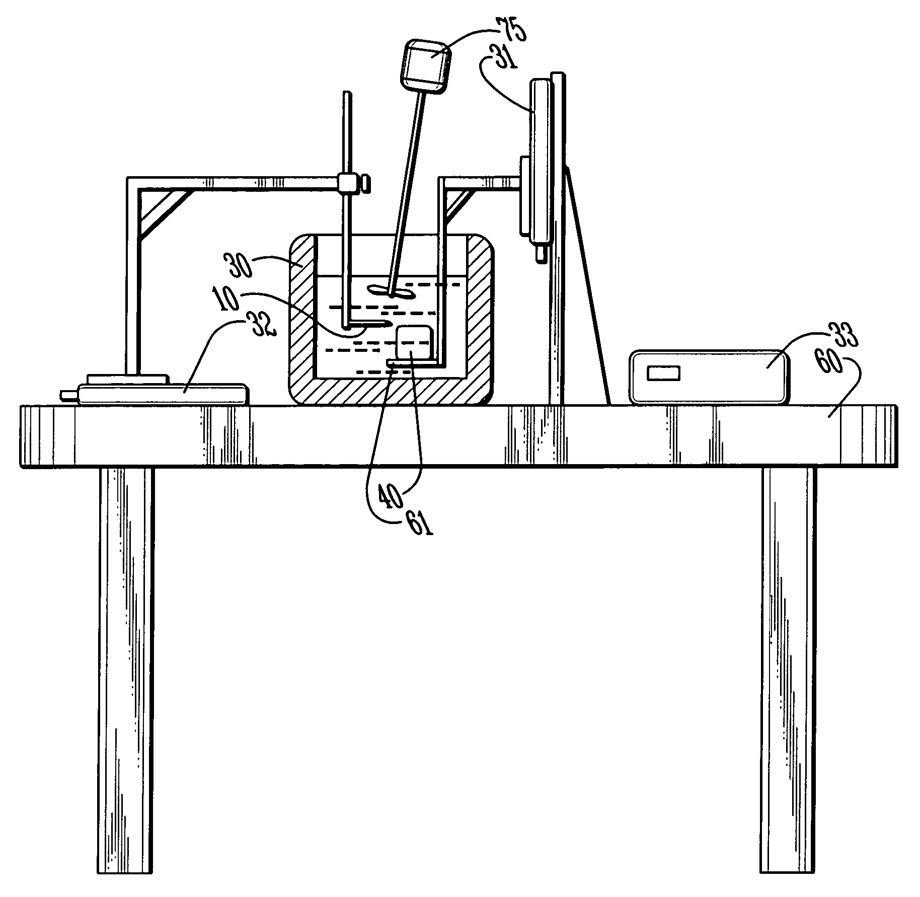
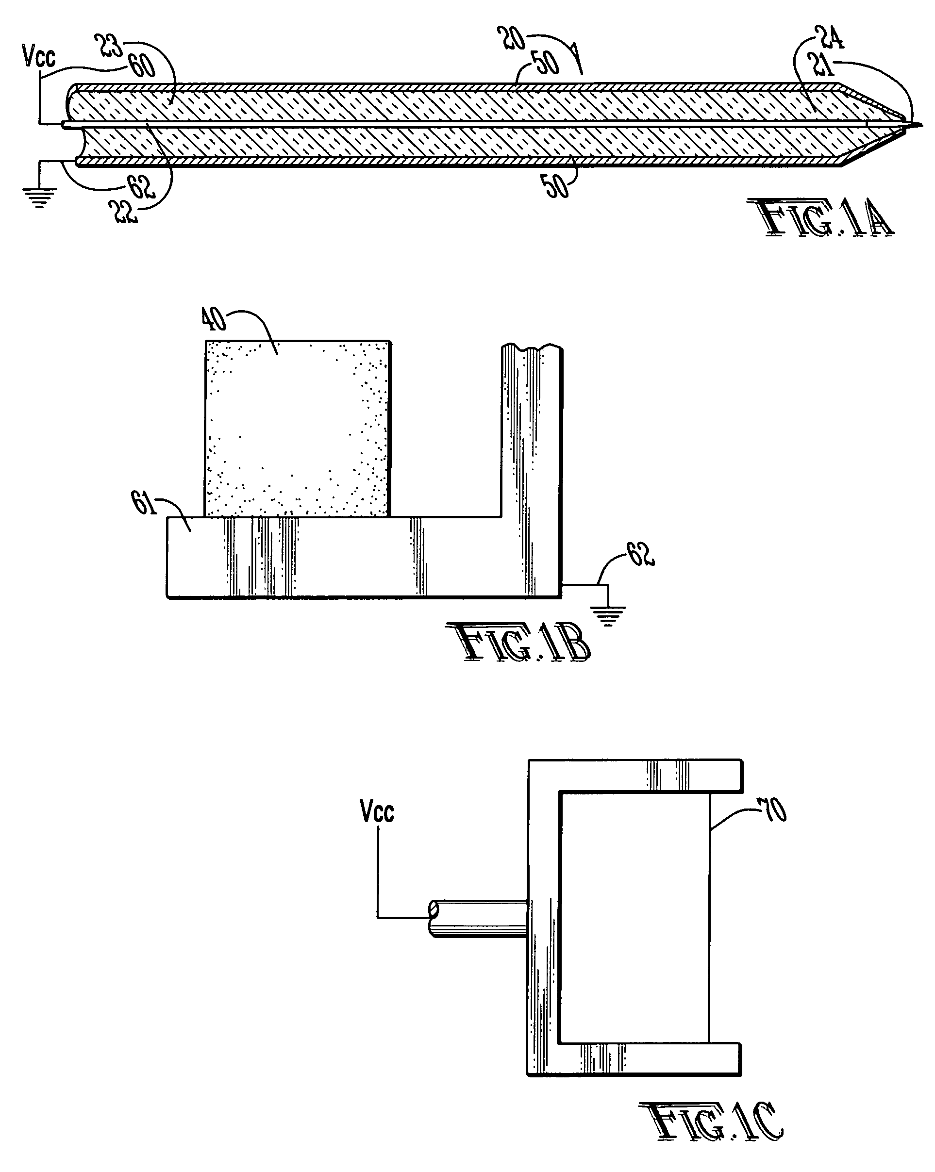
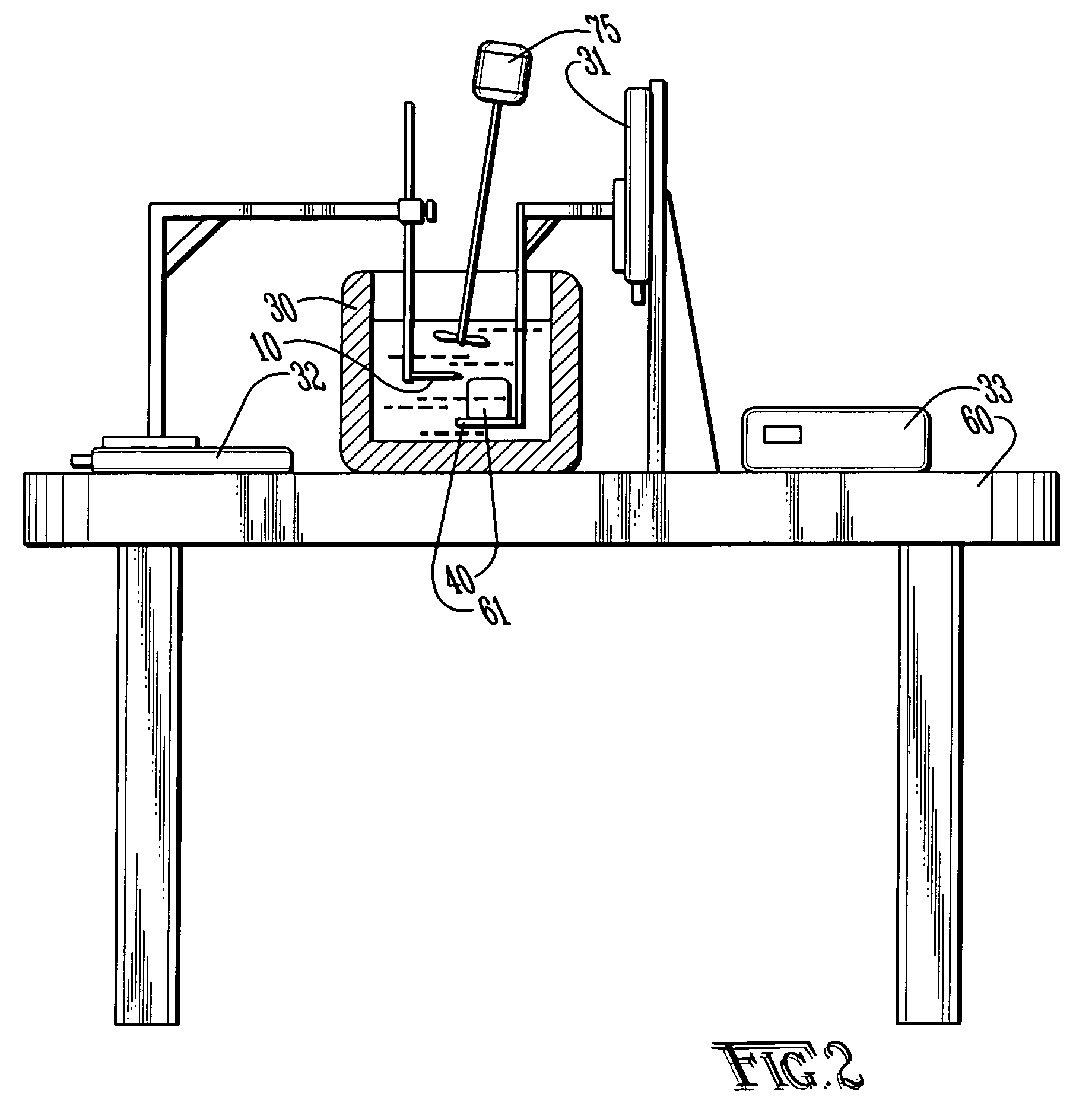
[0065] With reference to FIGS. 1A-C and 2, the preferred embodiments of the present invention may be described. The present invention is directed to satisfying the need to produce thin (4-10 μm) serial sections of large fresh tissue specimens that are suitable for high-resolution in situ protein / gene expression studies without ice artifacts or fixation-induced molecular damage.

[0066] Limitations of the existing sectioning techniques result from the fact that they rely on mechanical cutting, which in turn requires the tissue to be rigid. The present invention is a new approach to section tissue via an electro-sectioning process. In one embodiment, the sectioning tool is electrically biased with respect to the tissue sample which is submerged in a cooling bath. The sectioning tool may use focused radio frequency (RF) energy or pulsed DC. The present invention is directed to a method of using electro-sectioning to produce consecutive thin sections of fresh tissue for immunohistochemic...


PUM

PropertyMeasurementUnit
cryogenic temperaturesaaaaaaaaaa
thicknessaaaaaaaaaa
thicknessaaaaaaaaaa

Abstract

An apparatus for sectioning fresh unfixed tissue into very thin layers with preserved tissue architecture, antigenicity, mRNA content, and amenable to 3-D computer reconstruction without mechanical or thermal damage by employing a sectioning tool having an electrode with an intense focused electrical field at an edge. A computer controlled x-y-z translation stage moves the sectioning tool through the tissue as defined by a predetermined program. The sectioning tool produces consecutive thin sections of fresh tissue for immunohistochemical and nucleic acids analyses without mechanical or thermal damage, ultimately allowing high-resolution volumetric reconstruction of gene and protein expression patterns of large tissue specimens. The geometry of the sectioning tool is selected so as to produce a spatially localized electrical field of sufficient intensity to sever molecular bonds or propagate flaws in tissue without mechanical cutting.

Description

CROSS-REFERENCE TO RELATED APPLICATIONS [0001] This application is a continuation-in-part of co-pending U.S. patent application Ser. No. 10 / 816,016 filed Apr. 1, 2004, the disclosure of which is incorporated herein by reference.STATEMENT REGARDING FEDERALLY SPONSORED RESEARCH OR DEVELOPMENT [0002] Not applicable. BACKGROUND OF THE INVENTION [0003] 1. Field of the Invention [0004] The present invention relates to the automated sectioning or separating of consecutive thin sections of fresh tissues by electro-dissociation without mechanical force or thermal damage to the tissue. By shearing molecular bonds, damage to collateral tissue is avoided. Also, the need for tissue pre-processing along with the adverse effects associated with these preparation techniques is eliminated. The apparatus of the present invention may also be used for sectioning tissues in various surgical applications. [0005] 2. Brief Description of the Related Art [0006] The preparation of tissue for histopathologica...

Claims


Application Information

Patent Timeline
Patent Type & Authority Applications(United States)
IPC IPC(8): B01L3/00B23Q15/24C12M3/08G01N1/04G01N1/06
CPCC12M45/07G01N1/06G01N2001/045
Inventor SHAFIRSTEIN, GALFERGUSON, SCOTT L.FINK, LOUIS M.ULRICH, RICHARD K.
Owner THE BOARD OF TRUSTEES OF THE UNIV OF ARKANSAS
Why Eureka
Try Eureka