Neoepitope detection of disease using protein arrays

a protein array and disease technology, applied in the field of immunoassays for cancer diagnosis, can solve the problems of limited therapeutic options and survival rates, malignant and benign breast disease, and few advancements which can repeatably be used in diagnosing cancer, and achieve the effect of reducing morbidity and cost, and being sensitiv

Active Publication Date: 2005-10-27
WAYNE STATE UNIV
View PDF1 Cites 10 Cited by
  • Summary
  • Abstract
  • Description
  • Claims
  • Application Information

AI Technical Summary

Benefits of technology

[0018] Methods for detecting and measuring cancer markers have been recently revolutionized by the development of immunological assays, particularly by assays that utilize monoclonal antibody technology. Previously, many cancer markers could only be detected or measured using conventional biochemical assay methods, which generally require large test samples and are therefore unsuitable in most clinical applications. In contrast, modern immunoassay techniques can detect and measure cancer markers in relatively much smaller samples, particularly when monoclonal antibodies that specifically recognize a targeted marker protein are used. Accordingly, it is now routine to assay for the presence or absence, level, or activity of selected cancer markers by immunohistochemically staining tissue specimens obtained via conventional biopsy metho...

Problems solved by technology

However, there have been few advancements which can repeatably be used in diagnosing cancer prior to the existence of a tumor.
All too often, breast cancer is discovered at a stage that is too far advanced, when therapeutic options and survival rates are severely limited.
In addition to the problem of early detection, there remain serious problems in distinguishing between malignant and benign breast disease, in staging known breast cancers, and in differentiating between different types of breast cancers (eg. estrogen dependent versus non-estrogen dependent tumors).
Additionally, ovarian cancer has the highest mortality rate of all gynecological cancers and yet there is still no reliable and easy to administer screening test.
Despite advances in molecular biology, surgical oncology, and chemotherapy, the overall prognosis for ovarian cancer patients diagnosed at Stages II-IV remains poor.
There have been no effective and reliable tests developed to date.
Due to the low prevalence of ovarian cancer in the general population, this level of specificity is unacceptable for a realistic ovarian cancer diagnostic test.
Furthermore, some issues have arisen regarding the mass spectroscopy technology of protein profiling.
It has been reported that the data obtained by this technology are difficult to reproduce and that they may be biased by artifacts in sample preparation, storage and processing, and patient selection.
Previously, many cancer markers could only be detected or measured using conventional biochemical assay methods, which generally require large test samples and are therefore unsuitable in most clinical applications.
For the purpose of cancer evaluation, the use of conventional or needle biopsy samples for cancer marker assays is often undesirable, because a primary goal of such assays is to detect the cancer before it progresses to a palpable or detectable tumor stage.
Prior to this stage, biopsies are generally contraindicated, making early screening and low risk monitoring procedures employing such samples untenable.
Efforts to utilize serum samples for cancer marker assays have met with limited success, largely because the targeted markers are either not detectable in serum, or because telltale changes in the levels or activity of the markers cannot be monitored in serum.
In addition, the presence of cancer markers in serum probably occurs at the time of micro-metastasis, making serum assays less usef...

Method used


Image

  • Neoepitope detection of disease using protein arrays
  • Neoepitope detection of disease using protein arrays
  • Neoepitope detection of disease using protein arrays

Examples


example 1

[0111] The purpose of this study is to clone epitopes that are recognized by sera from women with ovarian cancer but not recognized by normal sera from unaffected women. As these epitopes are cloned, protein array assays are developed capable of detecting ovarian cancer at an early stage by analyzing antigens recognized in the sera of at risk women. Toward this end, individual sera were screened using these protein biochips to determine the antibody reactivity to each protein epitope. Antibody reactivity is detected that does not appear in control sera. The patients and control sera obtained for this study were used to calibrate the protein biochips and identify the most informative epitope-clones. The women were monitored for the appearance or reappearance of antibody reactivity and its correlation with tumor burden. By following the serum reactivity to tumor reactive new epitopes on the arrays of the phage display cDNA clones, the analysis of sera from women after their initial di...

example 2

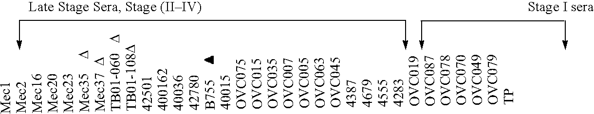
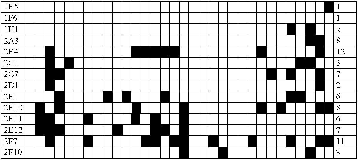
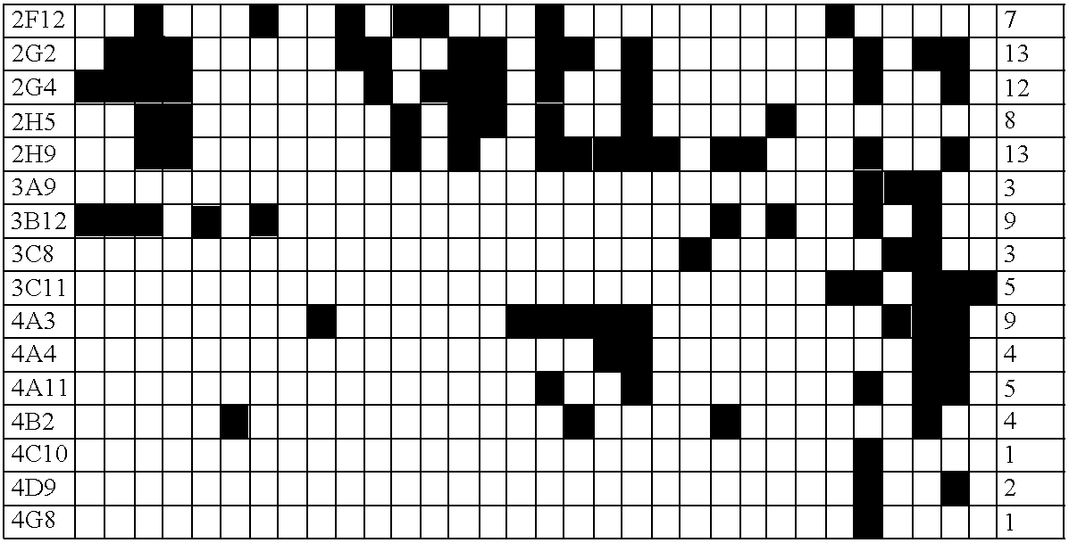
[0177] A strategy was developed for serological detection of large numbers of antigens indicative of the presence of cancer, thereby using the humoral immune system as a biosensor. The high-throughput selection strategy involved biopanning of an ovarian cancer phage display library using serum immunoglobulins from an ovarian cancer patient as bait. Protein macroarrays containing 480 of these selected antigen clones revealed 44 clones that interacted with immunoglobulins in sera from all (32 / 32) ovarian cancer patients, but not with sera from either healthy women (0 / 25) or patients having other benign or malignant gynecological diseases (0 / 14). An informative subset of 26 antigen clones was chosen based on the criterion that the serum from each of a group of 16 patients interacted with at least one of the clones. When another, independent group of 16 serum samples was used, all 16 samples interacted with one or more of the 26 clones, and none from 12 healthy women. The process ...


PUM

PropertyMeasurementUnit
Volumeaaaaaaaaaa
Volumeaaaaaaaaaa
Volumeaaaaaaaaaa

Abstract

A biosensor for use in detecting the presence of diseases, the biosensor comprising a detector for detecting a presence of at least one marker indicative of a specific disease. A method of determining efficacy of a pharmaceutical for treating a disease or staging disease by administering a pharmaceutical to a sample containing markers for a disease, detecting the amount of at least one marker of the disease in the sample, and analyzing the amount of the marker in the sample, whereby the amount of marker correlates to pharmaceutical efficacy or disease stage. Markers for gynecological disease selected from the list in Table 8. An immuno-imaging agent comprising labeled antibodies, whereby the labeled antibodies are isolated and reactive to proteins overexpressed in vivo. Informatics software for analyzing the arrays of claim 17, the software including analyzing means for analyzing the arrays.

Description

CROSS-REFERENCE TO RELATED APPLICATIONS [0001] This Continuation-In-Part Application claims priority to U.S. patent application Ser. No. 10 / 004,587, filed Dec. 4, 2001, which is incorporated herein by reference.GRANT INFORMATION [0002] Research in this application was supported in part by a grant from the National Institute of Health (NIH Grant No. IR21CA100740-01). The Government has certain rights in the invention.BACKGROUND OF THE INVENTION [0003] 1. Technical Field [0004] The present invention relates to an assay and method for diagnosing disease. More specifically, the present invention relates to an immunoassay for use in diagnosing cancer. [0005] 2. Background Art [0006] It is commonly known in the art that genetic mutations can be used for detecting cancer. For example, the tumorigenic process leading to colorectal carcinoma formation involves multiple genetic alterations (Fearon et al (1990) Cell 61, 759-767). Tumor suppressor genes such as p53, DCC and APC are frequently i...

Claims


Application Information

Patent Timeline
IPC IPC(8): G01N33/68
CPCG01N33/57411G01N33/57415G01N33/57449G01N2800/364G01N33/689G01N2800/36G01N33/68Y02A90/10
Inventor TAINSKY, MICHAELDRAGHICI, SORINCHATTERJEE, MADHUMITA
Owner WAYNE STATE UNIV
Why Eureka
Try Eureka