
Genotype specific detection of Chlamydophila psittaci

a technology of chlamydia psittaci and genotype, applied in the field of quantitative detection of chlamydia psittaci genotypes, can solve the problems of inability to meet requirements, poor specificity and sensitivity of cytological staining, and inability to use as a rapid preliminary investigation method

Inactive Publication Date: 2006-01-12
UNIV GENT
View PDF3 Cites 2 Cited by
  • Summary
  • Abstract
  • Description
  • Claims
  • Application Information

AI Technical Summary

Benefits of technology

[0006] In a first aspect, the invention relates to an ex vivo or in vitro method for the identification of the presence of one or more genotypes of Cp. psittaci in a sample. Thus the present invention provides a method for the determination of the presence of Cp. psittaci in a sample as well as a method to specifically identify amongst the different Cp. psittaci genotypes, which genotype is present in the sample, thus allowing the determination of the actively infecting agent, even when the samples is taken from an animal or human subject which has previously been infected with Cp. psittaci.
[0021] The methods and kits of the present invention, provide several advantages over the current detection and / or identification methods, such as easy sample collection methods, simple transport and storage requirements of the bacterial sample, rapid results, the possibility for automatisation, and a high sensitivity and specificity.
[0022] The present invention allows the genotype-specific detection of Cp. psittaci which is based on the identification of the presence of DNA of the bacterium. It allows the detection of the presence or absence of Cp. psittaci bacteria in a sample, independently of whether or not that sample comprises antibodies against bacteria of a previous infection. Thus, contrary to serotypic detection methods, the methods and kits of the present invention allow the detection and / or identification of an active infection.
[0024] The present invention further makes it possible to determine or to confirm and follow-up the relationship between the occurrence of a certain genotype and the pathogenicity thereof. DETAILED DESCRIPTION Definitions
[0040] According to a specific embodiment, the methods of the invention are quantitative real-time PCR assays. It is demonstrated herein that the assays of the invention meet the criteria proposed for a validated assay, as both new real-time PCR assays were compared with other assays such as ompA sequencing, ompA RFLP and MOMP serotyping. Real-time PCR technology offers a new diagnostic approach which allows amplicon quantification in one step via specific hybridisation, without the need to open tubes, minimising the risk of cross-contamination for further experiments in this way.
[0050] As detailed above, according to a particular embodiment, the method of detection of the binding is PCR. In a preferred embodiment the binding of a first and / or binding of a second oligonucleotide is determined by PCR amplification with a forward and a reverse primer, wherein the forward and reverse primer are located about 1, 5, 10, 20, 50 to 100 bp 3′ and 5′ from the first or second oligonucleotide. The PCR may be real-time PCR. Multiplexing can be used to reduce time.

Problems solved by technology

Cytological stainings have poor sensitivity and specificity and can only be used as a rapid preliminary investigation method.
The main disadvantage of isolation is the need for viable bacteria.
This means special requirements for collection and storage of samples, requirements that cannot always be fulfilled when collecting field samples.
In addition, isolation is time-consuming and costly and can only be performed in laboratories with a specific biosafety level since Cp. psittaci is a zoonotic agent which spreads by aerosol.
The current rapid antigen-detection methods are not recommended for demonstrating Cp. psittaci in individual birds because of shortcomings in either sensitivity or specificity.
Serology is not particularly useful in diagnosing an active Cp. psittaci infection in birds because of the high prevalence of this infection in birds and the long-term (up to several months) persistence of anti-Cp. psittaci antibodies.
In addition, antibody detection based on using whole organisms, LPS (LipoPolySaccharides) or outer membrane fractions can generate false positives due to the presence of antibodies cross reactive to the Cp. psittaci LPS or heat shock proteins.
Importantly, current Cp. psittaci antibody detection tests cannot be used for demonstrating a Cp. psittaci infection in man, as humans can also become infected with other members of the Chlamydiaceae as Chlamydia trachoinatis, Chlamydophila pneumonieae (formerly Chlamydia pneumoniae) and Chlamydophila abortus (formerly psittaci serotype 1) which can cause false-positive results.
Diagnosis of infection with Cp. psittaci has been difficult and cumbersome.
While RFLP analysis of the ompA gene encoding the MOMP is allows specific detection of the Cp. psittaci genotypes, restriction patterns in RFLP are sometimes difficult to analyse, and ompA amplification cannot always be carried out directly on clinical samples.
Moreover, this method requires the amplification of the entire 1200 bp OmpA gene which often fails when a limited amount of DNA is available.
Indirect micro-immunofluorescence (IMIF) with monoclonal antibodies always requires culturing, and is therefore expensive and labour-intensive and is definitely less sensitive then genotyping by means of RFLP or whole ompA sequence analysis.
Besides the interspecies diagnosis problems in the serological assays and the intraspecies difficulties when dealing with mixed infections in RFLP or serotyping, these tests all have the problem that they do not provide information about the actual number of infectious particles in the specimen, making it also difficult or impossible to follow up a treatment or to track down the origin of an infection.

Method used


Image

  • Genotype specific detection of Chlamydophila psittaci
  • Genotype specific detection of Chlamydophila psittaci
  • Genotype specific detection of Chlamydophila psittaci

Examples


example 1

General Methodology

[0067] Isolates and cell cultures. Cp. psittaci genotype A to F plus E / B reference strains 90 / 1051, 41A12, GD, 7344 / 2, 3759 / 2, 7778B15 and WS / RT / E30 (Table 1), were grown in Buffalo Green Monkey (BGM) cells. Infected monolayers were disrupted by freezing and thawing followed by ultrasonic treatment for 1 minute in a tabletop sonicator (Bransonic 12, BIOMEDevice, San Pablo, Calif., USA). The cell culture harvest was centrifuged for 10 min (1,000×g, 4° C.) to remove cellular fragments and subsequently concentrated by ultracentrifugation for 1 hour (45,000×g, 4° C.). Bacterial pellets were resuspended in Sucrose Phosphate Glutamate buffer (SPG) (218 mM sucrose, 38 mM KH2PO4, 7 mM K2HPO4, 5 mM L-glutamic acid) at a volume of 1 to 100 of the original culture volume and stored at −80° C. until use.

[0068] DNA extraction. Genomic DNA was prepared as follows. 200 μl cell culture harvest was centrifuged for 30 min at RT (16,000×g). The supernatant was discarded and the pe...

example 2

Genotype Specific Identification of Cp. psittaci

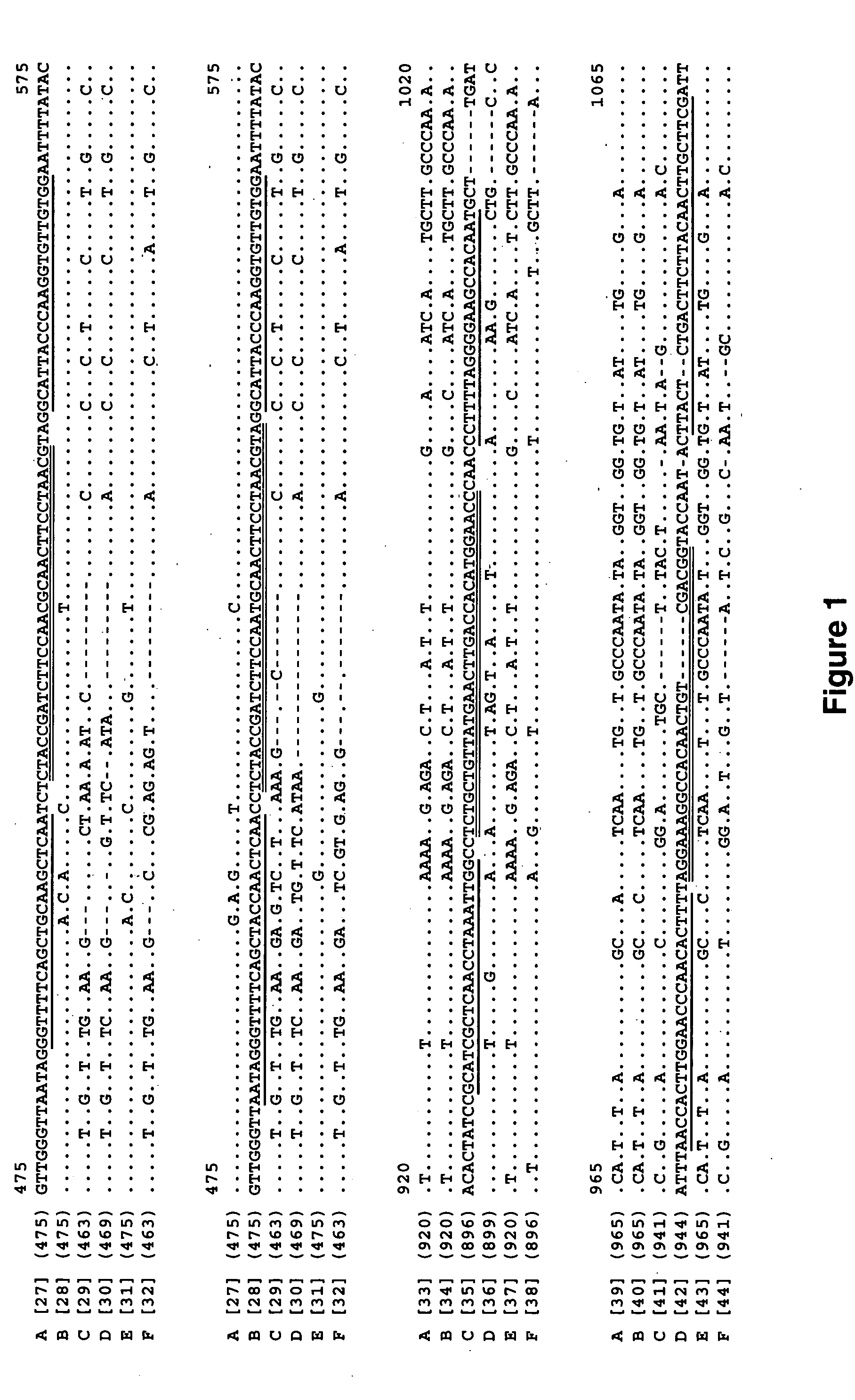
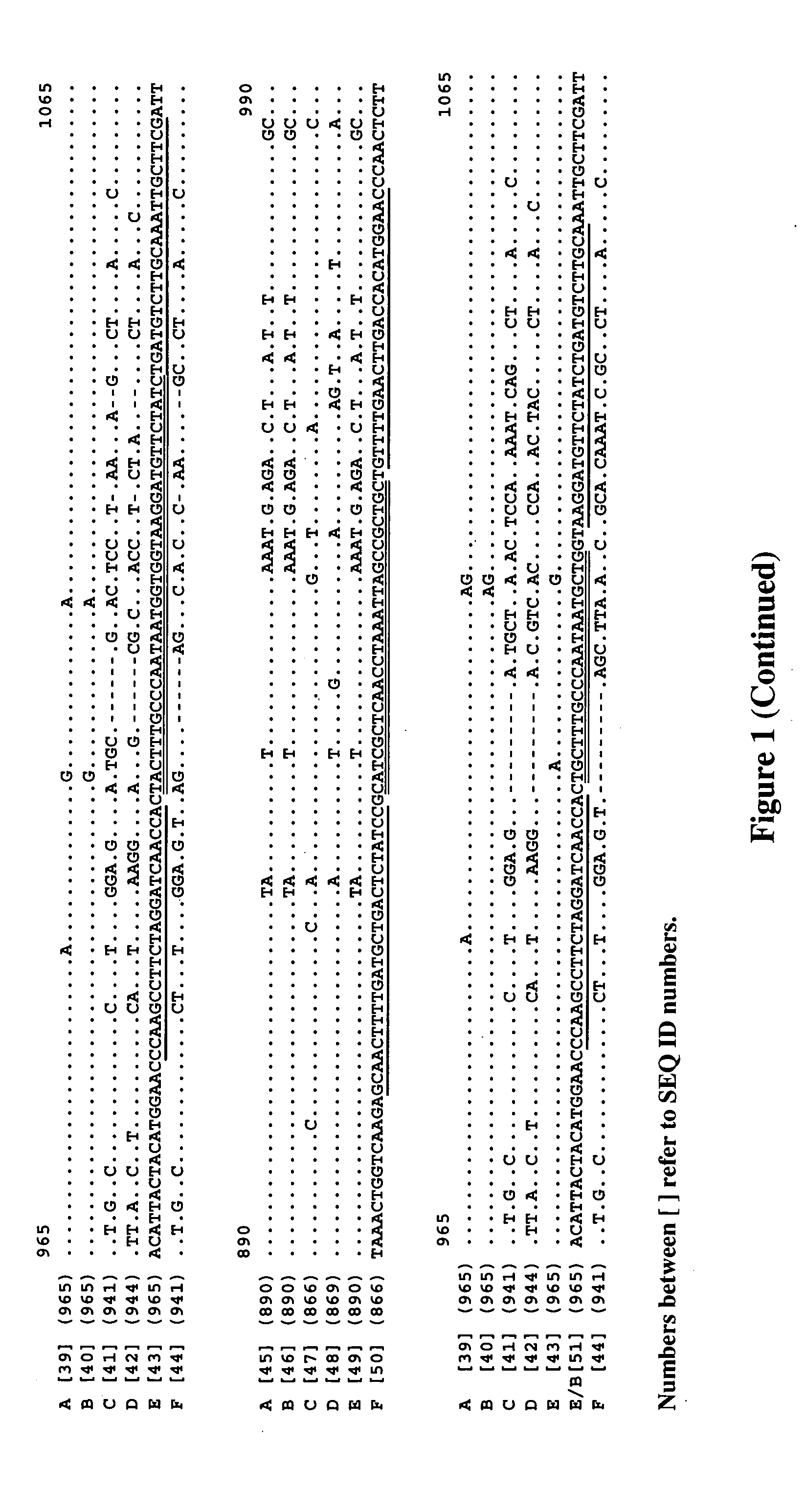
[0078] The present invention demonstrates for the fist time the use of real time PCR technology to detect seven different avian Cp. psittaci genotypes in human and animal samples and offers the possibility to discover new Cp. psittaci genotypes.

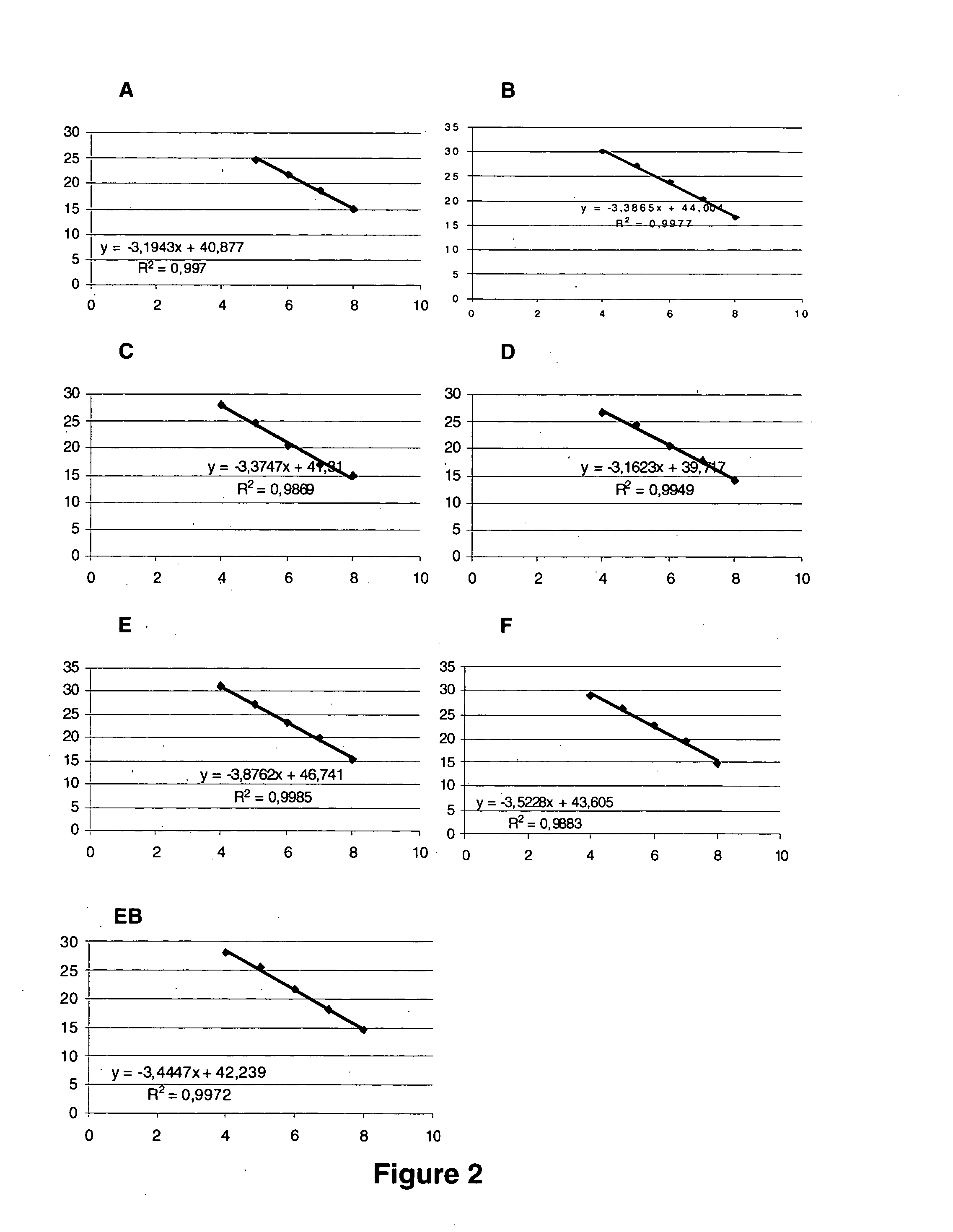
[0079] Using genotype specific reference plasmids, all seven PCR's (A to EB) are able to detect 10 copies of plasmid per μl. Standard curves could be made from 108 to 105 copies per μl with almost ideal slopes around −3,3 and correlation coefficients higher then 98,5% (FIG. 2). The highest dilutions were not taken into account for the regression because the reproducibility was too low, they reached the threshold around the same cycle or only after cycle 40.

[0080] The competitors which have been used in the PCR methods of the present invention are oligonucleotides without a fluorescent signal that go in competition with probes that bind to the target sequence. In Fluorescence In Situ Hybridiza...

example 3

Genotype Determination

[0082] Genotype A. The Chlamydophila psittaci genotype A specific competitor for binding on genotype B (CpPsGAScomB) [SEQ ID NO:8] has to be added to the reaction mixture to prevent false positive results if genotype B is possibly present in the sample. When added, the competitor will bind the genotype B DNA, leaving the probe only the binding site on genotype A, if present. As the competitor sequence is complementary to the genotype B sequence, the affinity is higher for this genotype, while the probe off course preferentially binds genotype A.

[0083] Genotype B. In genotype B determination, an elevated temperature can enhance the probe specificity: a specific reactions with genotypes E and EB disappear when the reaction is carried out at 63° C. in stead of 60° C. Addition of the competitor for genotype A material CpPsGBScomA [SEQ ID NO:7] will prevent false positive reactions if genotype A material is present.

[0084] Genotype E. Addition of both CpPsGEScomA / ...


PUM

PropertyMeasurementUnit
temperaturesaaaaaaaaaa

Abstract

The present invention describes novel methods for the specific detection and identification of Chlamydophila psittaci genotypes. According to one embodiment the method makes use of quantitative PCR with internal probes and optionally competitor probes which increase specificity. The invention also describes a strain of Cp. psittaci with a novel genotype EB and methods to distinguish said novel genotype from previously identified genotypes.

Description

CROSS-REFERENCE TO RELATED APPLICATIONS [0001] This application claims benefit of U.S. provisional patent application Ser. No. 60 / 584,725, filed Jun. 30, 2004, the disclosure of which is hereby incorporated by reference.FIELD OF THE INVENTION [0002] The present invention relates to the qualitative and quantitative detection of genotypes of Chlamydiaceae as well as to the detection and diagnosis of bacterial infections in mammals, including humans and birds. The invention further relates to the detection of a novel strain of an infectious bacterium. BACKGROUND OF THE INVENTION [0003] Bacteria in the family of the Chlamydiaceae are obligate intracellular parasites of eukaryotic cells. In animals, Chlamydophilae are capable of inducing a broad spectrum of symptoms like enteritis, urogenital infection, abortion, pneumonia, polyarthiritis, polyserositis, encephalitis and mastitis. Chlamydophila (Cp.) psittaci (formerly Chlamydia psittaci) causes respiratory diseases in birds and psittaco...

Claims


Application Information

Patent Timeline
Patent Type & Authority Applications(United States)
IPC IPC(8): C12Q1/68
CPCC12Q1/689
Inventor VANROMPAY, DAISY
Owner UNIV GENT
Learn More