DNA sequences encoding dystrophin minigenes and methods of use thereof
a dystrophin minigene and dna sequence technology, applied in the field of new dystrophin minigenes, can solve the problems of loss of dap complex, degenerative pathology, chronic muscle damage, etc., and achieve the effects of reducing size, efficient and stable correction of both biochemical and physiological defects, and convenient and less time-consuming
- Summary
- Abstract
- Description
- Claims
- Application Information
AI Technical Summary
Benefits of technology
Problems solved by technology
Method used
Image
Examples
example 1
Dystrophin Minigenes and AAV Vectors Carrying the Minigenes
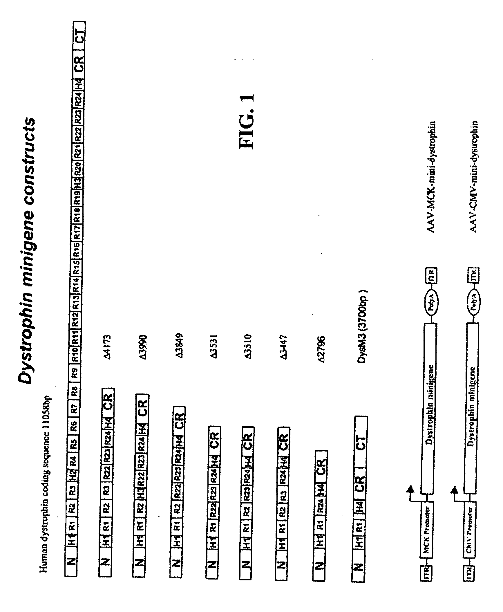
[0069] This example describes the construction of highly truncated dystrophin minigenes and AAV vectors carrying the minigenes. The dystrophin minigene constructs were made mainly by PCR cloning method using Pfu polymerase (Stratagene, Calif.) and human dystrophin cDNA (GenBank # NM 004006) as the template. For consistency, the numbering of the nucleotide only includes the 11,058 bp dystrophin protein coding sequence (SEQ ID NO: 1).
[0070] As depicted in FIG. 1, dystrophin minigene .DELTA.4173 (SEQ ID NO:2) contains nucleotides 1-1992 (N-terminus, hinge H1 and rods R1, R2 & R3, SEQ ID NO:3) and 8059-10227 (rods R22, R23 & R24, hinge H4 and CR domain, SEQ ID NO:4) and 11047-11058 (the last 3 amino acids of dystrophin, SEQ ID NO:5).
[0071] Dystrophin minigene .DELTA.3990 (SEQ ID NO:6) contains nucleotides 1-1668 (N-terminus, hinge H1 and rods R1 & R2, SEQ ID NO:7), 7270-7410 (hinge H3, SEQ ID NO:8), 8059-10227 (rods R22, R23...
example 2
[0080] Restoration of DAP Complexes
[0081] This example describes whether dystrophin minigene products still retain the major biochemical functionality including submembrane localization and interaction with dystrophin associated protein (DAP) complexes. Healthy C57 / B10 mice and dystrophic mdx mice were purchased from The Jackson Laboratory (Bar Harbor, Me.). Ten-day old mdx pups or 50-day old mdx adult mice were injected into the hindleg gastrocnemius muscle with 50.mu.l (5.times.10.sup.10 GC) of different AAV mini-dystrophin vectors.
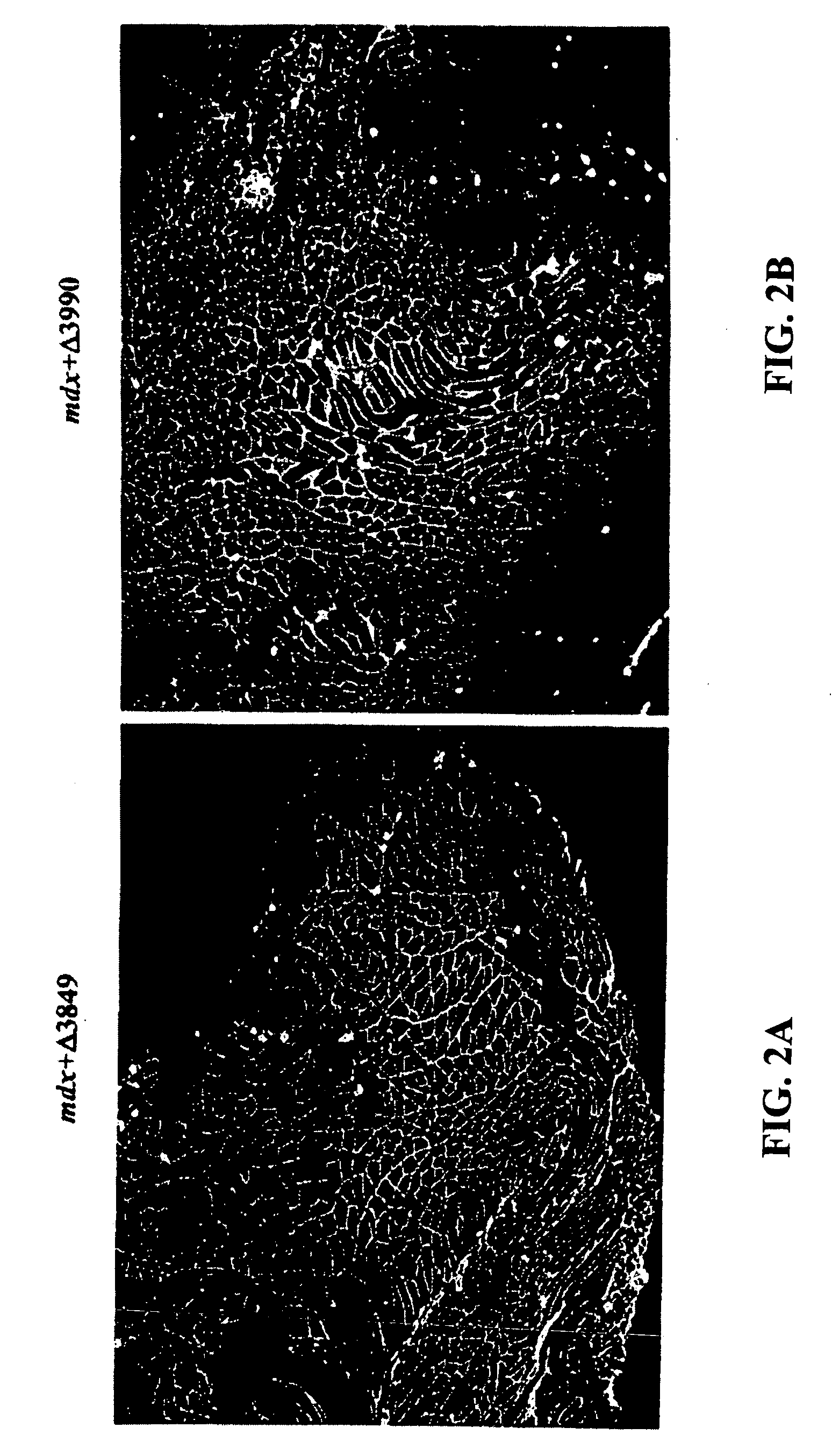
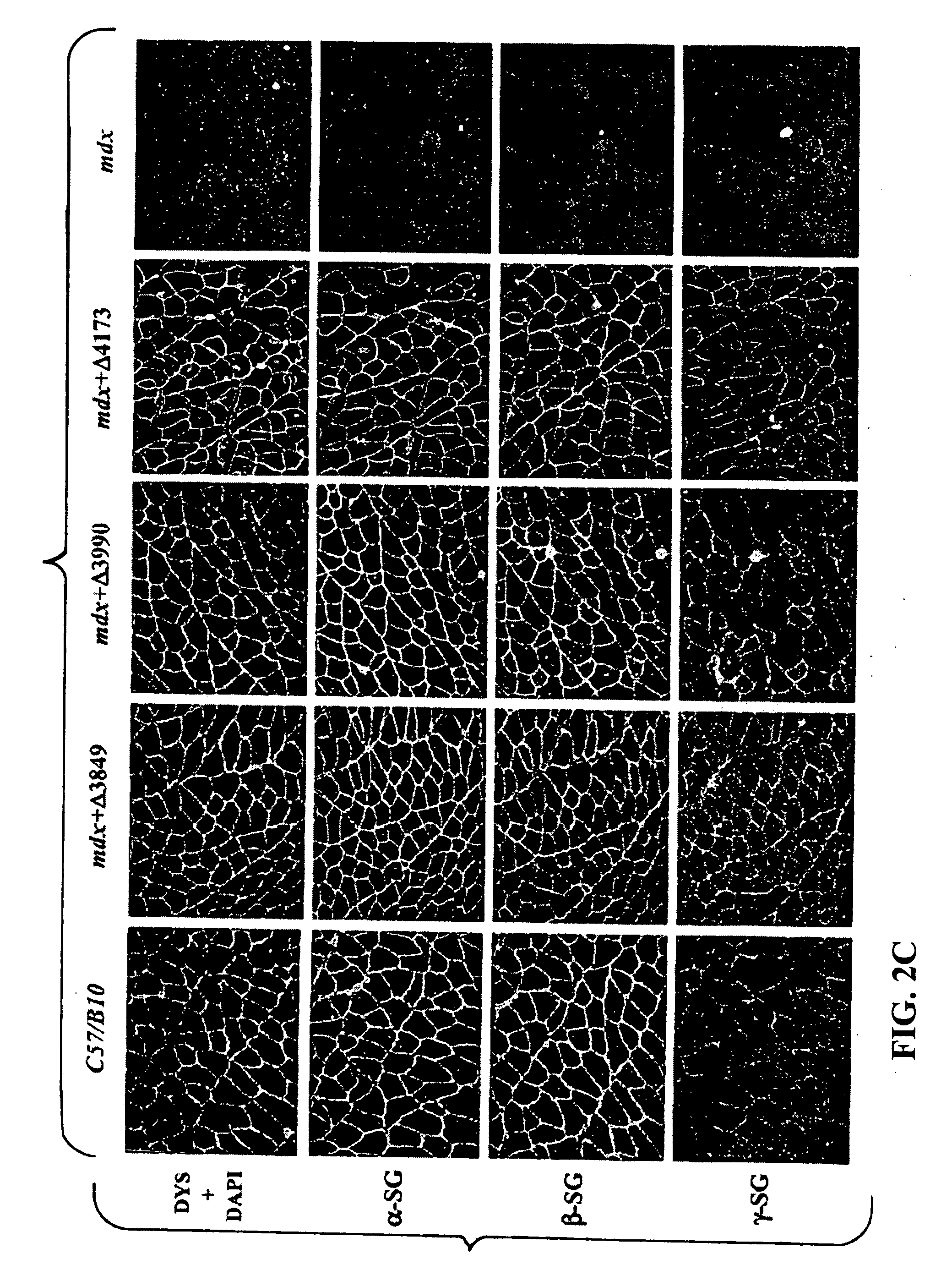
[0082] At three months and six months after vector injection, the muscles were collected for evaluation of mini-dystrophin expression and biochemical restoration of the DAP complexes, which were absent due to the primary deficiency of dystrophin. IF staining on thin sections of AAV treated muscles, using an antibody (Dys3) specific to human dystrophin, revealed widespread vector transduction and correct submembrane location of the mini-dystrophins in ...
example 3
[0084] Amelioration of Dystrophic Pathology
[0085] This example demonstrates that dystrophin minigene products can protect muscle from the pathological phenotypes. The onset of the pathology in mdx mice starts at around three weeks of age with massive waves of myofiber degeneration / regeneration. This process is characterized by the presence of central nuclei in myofibers, a primary pathological sign of muscular dystrophies. The absence or reduction of central nucleation after gene therapy would suggest that the therapy is successful. Therefore, we initially chose to test the AAV mini-dystrophin constructs in young mdx mice (10-day old) before the onset of central nucleation, to see whether muscle degeneration / regeneration can be prevented.
[0086] Histological examination of the mdx muscles at 3 and 6 months after AAV mini-dystrophin (containing more than 2 rod domains) treatment, which was prior to the onset of central nucleation, showed nearly exclusive (.about.98%) peripheral nucl...
PUM
 Abstract
Description
Claims
Application Information
 - R&D
- Intellectual Property
- Life Sciences
- Materials
- Tech Scout
- Unparalleled Data Quality
- Higher Quality Content
- 60% Fewer Hallucinations
Browse by: Latest US Patents, China's latest patents, Technical Efficacy Thesaurus, Application Domain, Technology Topic, Popular Technical Reports.
© 2025 PatSnap. All rights reserved.Legal|Privacy policy|Modern Slavery Act Transparency Statement|Sitemap|About US| Contact US: help@patsnap.com


