Substrate recognition by differentiable human mesenchymal stem cells

a mesenchymal stem cell and subset technology, applied in the field of three-dimensional nanofiber matrix, can solve the problems of poor quality reparative tissue, hyaline cartilage formation, and failure to restore a normal articular surface, and achieve the effect of tissue engineering therapy

Inactive Publication Date: 2006-06-15
NEW JERSEY INSTITUTE OF TECHNOLOGY
View PDF0 Cites 51 Cited by
  • Summary
  • Abstract
  • Description
  • Claims
  • Application Information

AI Technical Summary

Benefits of technology

[0020] The composition and methods of the invention provide microscale and nanoscale fibrous scaffolds as a substrate for human mesenchymal cells that have the potential to differentiate in situ into mature cell phenotypes. This combination may provide an effective tissue engineering therapy.

Problems solved by technology

Orthopedic management of lesions to articular cartilage remains a persistent problem for the orthopedist and patient because articular cartilage has a limited intrinsic ability to heal.
Implantation of scaffolds alone leads to a poor quality reparative tissue.
Chondrocytes implanted either alone or in combination with a scaffold have failed to restore a normal articular surface, and the hyaline cartilage formed early on in response to chondrocyte-containing scaffolds seems to deteriorate with time.
However, the use of MSCs in combination with biomaterials of varying architectures that may closely mimic the physical architecture of the native extracellular matrix during development to direct chondrogenic differentiation has yet to be investigated.
Most work to develop scaffold materials for tissue engineering, however, has relied on large diameter fibers, which do not mimic the morphological characteristics of the native fibrils of the extracellular matrix.
While the patent teaches that isolated cells must be undifferentiated and suggests that the cell delivery system when positioned at a desired location for cell delivery to a mammal can guide the development and shape of new tissue, it provides no guidance on how to do so.

Method used


Image

  • Substrate recognition by differentiable human mesenchymal stem cells
  • Substrate recognition by differentiable human mesenchymal stem cells
  • Substrate recognition by differentiable human mesenchymal stem cells

Examples


example 1

Substrate Recognition by Differentiable Human MSC Cells

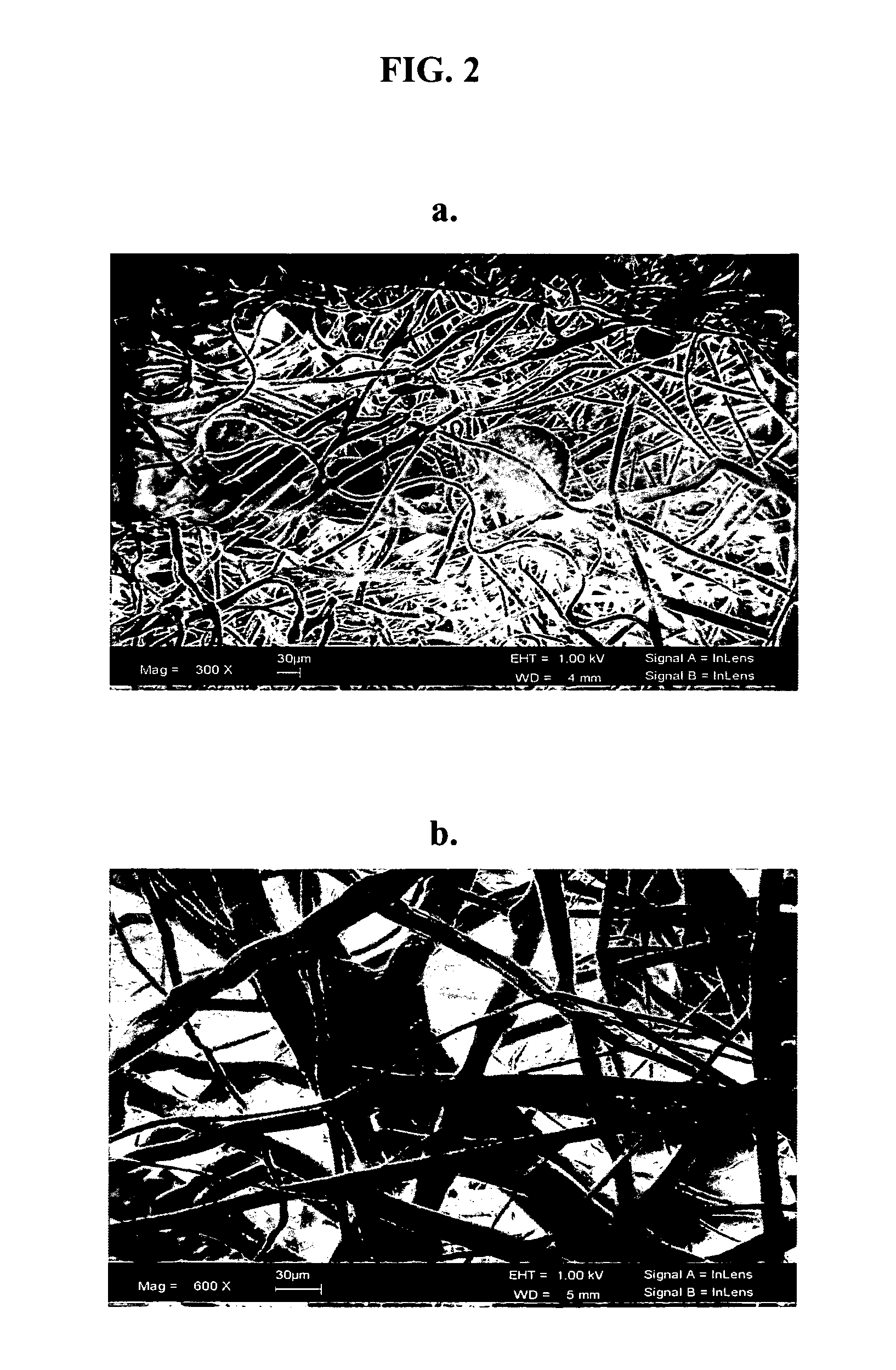
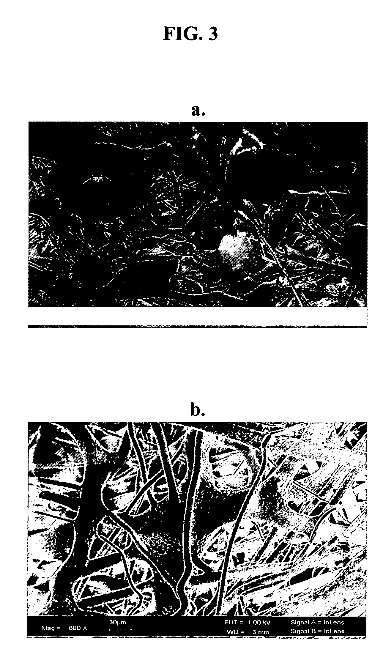
[0035] We have evaluated two commonly used polymeric compositions in the field of tissue engineering, namely poly-L-lactic acid (PLLA) and poly-D,L-lactide glycolide (PLGA) at the nano- and microscale fiber diameter range for their ability to support mesenchymal stem cell attachment. We then compared the morphology and growth characteristics of the attached cells on these substrates.

[0036] The term “nanoscale fiber” generally refers to fibers whose diameter ranges from about 1 to about 1000 nanometers. Nanoscale fibers whose average diameter ranges from about 400 to about 500 nanometers are most preferred. The term “microscale fiber” generally refers to fibers whose diameter ranges from about 1 to about 1000 micrometers. Microscale fibers whose diameter ranges from about 1 to about 100 micrometers are preferred, and microscale fibers whose diameter averages from about 10 micrometers to about 20 micrometers are most preferred. ...

example 2

Cell Proliferation

[0061] Human MSCs were isolated from adult, human whole bone marrow according to standard techniques and were seeded onto polymer scaffolds having the composition of PLLA or PLGA, each having fiber diameters on the micron scale (LF) or nano scale (SF) and grown in standard growth medium (DMEM, 10% fetal bovine serum, 1% antibiotic / antimycotic) for 14 days. Cell proliferation was assessed using Vybrant's MTT Cell Proliferation Assay Kit (Molecular Probes, Inc.).

[0062] The growth curves for cells seeded onto PLLA micron scale fibers (PLLA-LF), PLLA nano-scale fibers (PLLA-SF), PLGA micron scale fibers (PLGA-LF) and PLGA nano-scale fibers (PLGASF) are shown in FIG. 4. Cells grown on PLLA and PLGA micron scale and nanoscale fibers showed good comparable growth characteristics as measured by the MTT assay. No significant differences in human MSC proliferation were detected between PLLA and PLGA micron and nanoscale fibers.

example 3

PLLA / PLGA Micron and Nano Fiber Diameter Scaffolds Support Osteogenic Differentiation

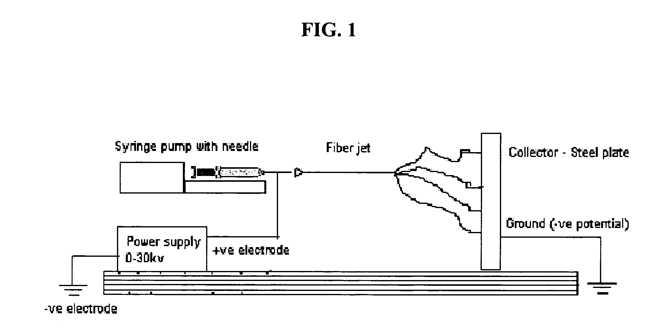
[0063] Scaffolds were created by the process of electrospinning, and human mesenchymal stem cells were grown on the scaffolds to determine whether PLLA / PLGA micron and nano-sized scaffolds support osteogenic differentiation.

[0064] Materials and Methods

[0065] hMSCs were grown in control medium (DMEM, 10% FBS, 1% antibiotic) or osteogenic inducing medium (OS) (Control medium with 100 nM dexamethasone, 10 mM b-glycerophosphate, 0.05 mM L-ascorbic acid-2-phosphate) on PLLA or PLGA scaffolds having micron or nano sized fiber diameters.

[0066] The four scaffolds, PLLA microfiber (“PLF”) scaffolds, PLLA nanofiber scaffolds (“PSF”), PLGA microfiber scaffolds (“GLF”), and PLGA nanofiber scaffolds (“GSF”), were created by electrospinning.

[0067] On the day of cell seeding, scaffolds were soaked first in 100% ethanol for 20 minutes, then three times in PBS, 20 minutes each, for sterilization. Scaffolds then...


PUM

PropertyMeasurementUnit
sizeaaaaaaaaaa
sizeaaaaaaaaaa
diameteraaaaaaaaaa

Abstract

The invention described herein provides a structure for growing isolated differentiable human mesenchymal cells, which includes a three-dimensional matrix of fibers. The matrix serves as an implantable scaffolding for delivery of differentiable human mesenchymal cells in tissue engineering. The invention further provides compositions that contain the three-dimensional matrix of fibers seeded with isolated differentiable human mesenchymal cells, wherein the matrix forms a supporting scaffold for growing the isolated differentiable human mesenchymal cells, and wherein the differentiable human mesenchymal cells differentiate into a mature cell phenotype. The invention further provides methods of preparing the implantable nanofiber matrix scaffolding seeded with differentiable human mesenchymal cells for use in tissue engineering.

Description

CROSS REFERENCE TO RELATED APPLICATION [0001] This application claims the benefit of U.S. Provisional Patent Application Ser. No. 60 / 633,233, filed Dec. 3, 2004, and entitled Substrate Recognition by Differentiable Human Mesenchymal Stem Cells, the entire disclosure of which is incorporated herein by reference.GOVERNMENT SUPPORT [0002] This work is supported at least in part by grants from the National Science Foundation to Dr. Arinzeh. The government may have certain rights in this invention.FIELD OF THE INVENTION [0003] The present invention relates to compositions and methods comprising a three-dimensional nanofiber matrix of synthetic polymers. The synthetic nanofiber matrix can serve as a scaffold / substrate for delivery of differentiable human mesenchymal cells for tissue engineering applications. BACKGROUND OF THE INVENTION [0004] Orthopedic management of lesions to articular cartilage remains a persistent problem for the orthopedist and patient because articular cartilage has...

Claims


Application Information

Patent Timeline
Patent Type & Authority Applications(United States)
IPC IPC(8): C12N5/08C12N5/02C12N5/077C12N5/0775
CPCA61K2035/124A61L27/18A61L27/3633A61L27/3821A61L27/3852A61L27/3895A61L27/56C12N2533/40D01D5/0038H03L7/093C08L67/04C12N5/0654C12N5/0655C12N5/0663C12N2506/1346C12M25/14
Inventor ARINZEH, TREENAJAFFE, MICHAELSHANMUGASUNDARAM, SHOBANA
Owner NEW JERSEY INSTITUTE OF TECHNOLOGY
Why Eureka
Try Eureka