
Wound dressing with a bacterial adsorbing composition

a technology of adsorption composition and wound dressing, applied in the field of wound dressing products, can solve the problems of high tissue count of microorganisms, delay wound healing, increase entropy, etc., and achieve the effect of reducing the number of pathogenic microorganisms

Active Publication Date: 2006-06-15
ABIGO MEDICAL AB
View PDF20 Cites 37 Cited by
  • Summary
  • Abstract
  • Description
  • Claims
  • Application Information

AI Technical Summary

Benefits of technology

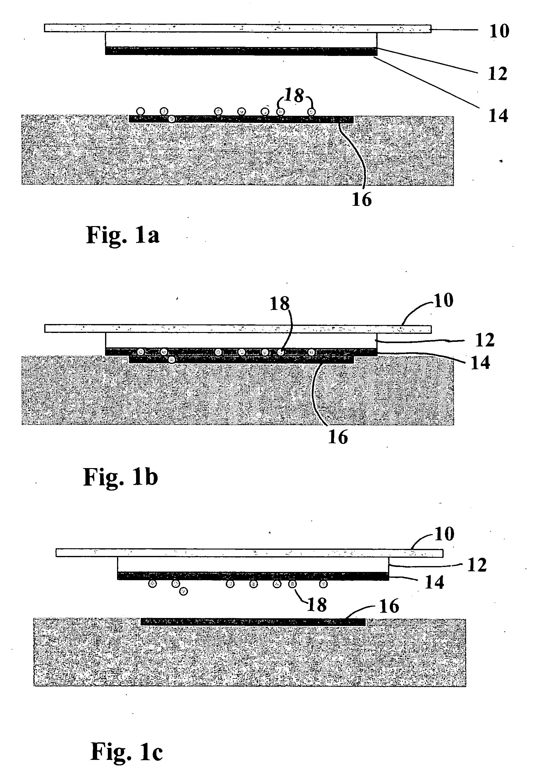
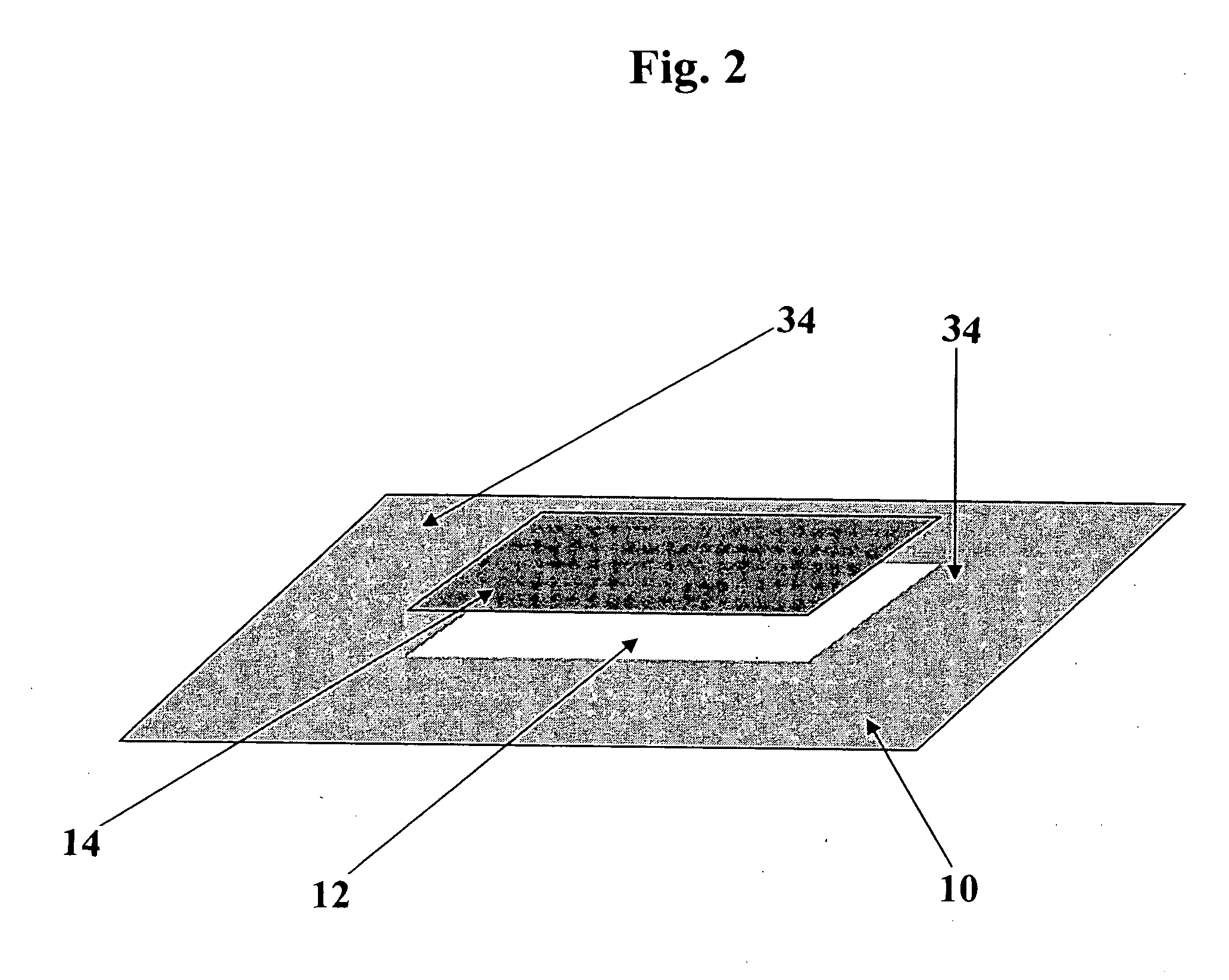
[0033] The invention herein relates to a wound dressing product, combining both a traditional dressing with a mounted absorbent material and a hydrophobic fabric attached thereto, which is to be placed towards the wound, and is capable of binding unwanted micro organisms. The product of the invention is ideal for treatment of wounds. It protects the wound, absorbs exudate and reduces the number of pathogenic microorganisms, without using antimicrobial substances. It consists basically of three bonded layers: hydrophobic cellulose acetate fabric which binds microorganisms, an attached efficient absorbent material, and a backing (cover) which in the preferred embodiment is visually transparent, semi-permeable and self-adhesive. These properties of the one-piece product of the invention allow passage of oxygen and moisture and inspection of the surrounding skin at the same times as the product binds existing pathogens and prevents the entry of new ones from the outside. The dressing is waterproof and allows the user to shower.

Problems solved by technology

The problem in healing of wounds associated with bacterial loads is for example tissue damage by release of toxins and enzymes, and possible spread of infections to the blood stream.
Studies have shown that high tissue counts of microorganisms delay wound healing.
When two water repellent molecules come into collision with each other they will increase the entropy and create disorder.
In wound treatment, after tissue colonization wound microbes multiply, cause tissue damage by release of toxins and enzymes, and even spread to the blood stream.
Numerous studies have shown that high tissue counts of microorganisms delay wound healing.
This is of importance since debris hampers wound healing.
A number of disadvantages to these methods can be mentioned.
For example, bacteria which have died remain in the wound, and wounds cannot be cleaned to remove the active compound reliably after the application, since it spreads in the entire wound.
Microbiologists disapprove of the use of local antibiotics since this is known to induce antibiotic resistance.
It has shown to be very difficult to adhere a strongly hydrophobic layer to hydrophilic liquid absorbent layers, and hence products made according to U.S. Pat. No. 4,617,326 have so far only been possible to manufacture by means of folding a separate layer in and around the hydrophilic absorbent layer and hydrophobic layer in order to keep the layers together.
This is an important drawback, limiting the design and production of such wound pads.
However, the so produced sponge is not using a hydrophobic material binding bacteria to any larger extent, nor does it have any backing which is visually transparent, semi-permeable and self-adhesive.
Even if the traditional Sorbact pad product solves an important problem of reducing the number of microorganisms in a wound without using chemicals or antibiotics, it has several disadvantages when used as a wound dressing.
Firstly, it will need to be attached to the wound surface with a bandage using such materials as a cotton gauze wrap, surgical tape and the like.
This means that there are several steps and products involved for the user to apply the Sorbact pad, which among other things leads to increased costs and time to handle.
Depending on the location on the body of the wound, this is not always easy to achieve with the loose pad and a bandage.
Further, it is difficult to inspect the status of the wound in terms of inflammation etc. through a bandage.

Method used


Image

  • Wound dressing with a bacterial adsorbing composition
  • Wound dressing with a bacterial adsorbing composition
  • Wound dressing with a bacterial adsorbing composition

Examples


example 1

Manufacture of Wound Dressing Product

[0059] In this example we manufacture a standard wound dressing based on the invention in the following manner:

[0060] Materials: (From Inside-Out See FIG. 4)

COMMERCIALLAYERPRODUCT NAMEMANUFACTURER1.HydrophobicGreen CelluloseABIGO Medical ABlayerAcetate wovenSwedenprepared accordingto U.S. Pat. No.4,617,3262.AdhesiveNational 505ENational Starch &Chemical Ltd.,United Kingdom3.Absorbent(Airlaid) ConcertConcert GmbH,materialMH080.104.P000Falkenhagen, Germany4.AdhesiveNational 505ENational Starch &Chemical Ltd.,United Kingdom5.CarrierPolyurethanefilm,3M, St Paul, MN, U.S.A.layer3M, 9482 no. ID70-0000-6538-66.Adhesive:DURO-TAK 380-295National Starch &Chemical Ltd.,United Kingdom7.SiliconizedLoparexLoparex OYrelease paper,ESP35TEELohja, Finland

A. The hydrophobic layer is preferably produced according to U.S. Pat. No. 4,617,326 by applying to a cellulose acetate fabric an amount of dioctadecyl carbamoyl chloride as disclosed in this patent making a ...

example 2

Use of the New Dressing Product to Bind Pathogens

[0066] Material: Wound dressing as described in Example 1 [0067] Bacterial strains: Staphylococcus aureus Newman, Pseudomonas aeruginosa 510, Enterococcus faecalis, Candida albicans

[0068] Isolates were cultured on agar with 5% horse erythrocytes in 5% CO2 atmosphere at 37° C. Suspensions were made in phosphate-buffered saline (PBS, 0.02 M sodium phosphate and 0.15 M sodium chloride, pH 7.2) at 109 bacterial cells / ml, 107 fungal cells / ml or indicated concentration.

[0069] The dressing was cut in 1 cm2 pieces. Incubation was made in 24 well polymer plates. 1 ml of suspension was added to each dressing. The plates were placed on a rotary shaker at very low speed. Incubation was performed at room temperature for the indicated time. After incubation, dressings were rinsed in PBS several times, and then put in 2.5% TCA (tricarboxylic acid).

[0070] The ATP content was measured in a luminometer (LKB Wallac). Controls: Number of adhered bact...

example 3

Manufacture of a Vein Catheter Dressing

[0075] In this example, a vein catheter dressing based on the invention was manufactured. The manufacture is made according to Example 1 for the first steps and then with a revised Step C as follows:

C. Cutting and Adding of Carrier Layer.

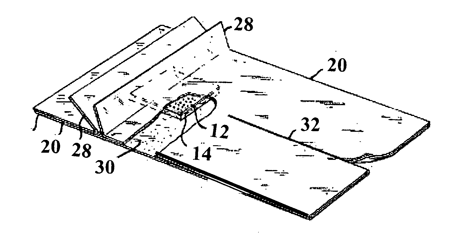
[0076] The now bonded hydrophobic layer and absorbing layer is cut into suitable pieces for a vein catheter product, in this case 20 mm×20 mm. The cut pieces are placed on carrier-film, prepared with the adhesive and two release papers 20 with folded gripping tabs 28 (see FIG. 3), and in strips of 100 mm. The bonded pieces are centered on the carrier strips and with a distance from center to center of 100 mm. A release paper of the same width is then applied on inner side. The now completely assembled dressing, is cut into pieces of 100 mm×60 mm, cutting 20 mm from the pad of attached layers in the top end and cutting a slit for the catheter from the opposite side to a distance close to the pad preferably 5...


PUM


Abstract

A wound dressing product, combining both a traditional dressing with a mounted absorbent material and a hydrophobic fabric attached thereto, which is to be placed towards the wound, and is capable of binding unwanted microorganisms. The wound dressing product protects the wound, absorbs exudate and reduces the number of pathogenic microorganisms, without using antimicrobial substances, and consists of three bonded layers: hydrophobic cellulose acetate fabric which binds microorganisms, an attached efficient absorbent material, and a backing (cover) which is preferably visually transparent, semi-permeable and self-adhesive.

Description

BACKGROUND OF THE INVENTION [0001] 1. Field of the Invention [0002] The invention herein relates to a wound dressing product, combining both a traditional dressing including a mounted absorbent material on a film backing and a hydrophobic fabric attached thereto, facing towards the wound and being capable of binding unwanted micro organisms. [0003] 2. Description of the Related Art [0004] The problem in healing of wounds associated with bacterial loads is for example tissue damage by release of toxins and enzymes, and possible spread of infections to the blood stream. Studies have shown that high tissue counts of microorganisms delay wound healing. [0005] Numerous studies during the last few decades have also shown that bacteria, such as Staphylococcus aureus and Group A streptococci, both common wound pathogens, and the yeast Candida albicans commonly express profound cell surface hydrophobicity. Several structures which render the cell surface hydrophobic have been defined, like t...

Claims


Application Information

Patent Timeline
Patent Type & Authority Applications(United States)
IPC IPC(8): A61F15/00A61F13/00
CPCA61F13/00042A61F13/00063A61F2013/00182A61F2013/00263A61F2013/00412A61F2013/00582A61F2013/00604A61F2013/00727A61F2013/00846A61F2013/00876A61L15/44A61L15/46A61L15/58A61F13/01042
Inventor BJORNBERG, STEN G.SMITH, JAN G.
Owner ABIGO MEDICAL AB
Learn More