Matrix composed of a naturally-occurring protein backbone cross linked by a synthetic polymer and methods of generating and using same

a technology of naturally occurring protein and polyethylene glycol, which is applied in the direction of cell culture supports/coatings, peptide/protein ingredients, depsipeptides, etc., can solve the problems of uneven cell seeding of prefabricated materials, limited cell migration into the depth of synthetic scaffolds, and the possibility of prefabricated tissue constructs

Active Publication Date: 2006-10-19
REGENTIS BIOMATERIALS
View PDF27 Cites 75 Cited by
  • Summary
  • Abstract
  • Description
  • Claims
  • Application Information

AI Technical Summary

Benefits of technology

[0064] Unless otherwise defined, all technical and scientific terms used herein have the same meaning as commonly understood by one of ordinary skill in the art to which this invention belongs. Although methods and materials similar or equivalent to those described herein can be used in t

Problems solved by technology

However, cell migration into the depth of synthetic scaffolds is limited by the lack of oxygen and nutrient supply.
However, creating pores into these materials is an aggressive procedure involving the use of toxic reagents which eliminate the possibility of pre-casting tissue constructs with living cells.
Therefore, many of the prefabricated materials are subject to uneven cell seeding and non-homogeneous populations of cells within the constructs.
Furthermore, the materials are generally degraded unevenly during the tissue cultivation process, creating a highly anisotropic tissue with altered growth kinetics.
However, while the abovementioned synthetic polymers enable precise control over the scaffold material, they often provide inadequate biological information for cell culture.
As a result, these materials are unsuitable for long-term tissue culture or in vivo tissue regeneration.
However, because such biological materials exhibit multiple inherent signals (e.g., regulation of cell adhesion, proliferation, cellular phenotype, matrix production and enzyme activity), their use as scaffolds in tissue regeneration often results

Method used


Image

  • Matrix composed of a naturally-occurring protein backbone cross linked by a synthetic polymer and methods of generating and using same
  • Matrix composed of a naturally-occurring protein backbone cross linked by a synthetic polymer and methods of generating and using same
  • Matrix composed of a naturally-occurring protein backbone cross linked by a synthetic polymer and methods of generating and using same

Examples


example 1

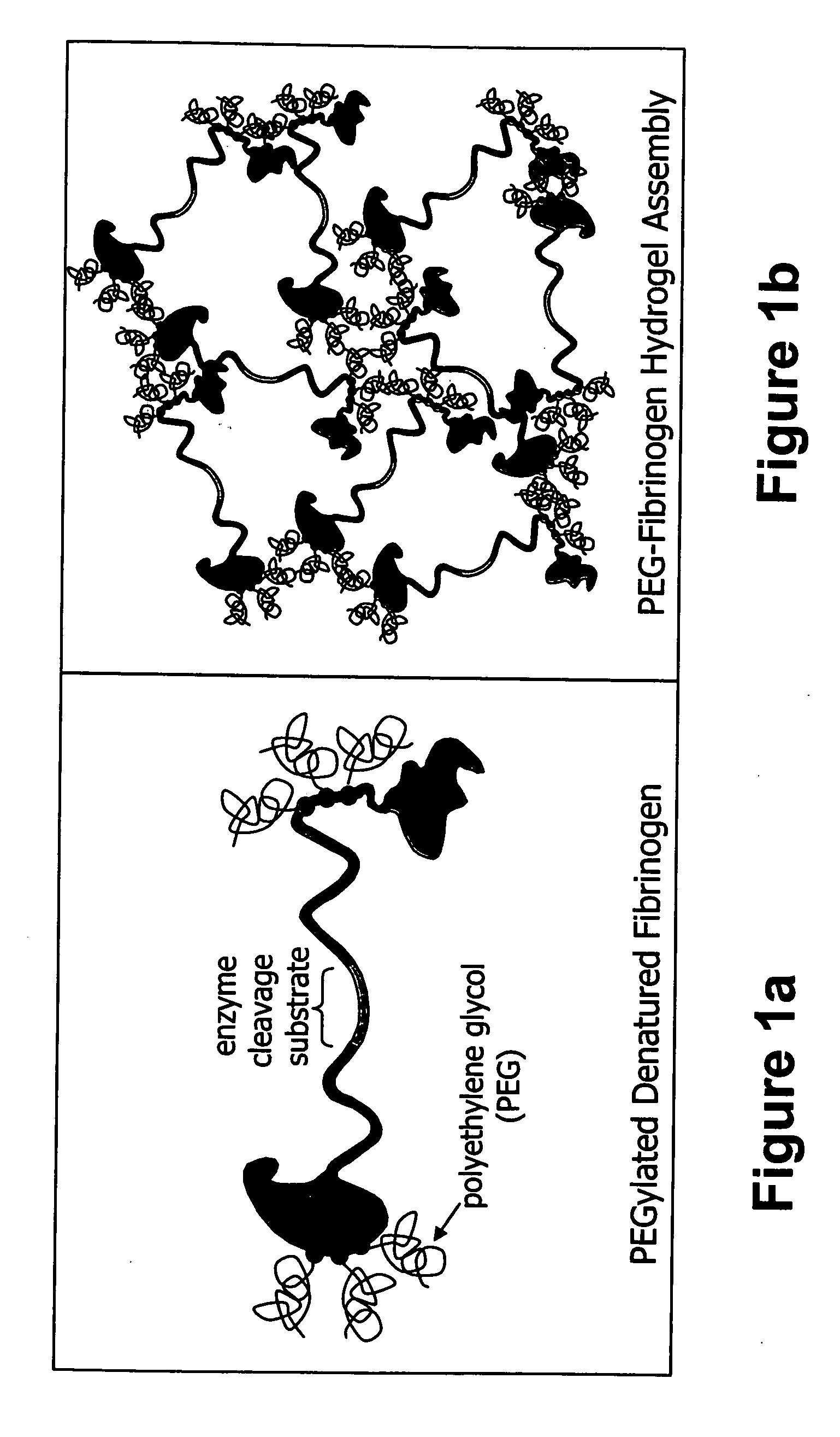
Generation of PEG-Fibrinogen Hydrogels

[0197] Tissue engineering scaffolds with controllable mechanical properties and adequate biofunctional signals were generated from PEG and fibrinogen. Briefly, denatured fibrinogen fragments were PEGylated with PEG-diacrylates, mixed with photoinitiator and exposed to UV light to form a hydrogel material in the presence of a cell suspension. The degradability of the PEG-fibrinogen scaffold was further tested by enzyme-mediated proteolysis, as follows.

[0198] Materials and Experimental Methods

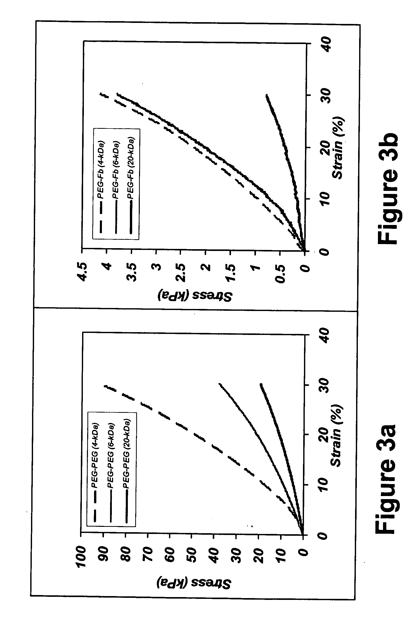
[0199] Synthesis of PEG Diacrylate (PEG-DA)—PEG-diacrylate (PEG-DA) was prepared from linear PEG, MW=4-kDa, 6-kDa, and 20-kDa (Fluka, Buchs, Switzerland), essentially as described elsewhere (Lutolf and Hubbell, 2003; Elbert D L., et al., 2001). Briefly, acrylation of PEG-OH was carried out under Argon by reacting a dichloromethane (DCM) (Aldrich, Sleeze, Germany) solution of the PEG-OH with acryloyl chloride (Merck, Darmstadt, Germany) and triethylamine (F...

example 2

The PEG-Fibrinogen Hydrogels Support Cell Spreading and Extension

[0228] To test the capacity of the PEG-fibrinogen hydrogels to promote the expansion and differentiation of cell cultures, cell of bovine aortic endothelial cells and smooth muscle cells were cultured on various PEG-based hydrogels, as follows.

[0229] Materials and Experimental Methods

[0230] In vitro cell-culture studies—Bovine aortic smooth muscle cells (BSMCs) from young donors were isolated and cultured according to a modified protocol of Oakes et al., 1982. The BSMCs were cultured up to 6th passage in Dulbecco's Modified Eagle Medium (DMEM) (Gibco, U.K.) containing 10% fetal bovine serum (FBS) (Biological Industries, Israel), 1% penicillin-streptomycin (Biological Industries), and 1% L-glutamine (Gibco). PEG hydrogels containing BSMCs were made by mixing a PBS cell suspension and PEGylated fibrinogen precursor solution containing Igracure™ photoinitiator to make a 10% (w / v) solution with 1.5×10⁶ cells / ml. Aliquot...

example 3

In Vivo Regeneration of Bone Using the PEG-Fibrinogen Gelrin™ Scaffold

[0238] To test the potential of the PEG-fibrinogen scaffold material to facilitate tissue regeneration, a critical size tibial defect was introduced in rats and the Gelrin™ scaffold was implanted at the site of surgery (tibia diaphysis).

[0239] Materials and Experimental Methods

[0240] Animals—Female Sprague-Dawley rats (age 3-4 months) were adapted to animal cage life for 5 days prior to the experiment. The weight of the animal was monitored during this period to ensure stability and proper adaptation. The animals were fed and watered daily without restrictions.

[0241] Introduction of a critical size tibia defect—The animals were anesthetized with a combination of Ketamine (120 mg / kg) and Xylazine (17 mg / kg). During the surgical procedure the animals were placed on a warm plate to maintain body temperature (and prevent hypothermia). The right tibia was shaved and wiped with polydine tincture solution. The mid-po...


PUM

PropertyMeasurementUnit
Fractionaaaaaaaaaa
Fractionaaaaaaaaaa
Fractionaaaaaaaaaa

Abstract

The present invention relates to biodegradable scaffolds composed of a naturally-occurring protein backbone cross-linked by a synthetic polymer. Specifically, the present invention provides PEGylated-fibrinogen scaffold and methods of generating and using same for treating disorders requiring tissue regeneration.

Description

RELATED APPLICATIONS [0001] This is a continuation-in-part of PCT Patent Application No. PCT / IL2004 / 001136 filed Dec. 15, 2004, which claims the benefit of U.S. Provisional Patent Application No. 60 / 530,917 filed Dec. 22, 2003. The contents of the above applications are all incorporated by reference.FIELD AND BACKGROUND OF THE INVENTION [0002] The present invention relates to a matrix composed of a naturally-occurring protein backbone cross-linked by polyethylene glycol (PEG) and, more particularly, to methods of generating and using same in tissue regeneration. [0003] Tissue engineering, i.e., the generation of new living tissues in vitro, is widely used to replace diseased, traumatized or other unhealthy tissues. The classic tissue engineering approach utilizes living cells and a basic scaffold for cell culture (Langer and Vacanti, 1993; Nerem and Seliktar, 2001). Thus, the scaffold structure attempts to mimic the natural structure of the tissue it is replacing and to provide a te...

Claims


Application Information

Patent Timeline
IPC IPC(8): A61F13/00A61K38/18A61K38/21A61K38/48A61K47/48A61L24/10A61L27/22A61L27/38C07K14/75C12N5/00
CPCA61K47/48215C12Y203/02013A61K47/48784A61L27/18A61L27/227A61L27/24A61L27/38A61L27/52C12N5/0068C12N2533/30C12N2533/40C12N2533/50C12N2533/52C12N2533/54A61K47/48292A61L2300/418A61K38/363A61L2300/414A61L27/58A61L27/54A61K38/45A61K38/39A61L2430/34C08L71/02C12M25/14A61K47/60A61K47/6435A61K47/6903A61P17/00A61P19/08A61P21/00A61P25/00A61P9/00A61L27/225A61L27/26A61L2430/02A61L2430/06
Inventor SELIKTAR, DRORALMANY, LIORA
Owner REGENTIS BIOMATERIALS
Why Eureka
Try Eureka