Novel and practical serological assay for the clinical diagnosis of leishmaniasis

a serological assay and clinical diagnosis technology, applied in the field of leishmaniasis clinical diagnosis, can solve the problems of affecting patient management, affecting the treatment effect,

Inactive Publication Date: 2006-12-14
UNITED STATES OF AMERICA THE AS REPRESENTED BY THE SEC OF THE ARMY
View PDF2 Cites 0 Cited by
  • Summary
  • Abstract
  • Description
  • Claims
  • Application Information

AI Technical Summary

Benefits of technology

[0037]FIG. 11A shows that cells ex vivo can be maintained in plasma.

Problems solved by technology

Unfortunately, current acceptable diagnostic practices lack the means for efficiently and accurately identifying those infected or exposed to the disease-causing parasite as explained in Martin, S. et al.
As a result, the prevention of leishmanial epidemics is greatly hindered and patient management is difficult.
Additionally, there is an imminent threat of HIV and Leishmania co-infection, a more malignant infection that is difficult to diagnose and treat.
To date, there are no antigen-detection type diagnostic tests available for leishmaniasis.
However, assays developed with this antigen and others fail to consistently detect antibodies in other clinical syndromes associated with a predominately T-cell and muted B-cell response.
Moreover, antibody-detection assays have an inherent dependence on the immune response of the host to the parasite antigen which significantly diminishes its use.
For example, in HIV / AIDS and other immuno-compromised conditions, the infection may not produce proportional antibody production and thereby escape detection.
These serological tests, however, pose problems of non-specificity resulting in false positive results from reference samples of other infectious diseases.
Unfortunately, serological tests are rarely performed to diagnose cutaneous leishmaniasis, the most common form of the disease, because the sensitivities and specificities are disappointingly low for this clinical manifestation.
Thus, the presence of complex proteins or serum components required for growth of the parasites in the culture medium pose several problems in the prior art assays.
Furthermore, insufficient purification of parasite products may create problems with specificity as serum proteins contaminants in the antigen preparations, cause non-specific downstream reactions.
Generally, the prior art assays are limited in scope to one species complex or clinical manifestation and have never demonstrated combined sensitivity and specificity of more than 90%.

Method used


Image

  • Novel and practical serological assay for the clinical diagnosis of leishmaniasis
  • Novel and practical serological assay for the clinical diagnosis of leishmaniasis
  • Novel and practical serological assay for the clinical diagnosis of leishmaniasis

Examples


example 1

Sera Preparation

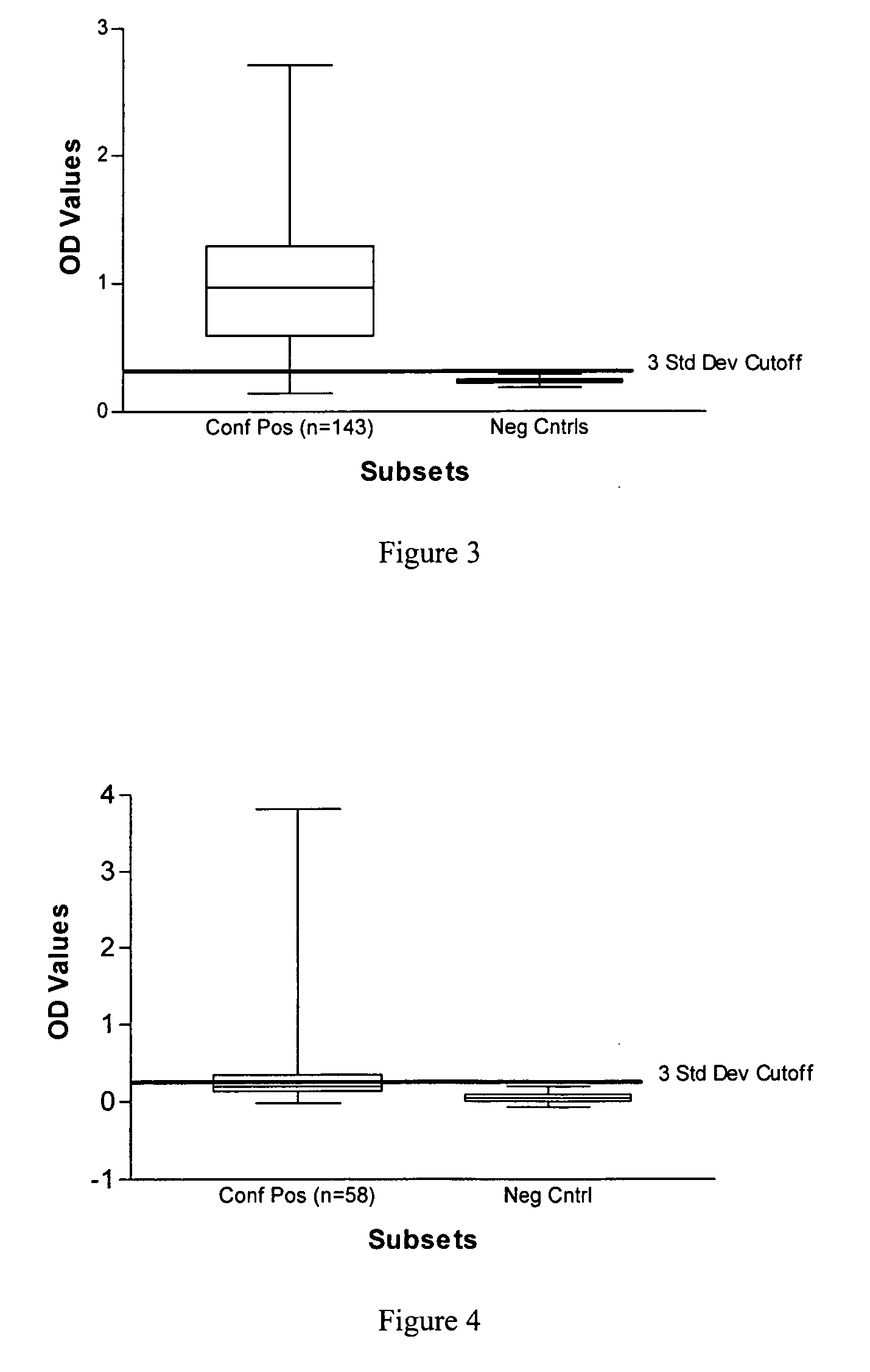
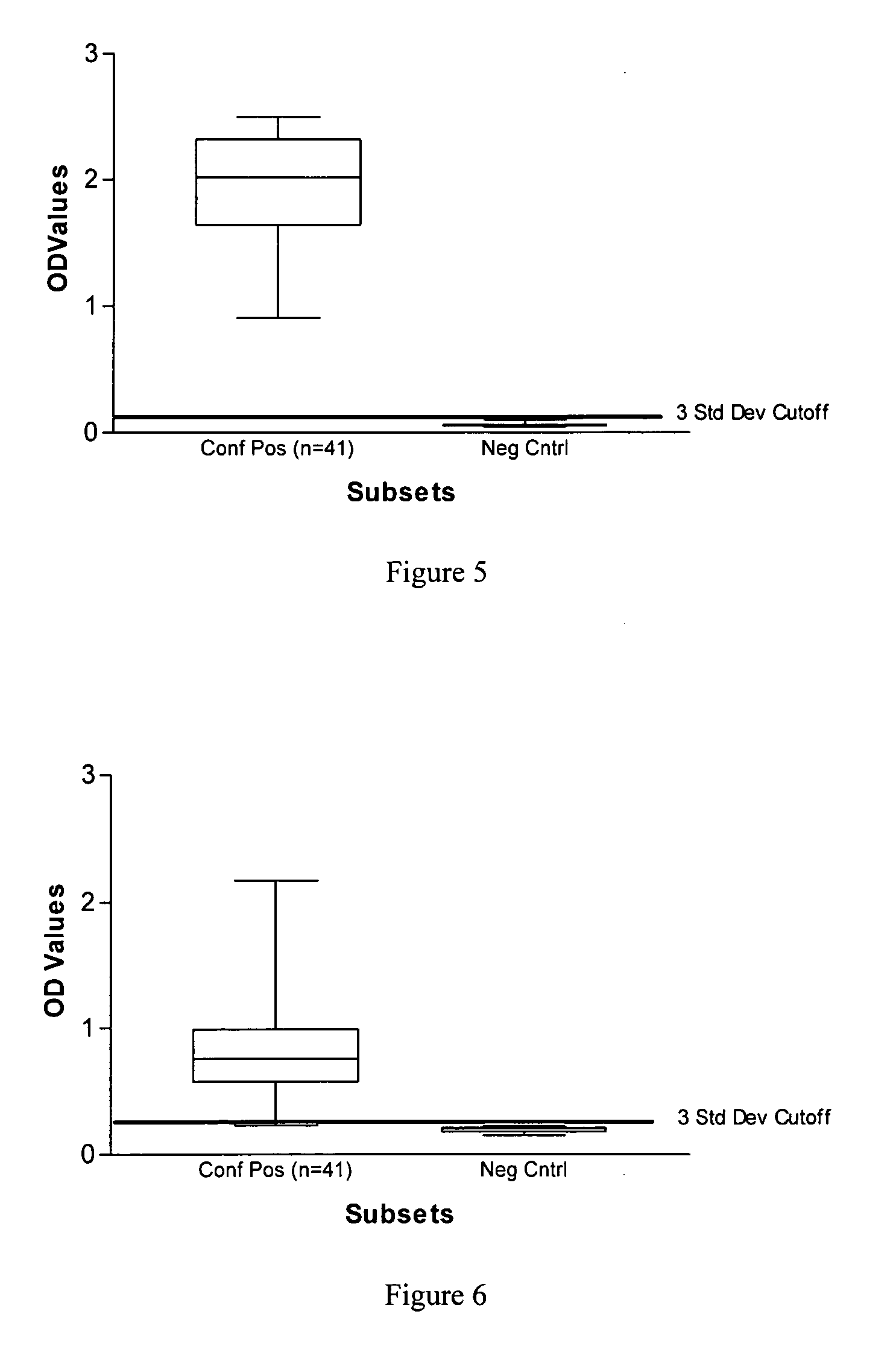
[0121] Serum was collected from human patients who were admitted to clinics in Brazil, Italy, North Africa, Nepal and Walter Reed Army Medical Center and who had either splenic aspirates or skin biopsies from lesions that tested positive for Leishmania parasites by culture and microscopy. In total, 129 visceral (Italy, Brazil, North Africa, and Nepal) and 143 cutaneous (Brazil) leishmaniasis patients (136—L. braziliensis-infected, and 7 L. panamensis-infected) with controls were tested.

[0122] Human negative controls were from 12 non-endemic area normal patients with no documented infection or exposure to Leishmania parasites. In addition to the human manifestations assayed, sera from 42 Brazilian dogs with a clinical diagnosis for canine leishmaniasis were tested against positive control sera from a commercial source (Bordier Affinity Products, S.A., Crissier, Switzerland) and 10 negative controls from a pathogen-free, canine research colony (College of Veterinary ...

example 2

Antigen Preparation

[0123] The Leishmania soluble antigen (exo-antigen) preparation was made by cultivating Leishmania promastigotes in normal supplemented media (RPMI, MEM plus FBS) at 26° C. until the culture reached mid-log phase at a density of about 109 cells / ml. Then the cells were pelleted and washed 6 times in a defined, protein-free medium such as XOM available from GIBCO BRL, formula number 96-0051DJ, RPMI Medium 1640 comprising D, xylose at 0.076 mM, Hepes buffer at 25 mM, L-glutamine, and sodium bicarbonate at 30 mM without phenol red.

[0124] The cells were then resuspended in protein-free medium such as XOM to a final density of 108 promastigotes / ml and incubated at 26° C. in a roller bottle with 0.01% Tween 80 (Sigma Chemical Co., St. Louis, Mo.) for 72 hours. The cells were pelleted by centrifugation at 9,000×g for 30 minutes and the supernatant was collected. The relative protein concentration of the soluble antigens was estimated by measuring the optical density at ...

example 3

Antibody and Conjugate Production

[0125] The leishmania soluble antigen preparation produced by the method explained in Example 2 was used without an adjuvant to immunize rabbits. The antiserum was pooled and affinity-purified on a Protein A column containing the antigen preparation of Example 2 above. Fractions of the polyclonal antibody (PAb) were conjugated with an appropriate reporter system such as horseradish peroxidase, fluorescein and colloidal gold. These tagged antibodies may then be used in antigen-detection immunoassays such as ELISA, histochemical stain and dipstick test formats. These test formats may be used to detect parasite antigens in tissues and body fluids of mammalian hosts and vectors.

[0126] The rabbit anti-leishmania polyclonal antibody preparation demonstrated high affinity and avidity for the immunogens used to immunize the rabbits. The PAb produced a very striking Western Blot pattern similar to that of kala azar patients. See FIG. 7 of U.S. patent applic...


PUM


Abstract

Methods for the diagnosis of visceral, cutaneous and canine leishmaniasis in a subject suspected of being infected with the parasitic protozoa Leishmania is disclosed. Disclosed are antibody-capture enzyme-linked immunosorbent assays (ELISAs) for the detection of antibodies to Leishmania parasite soluble antigens and antigen-capture ELISAs for the detection of Leishmania parasite soluble antigens in host samples. Also disclosed are immunodiagnostic kits for the detection of Leishmania parasite circulating antigens or IgM and IgG antibodies in a sample from subject having visceral, cutaneous or canine leishmaniasis. In these methods and kits, detection may be done photometrically or visually. The methods and kits also allow the visualization of Leishmania amastigotes or promastigotes in a sample.

Description

CROSS REFERENCE TO RELATED APPLICATIONS [0001] This application is a continuation-in-part of U.S. patent application Ser. No. 10 / 990,971, filed 18 Nov. 2004, which is a continuation of U.S. patent application Ser. No. 10 / 173,586, filed 18 Jun. 2002, which is a continuation of U.S. patent application Ser. No. 09 / 725,182, filed 29 Nov. 2000, now abandoned, which claims the benefit of U.S. Provisional Patent Application No. 60 / 168,300, filed 1 Dec. 1999, now abandoned, naming Jeffrey R. Ryan, Samuel K. Martin, and Anthony M. Smithyman as co-inventors, all of which are herein incorporated by reference.ACKNOWLEDGMENT OF GOVERNMENT SUPPORT [0002] This invention was made by employees of the United States Army. The government has rights in the invention.BACKGROUND OF THE INVENTION [0003] 1. Field of the Invention [0004] This invention relates generally to a method of diagnosing leishmaniasis in a subject suspected of being infected with the parasitic protozoa Leishmania. In particular, the ...

Claims


Application Information

Patent Timeline
Patent Type & Authority Applications(United States)
IPC IPC(8): A01N1/02
CPCA01N1/02G01N33/56905C12Q1/08A01N1/0221Y02A50/30
Inventor MARTIN, SAMUEL K.
Owner UNITED STATES OF AMERICA THE AS REPRESENTED BY THE SEC OF THE ARMY
Why Eureka
Try Eureka