
Devices and methods for enrichment and alteration of circulating tumor cells and other particles

a technology of tumor cells and particles, applied in the field of medical diagnostics and microfluidics, can solve the problems of many cancers going undetected and damaging healthy tissue, and achieve the effects of decreasing the hydrodynamic size of particles, increasing deformation, and decreasing volum

Inactive Publication Date: 2007-02-01
CELLECTIVE DX CORP +4
View PDF43 Cites 86 Cited by
  • Summary
  • Abstract
  • Description
  • Claims
  • Application Information

AI Technical Summary

Benefits of technology

[0016] The labeling reagent can include a quantum dot, an antibody, a phage, an aptamer, a fluorophore, an enzyme, or a bead which can include, e.g., an affinity reagent or polystyrene, can be neutrally buoyant, or can include an antibody. The labeling reagent can increase the size of said cells, e.g., by at least 10%, by at least 100%, or by at least 1000%.
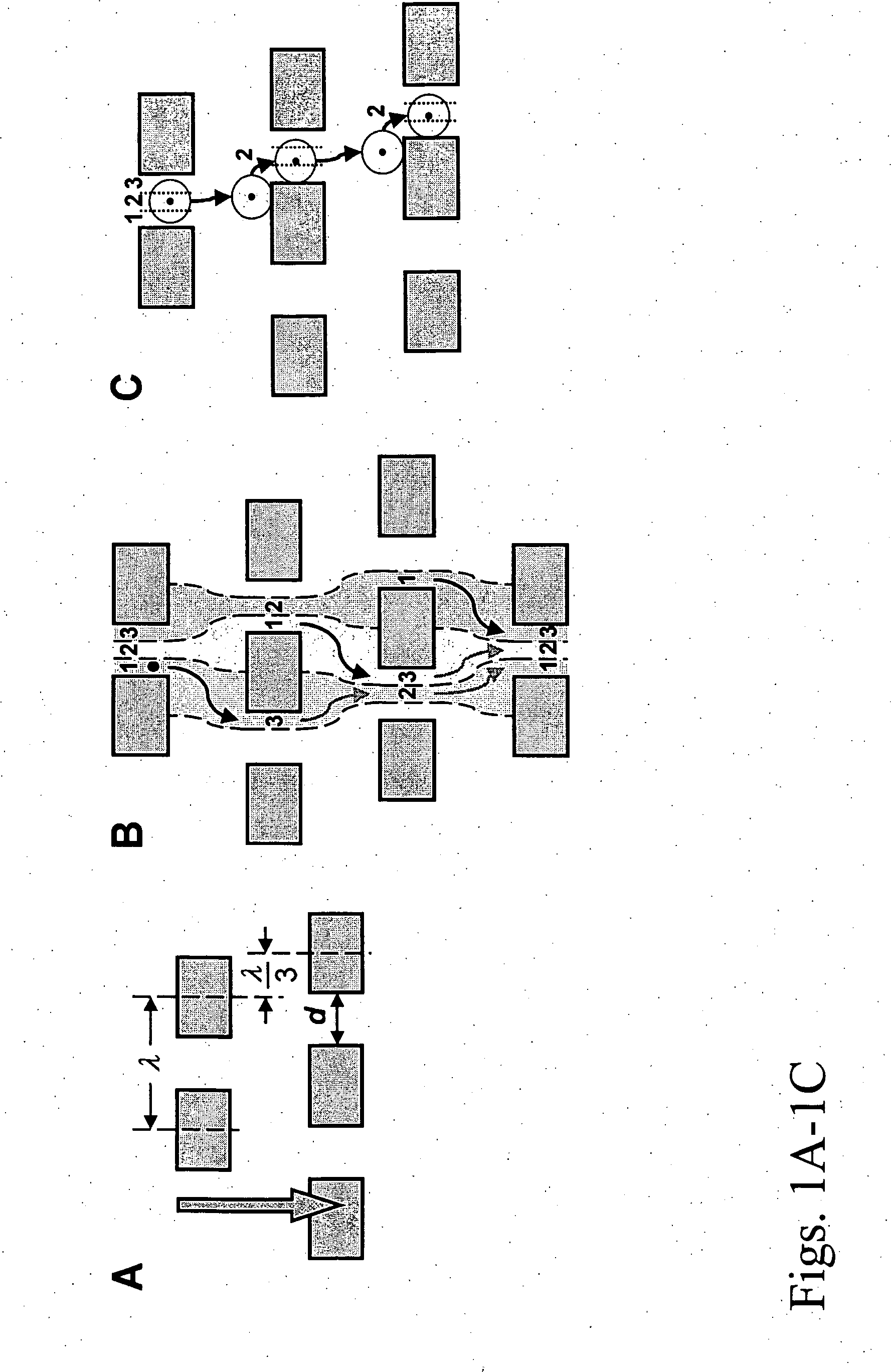
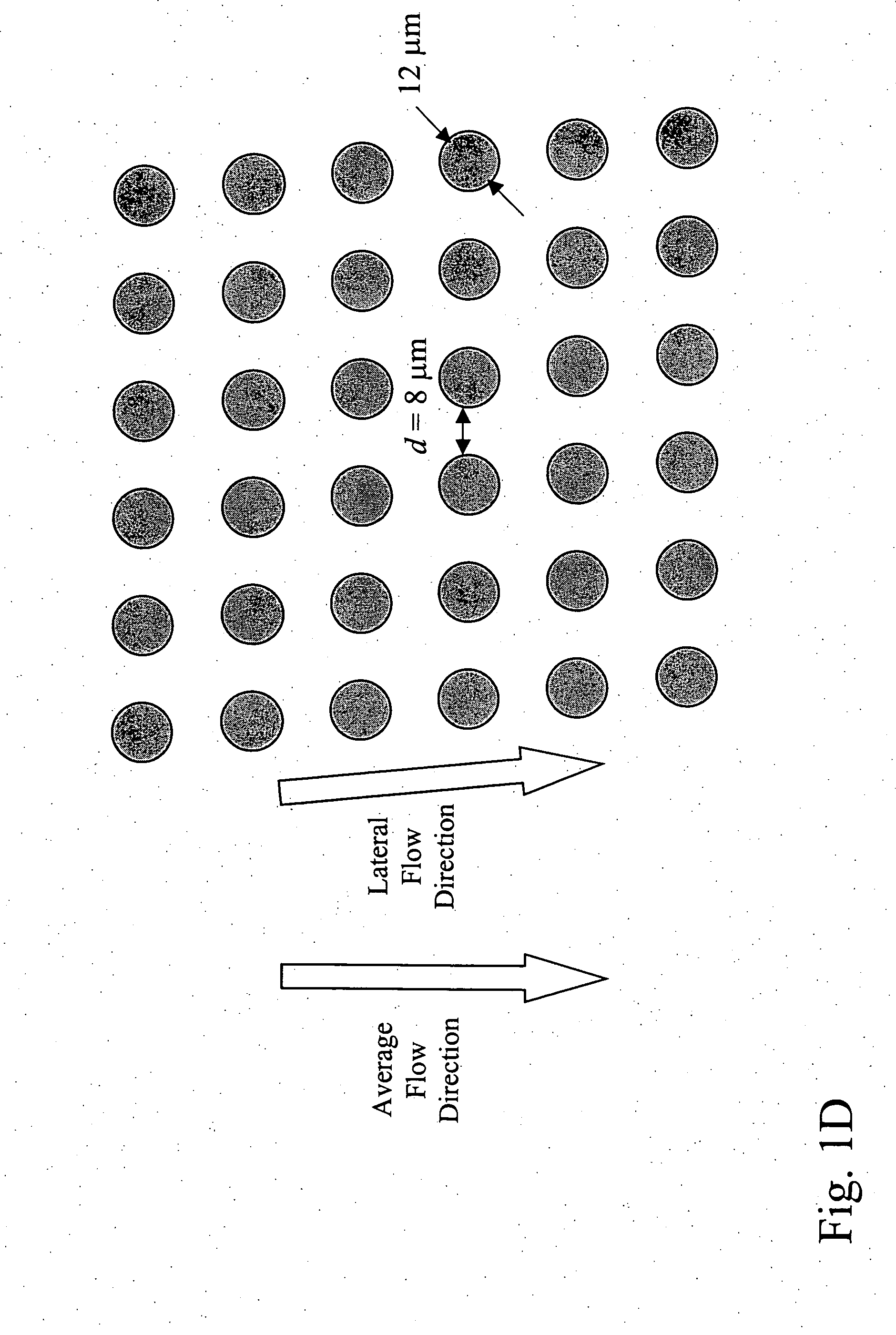
[0054] By “density” in reference to an array of obstacles is meant the number of obstacles per unit of area, or alternatively the percentage of volume occupied by such obstacles. Array density is increased either by placing obstacles closer together or by increasing the size of obstacles relative to the gaps between obstacles.
[0067] By “shrinking reagent” is meant a reagent that decreases the hydrodynamic size of a particle. Shrinking reagents may act by decreasing the volume, increasing the deformability, or changing the shape of a particle.
[0068] By “swelling reagent” is meant a reagent that increases the hydrodynamic size of a particle. Swelling reagents may act by increasing the volume, reducing the deformability, or changing the shape of a particle.

Problems solved by technology

Metastasis results in the formation of secondary tumors at multiple sites, damaging healthy tissue.
Despite decades of advances in cancer diagnosis and therapy, many cancers continue to go undetected until late in their development.

Method used


Image

  • Devices and methods for enrichment and alteration of circulating tumor cells and other particles
  • Devices and methods for enrichment and alteration of circulating tumor cells and other particles
  • Devices and methods for enrichment and alteration of circulating tumor cells and other particles

Examples


example 1

[0281] Microfluidic devices of the invention were designed by computer-aided design (CAD) and microfabricated by photolithography. A two-step process was developed in which a blood sample is first debulked to remove the large population of small cells, and then the rare target epithelial cells target cells are recovered by immunoaffinity capture. The devices were defined by photolithography and etched into a silicon substrate based on the CAD-generated design. The cell enrichment module, which is approximately the size of a standard microscope slide, contains 14 parallel sample processing sections and associated sample handling channels that connect to common sample and buffer inlets and product and waste outlets. Each section contains an array of microfabricated obstacles that is optimized to enrich the target cell type by hydrodynamic size via displacement of the larger cells into the product stream. In this example, the microchip was designed to separate red blood cells (RBCs) an...

example 2

Device Embodiments

[0284] A design for preferred device embodiments of the invention is shown in FIG. 57A, and parameters corresponding to three preferred device embodiments associated with this design are shown in FIG. 57B. These embodiments are particularly useful for enrich epithelial cells from blood.

example 3

Determining Counts for Non-Epithelial Cell Types

[0285] Using the methods of the invention, one may make a diagnosis based on counting cell types other than CTCs or other epithelial cells. A diagnosis of the absence, presence, or progression of cancer may be based on the number of cells in a cellular sample that are larger than a particular cutoff size. For example, cells with a hydrodynamic size of 14 microns or larger may be selected. This cutoff size would eliminate most leukocytes. The nature of these cells may then be determined by downstream molecular or cytological analysis.

[0286] Cell types other than epithelial cells that would be useful to analyze include endothelial cells, endothelial progenitor cells, endometrial cells, or trophoblasts indicative of a disease state. Furthermore, determining separate counts for epithelial cells, e.g., cancer cells, and other cell types, e.g., endothelial cells, followed by a determination of the ratios between the number of epithelial ce...


PUM

PropertyMeasurementUnit
Fractionaaaaaaaaaa
Fractionaaaaaaaaaa
Diameteraaaaaaaaaa

Abstract

The invention features devices and methods for detecting, enriching, and analyzing circulating tumor cells and other particles. The invention further features methods of diagnosing a condition, e.g., cancer, in a subject by analyzing a cellular sample from the subject.

Description

CROSS-REFERENCE TO RELATED APPLICATIONS [0001] This application claims the benefit of U.S. Provisional Application No. 60 / 703,833, filed Jul. 29, 2005, which is hereby incorporated by reference.BACKGROUND OF THE INVENTION [0002] The invention relates to the fields of medical diagnostics and microfluidics. [0003] Cancer is a disease marked by the uncontrolled proliferation of abnormal cells. In normal tissue, cells divide and organize within the tissue in response to signals from surrounding cells. Cancer cells do not respond in the same way to these signals, causing them to proliferate and, in many organs, form a tumor. As the growth of a tumor continues, genetic alterations may accumulate, manifesting as a more aggressive growth phenotype of the cancer cells. If left untreated, metastasis, the spread of cancer cells to distant areas of the body by way of the lymph system or bloodstream, may ensue. Metastasis results in the formation of secondary tumors at multiple sites, damaging h...

Claims


Application Information

Patent Timeline
IPC IPC(8): C12Q1/68G01N33/53G01N33/574C12M3/00
CPCB01L3/502746G01N2800/52B01L3/502761B01L2200/0647B01L2300/0636B01L2300/0816B01L2300/0864B01L2400/0409B01L2400/086B82Y5/00B82Y10/00G01N33/5005G01N33/54366G01N33/57492B01L3/502753
Inventor FUCHS, MARTINTONER, MEHMETWANG, YING-XINHUANG, YI-SHUIANKRUEGER, NEIL X.HUANG, LOTIEN RICHARD
Owner CELLECTIVE DX CORP
Learn More