Identification, diagnosis, and treatment of neuropathologies, neurotoxicities, tumors, and brain and spinal cord injuries using electrodes with microvoltammetry

a microvoltameter and neurotoxicology technology, applied in the direction of instruments, catheters, material electrochemical variables, etc., can solve the problems of insufficient linear diffusion, insufficient in vitro analysis techniques, and criticized techniques

Inactive Publication Date: 2007-02-01
RES FOUND THE CITY UNIV OF NEW YORK +1
View PDF12 Cites 78 Cited by
  • Summary
  • Abstract
  • Description
  • Claims
  • Application Information

AI Technical Summary

Benefits of technology

[0023] The present invention relates to devices and methods for microvoltammetric and/or chronoamperometric imaging of temporal changes in neurotransmitter concentrations in living humans and non-human animals comprising contacting cells with a Broderick probe or BRODERICK PROBE® sensor, applying a potential to said Broderick probe, and generating a temporally resolved microvoltammogram. The method may further comprise determining from said microvoltammogram the presence and...

Problems solved by technology

However, Wightman et al. observed that linear diffusion is not enough to describe the action that takes place at spherical microelectrodes (Dayton M A et al., 1980, Anal. Chem. 52:948-950).
Previous in vitro analysis techniques have yielded disappointing results.
However, this technique has been criticized because of the local gliosis caused by the dialysis probes and the perfusion process that can alter the biochemical parameters under study.
Neurotransmitter data from experimental epilepsy models and in vitro analysis of surgically resected specimens from patients with partial epilepsy have thus far yielded conflicting results.
These conflicting results may be due to significant variations between samples as well as choice of controls.
Additionally, highly localized changes in epileptic cortex are not detectable ...

Method used


Image

  • Identification, diagnosis, and treatment of neuropathologies, neurotoxicities, tumors, and brain and spinal cord injuries using electrodes with microvoltammetry
  • Identification, diagnosis, and treatment of neuropathologies, neurotoxicities, tumors, and brain and spinal cord injuries using electrodes with microvoltammetry
  • Identification, diagnosis, and treatment of neuropathologies, neurotoxicities, tumors, and brain and spinal cord injuries using electrodes with microvoltammetry

Examples


example 1

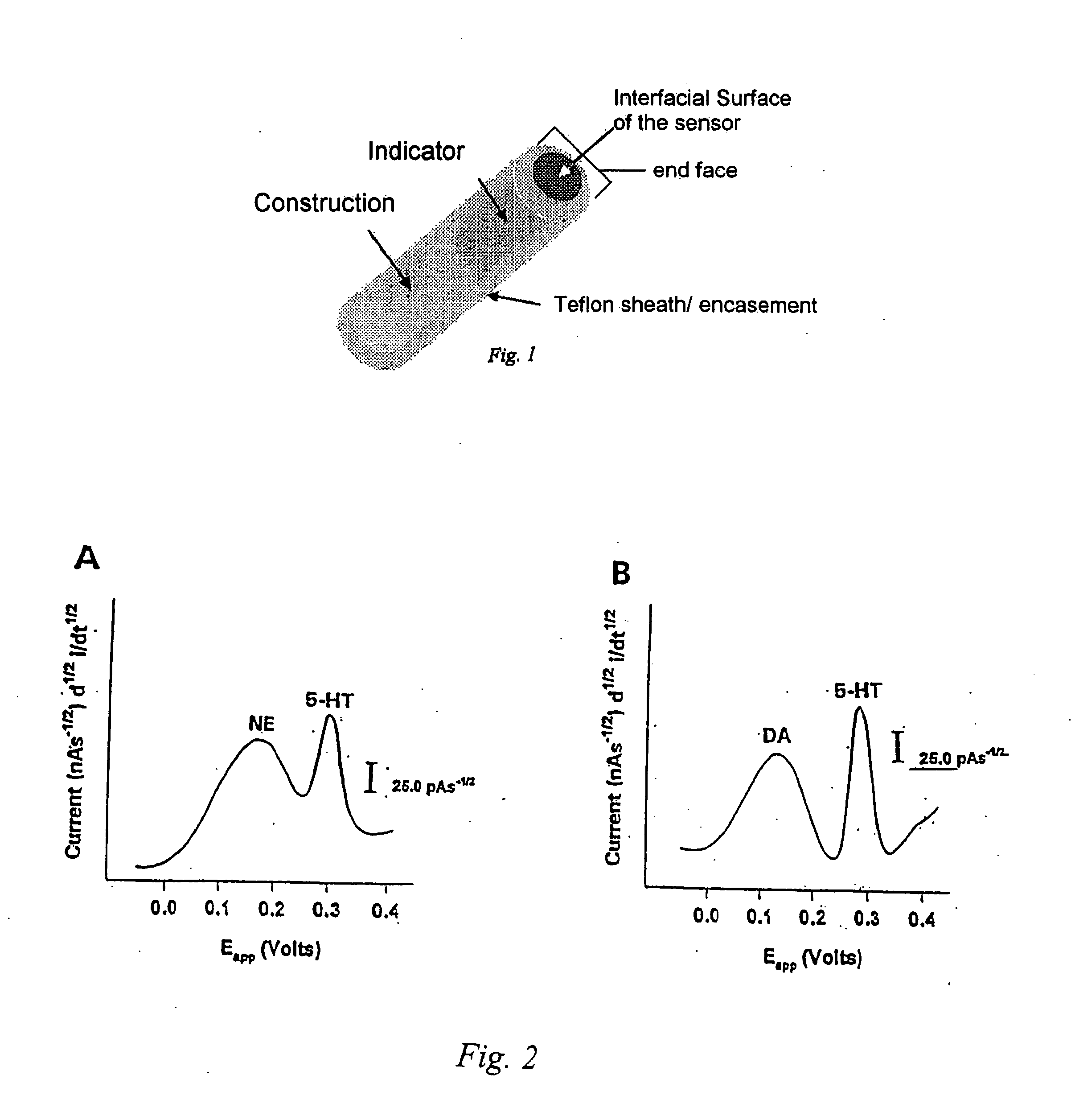
[0117] BRODERICK PROBE® sensors may have different formulations, e.g., cerebrosides and oleic acid; however, for these studies, the laurate sensor was used because it reaches steady state quickly. Although these sensors can be manufactured at any size or any length, for these studies, the laurate sensors comprising an indicator and construction. The construction portion was made of a polytetrafluoroethylene (PTFE; Teflon®) coated stainless steel wire having a diameter of 150 μm (Medwire, Mount Vernon, N.Y.). The coating was gently pulled 500 μm over the stainless steel tip to form a well. The well, or indicator portion, was packed with a graphite paste modified with (99+%) lauric acid. The graphite / lauric acid (Ultra Carbon, Bay City Mich.) (Sigma, St. Louis, Mo.) was admixed with mineral oil (a.k.a. Nujol) containing DL-alpha tocopherol as a stabilizer (Plough Inc., Memphis, Tenn.).

[0118] The reference microelectrode was constructed by electrochemically coating silver wire (Medwir...

example 2

Human Epilepsy

[0121] Fourteen patients who had temporal lobectomies for intractable seizures were studied. Patients underwent intraoperative surgery within the same time period and were studied in order of time, within the same time period. Patients were classified as having mesial temporal lobe epilepsy if pathologic examination of the resected temporal lobe revealed severe hippocampal neuronal loss and gliosis and if examination of the neocortex revealed no other etiology for the patient's epilepsy. Nine patients were classified as mesial temporal lobe epilepsy based on these features. Five patients were classified as having neocortical temporal lobe epilepsy based on the lack of hippocampal atrophy on magnetic resonance imaging (MRI) and demonstration of seizure onset in temporal neocortex during chronic intracranial EEG study with lateral temporal subdural grid electrodes and multiple baso-mesial temporal subdural strip electrodes.

[0122]FIG. 1 shows a schematic diagram of a Br...

example 3

Human Epilepsy

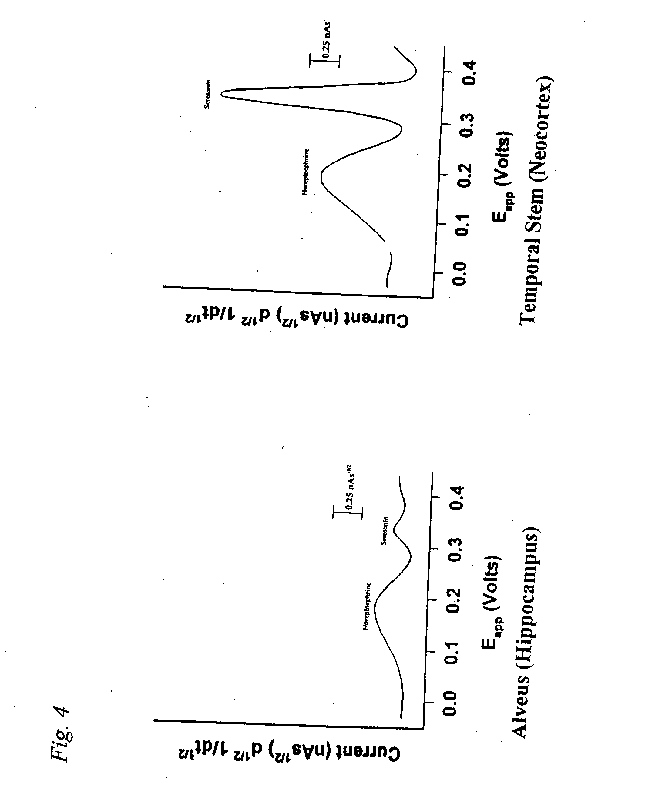
[0132] Significant differences in the monoamine signatures from the hippocampal subparcellations in patients with MTLE and NTLE have been observed. The alveus (hippocampal white matter) contains both efferent fibers from hippocampus that form the fornix and afferent pathways connecting entorhinal cortex and the CA1 region of the hippocampus. The neurochemistry of the alveus in patients with MTLE and NTLE was studied to determine whether similar neurotransmitter alterations exist.

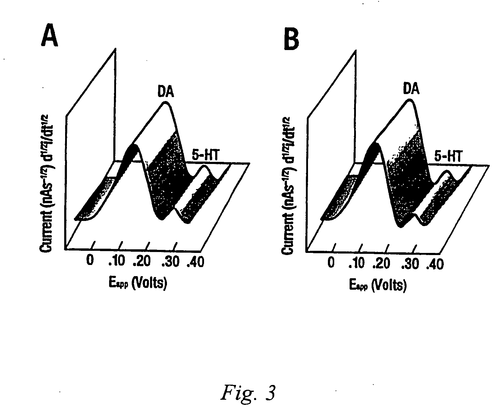
[0133] Microvoltammetry with Broderick probe stearic acid electrodes was used to detect norepinephrine (NE), dopamine (DA), ascorbic acid (AA), and serotonin (5-HT) in resected temporal lobes of 9 MTLE and 4 NTLE patients with temporal lobe epilepsy. Neurotransmitters were detected in separate signals within the same recording within seconds in alveus by experimentally derived oxidative potentials, determined in vitro in Ringers Lactate or PO4 buffer. Ag / AgCl reference and stainless steel aux...


PUM

PropertyMeasurementUnit
Massaaaaaaaaaa
Concentrationaaaaaaaaaa
Shapeaaaaaaaaaa

Abstract

The present invention relates to devices and methods of use thereof for detection of biomolecules, in vitro, in vivo, or in situ. The invention relates to methods of diagnosing and/or treating a subject as having or being at risk of developing a disease or condition that is associated with abnormal levels of one or more biomolecules including, but not limited to, inter alia, epilepsy, diseases of the basal ganglia, athetoid, dystonic diseases, neoplasms, Parkinson's disease, brain injuries, spinal cord injuries, and cancer. The invention also provides methods of differentiating white matter from gray matter. In some embodiments, regions of the brain to be resected or targeted for pharmaceutical therapy are identified using sensors. The invention further provides methods of measuring the neurotoxicity of a material by comparing microvoltammograms of a neural tissue in the presence and absence of the material using the inventive sensors.

Description

[0001] This application is a continuation-in-part application under 35 U.S.C. §120 of U.S. patent application Ser. No. 10 / 118,571, filed Apr. 8, 2002, now allowed, entitled, “Identification, Diagnosis, And Treatment Of Neuropathologies, Neurotoxicities, Tumors, And Brain And Spinal Cord Injuries Using Microelectrodes With Microvoltammetry,” which claims the benefit of U.S. provisional application Ser. Nos. 60 / 326,407, filed on Oct. 1, 2001; 60 / 297,276, filed on Jun. 11, 2001; and 60 / 282,004, filed on Apr. 6, 2001, all now expired, all of which are herein incorporated by reference.FIELD OF INVENTION [0002] This invention relates to the identification, diagnosis, and treatment of neuropathologies, neurotoxicities, tumors, and brain and spinal cord injuries using electrodes with microvoltammetry. BACKGROUND OF THE INVENTION [0003] The present invention relates to devices and methods of use thereof for determining the presence and concentration of chemicals in a cell, tissue, organ or o...

Claims


Application Information

Patent Timeline
IPC IPC(8): C12Q1/68G01N33/574A61B5/05
CPCA61B5/14542A61B5/1468C12Q1/6883A61B5/4094A61B5/1473G01N33/48728A61B34/20A61B5/14507A61B5/14546A61B5/14735A61B5/4041A61B5/4082A61B5/4845A61B2562/0209G01N33/5058G01N33/5091G01N2800/2857G01N2800/302
Inventor BRODERICK, PATRICIA A.PACIA, STEVEN V.
Owner RES FOUND THE CITY UNIV OF NEW YORK
Why Eureka
Try Eureka