Optical probe for arterial tissue analysis

an optical probe and arterial tissue technology, applied in the field of arterial tissue diagnosis, can solve the problems of blood clots, heart attacks or stroke, but in the rapid and reliable measurement of such plaques, and achieve the effect of precise color analysis and reliable identification of vulnerable plaques

Inactive Publication Date: 2007-02-15
NEWTON LAB
View PDF6 Cites 26 Cited by
  • Summary
  • Abstract
  • Description
  • Claims
  • Application Information

AI Technical Summary

Benefits of technology

[0004] Angioscopy is of value in visualizing atherosclerotic lesions in a vascular field flushed with saline or other clear fluid. Angioscopy has indicated that yellow color intensity of the plaque is strongly related with the prevalence of thrombosis on the plaque. The yellow color intens

Problems solved by technology

More specifically, these vulnerable plaques can result in the formation of blood clots that can cause heart attacks or s

Method used


Image

  • Optical probe for arterial tissue analysis
  • Optical probe for arterial tissue analysis
  • Optical probe for arterial tissue analysis

Examples


Embodiment Construction

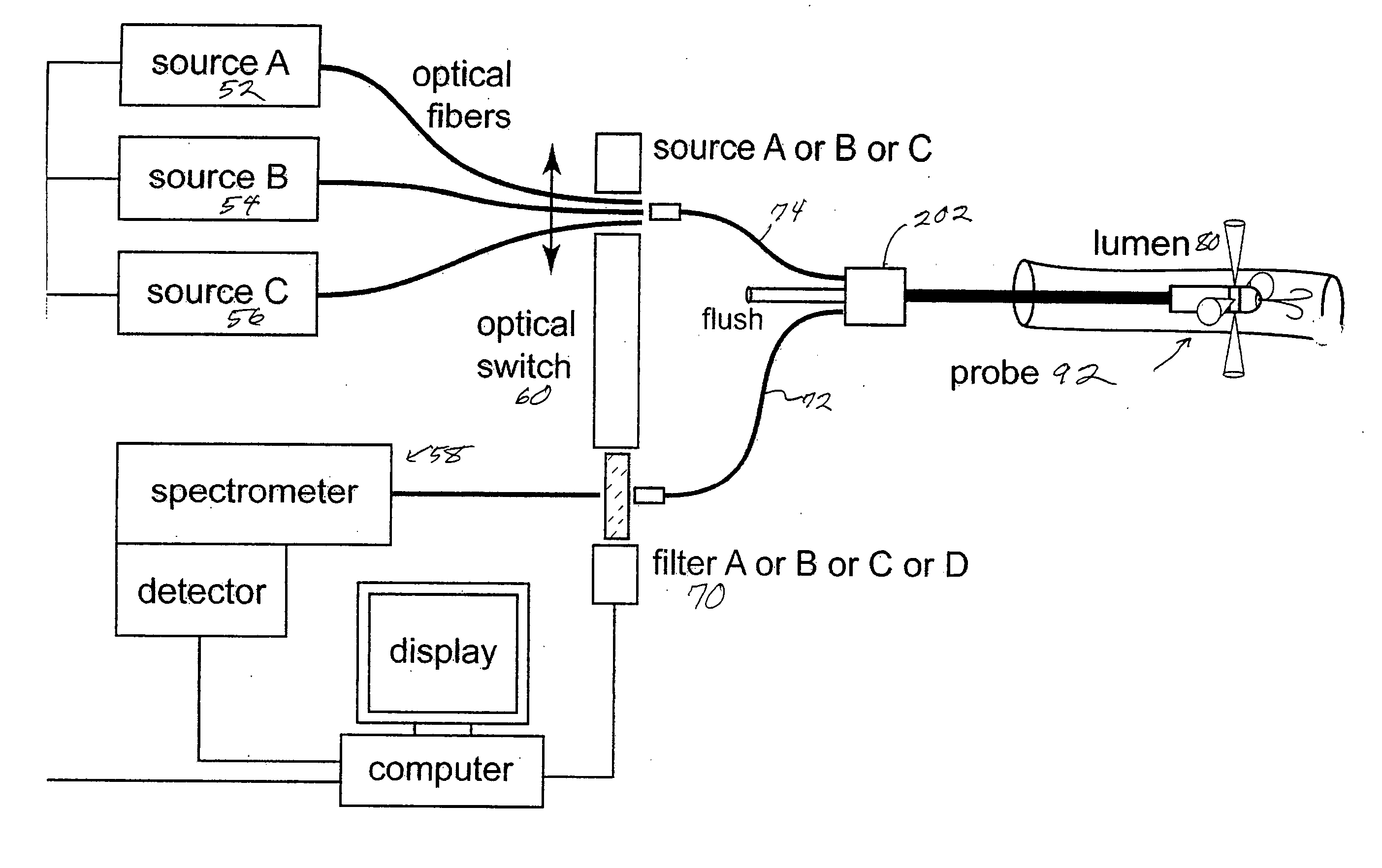
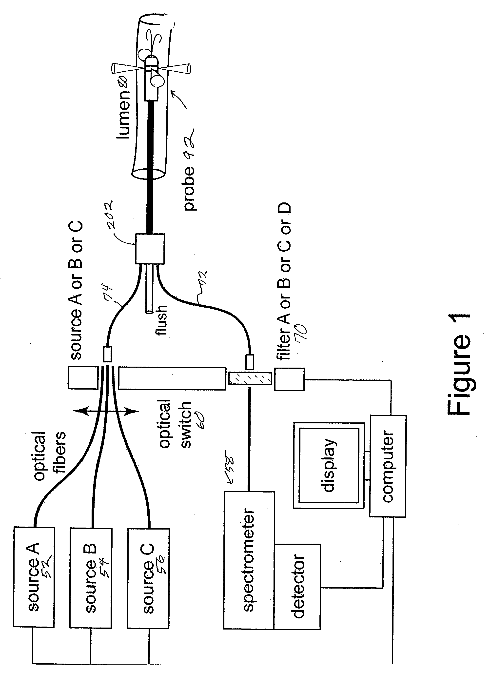
[0022]FIG. 1 shows a schematic of the diagnostic system. Three excitation sources can be used. An ultraviolet (UV) excitation source (A), operating at 337 nm from a nitrogen laser, for example, a visible excitation range in the range of 400 nm to 700 nm from a diode laser (B), for example, and a broadband white light excitation from a source (C) such as a xenon arc lamp or quartz tungsten halogen bulb for example. The UV and visible lasers are chosen to excite particular fluorophores in the tissue. The white light is used to determine the scattering characteristics of the tissue including its apparent color. An optical switch 60 can be used to select which of the light sources is coupled to the excitation delivery fibers 74 in the probe 92 at a given moment and simultaneously moves the appropriate optical filter 70 into the receiver fiber 72 path before the spectrometer 58.

[0023] In the case of the UV or visible fluorescence diagnostics these filters are used to block excitation li...


PUM


Abstract

The present invention relates to systems and methods used in the measurement of arterial tissue. Optical probes in accordance with the invention use optical fibers to deliver and collect light using a sidelooking catheter. Diffused white light and fluorescence scattering is collected and processed to provide for improved artery wall diagnosis.

Description

CROSS REFERENCE TO RELATED APPLICATIONS [0001] This application claims the priority of U.S. Provisional Application No. 60 / 686,600 filed Jun. 2, 2005 entitled, OPTICAL PROBE FOR ARTERIAL TISSUE ANAYLSIS, and U.S. Provisional Application No. 60 / 686,601 filed Jun. 2, 2005 entitled OPTICAL PROBE FOR RAMAN SCATTERING FROM ARTERIAL TISSUE. The entire contents of the above applications are being incorporated herein by reference.BACKGROUND OF THE INVENTION [0002] Methods and devices have been developed for the diagnosis of arterial tissue. These methods have included spectroscopic techniques such as tissue autofluorescence to measure characteristics of the tissue. Among those characteristics to be measured are included the presence of vulnerable plaques that contribute to the susceptability of individuals to heart disease or stroke. More specifically, these vulnerable plaques can result in the formation of blood clots that can cause heart attacks or stroke. [0003] Fiber optic catheters hav...

Claims


Application Information

Patent Timeline
IPC IPC(8): A61B6/00
CPCA61B5/0071A61B5/02007A61B5/0084A61B5/0075
Inventor FULGHUM, STEPHEN F. JR.THIMMARAJU, SUDHAFELD, JONATHAN
Owner NEWTON LAB
Why Eureka
Try Eureka