
Subcutaneous needle connection system

a subcutaneous needle and connection system technology, applied in the field of surgical implants, can solve the problems of significant health care hazards for both patients and healthcare workers associated with such traditional needle access methods, such as the death of esrd patients within days of total renal failure, and the inability to create a graft or fistula in some patients, so as to improve the fluid flow dynamics, improve the sealing effect, and improve the mechanical stability of the skin

Inactive Publication Date: 2007-04-12
ATRIUM MEDICAL
View PDF4 Cites 109 Cited by
  • Summary
  • Abstract
  • Description
  • Claims
  • Application Information

AI Technical Summary

Benefits of technology

[0011] The present invention provides a subcutaneous needle connection system for providing access to a vascular system of a patient that has enhanced mechanical stability under the skin, improved sealing, improved fluid flow dynamics, improved needle hole healing potential of the skin and improved needle edge interfacing to minimize flow disturbance and blood cell trauma during use. The subcutaneous needle connection system is not a port chamber device like other early access systems, as it comprises a housing having a shaped bottom surface designed to facilitate stability of the subcutaneous needle connection system with a straight bidirectional fluid path. The subcutaneous needle connection system is connected to at least one catheter in communication with a vascular system of a patient and further includes one or more needle access openings for receiving a needle for accessing blood from the patient or returning treated blood to a patient for a procedure such as hemodialysis. The needle access openings may include a set of inner valves configured to automatically open upon insertion of a needle and a set of outer valves that may require manual activation in order to open, thereby ensuring enhanced sealing of the fluid paths.
[0012] The needle access openings may be angled away from each other to facilitate needle insertion and reduce operator error by preventing accidental insertion of the wrong needle in an opening. In addition, the surgically implanted housing may be manually rotated within the body or manipulated by an operator to increase the needle penetration zone are of the skin, reducing the need for repeated penetration of one area of skin during subsequent needle insertions into the needle connection system. Furthermore, the subcutaneous needle connection system may provide a substantially turbulent-free fluid flow path between one or both needle access openings and a corresponding catheter to maximize blood flow through the subcutaneous connection device.

Problems solved by technology

Without needle or indwelling catheter organ access for dialysis, there is no physical connection means to conduct dialysis toxin removal, and the ESRD patients would die within days of total renal failure.
However, the skin surrounding the holes where the catheters enter the body have trouble sealing around the catheters and can become infected.
However, the creation of an AV fistula or graft can be difficult or impossible in some patients.
In addition, because both types of dialysis treatment techniques discussed above require trained medical personnel for needle or catheter access and the administration of the actual external dialysis filtration process, there are significant health care hazards for both the patient and healthcare worker associated with such traditional needle access methods.
There are a number of additional complicating issues relating to the process of repeatedly sticking and cannulating a patient's circulatory organ system and removing / returning blood back to the patient.
One significant complication is the need for maintenance of a sterile installation and connection technique for all components involved in establishing dialysis access through the patient's skin and into the hollow targeted organ, as well as connection to the dialysis tubing set, filter, and dialysis pump apparatus.
Another complication is the inability of a particular artery or vein to be repeatedly cannulated or punctured at substantially the same convenient needle or catheter access site, due to vessel trauma, exit wound inflammation, dialysis graft complications, and / or enlarged needle hole formation resulting in massive needle hole bleeding / hematoma formation.
Repeated dialysis needle punctures create unwanted scar tissue formation and infection.
Such large gauge needle cannulation complications are uncomfortable for the patient and the healthcare worker, due to the associated pain of additional needle sticks.
Infection complications of these needle access sites are difficult to treat, because of the constant migratory effects of nosocomial infections, which have been well documented to routinely originate from direct contact with topical skin sourced Staphylococcocus bacteria.
These frequently occurring needle access complications often require surgical intervention to repair, reconstruct, or remove the affected vessel organ, in addition to requiring extended hospital admission and costly medication treatment with IV medications.
However, prior vascular access systems used for percutaneous needle cannulation for removal and return of blood during dialysis suffer many clinically devastating problems.
Clinical problems include, but are not limited to, complications from poor tissue healing around the needle holes in the skin and subcutaneous needle tracks under the skin, caused by multiple puncturing of the same location, subcutaneous necrosis due to the proximity of the needle entrance to such devices, fluid contamination from leakage within the system and outside the needle entry, poor tissue coverage due to the shape and location of the indwelling permanent catheter connections as they exit the access system device, risk of infection caused by the compromised skin healing and leaking fluid during and after needle insertion, infection accumulation around the catheter connection and other complications.
The condition of the skin with such devices has been observed to often include a poor blood supply condition of the skin because of the stretched condition of the skin, and the lack of adequate vascularized subcutaneous tissue to help fight infection and promote healing of the injured skin and subdermal tissue.
Furthermore, the extremely short path of the needle through the skin to the opening of these port devices also provides less opportunity for the vascularized tissue to help control and prevent bacterial infection from occurring in and around the openings to the port opening.
Moreover, because the needle punctures are occurring in the same skin location each time, infection and localized fluid accumulation abound the skin and device is common.
The needle punctures create a clottable buttonhole over time, which is a skin injury that is not well controlled or completely sealed to the environment and therefore prone to repeatable, if not chronic infection.
A further difficulty of conventional port access systems is their fluid filling chamber design, open bevel needle exposure to flowing blood fluids, causing massive blood cell damage and turbulent disruption to the blood by requiring sharp changes in fluid flow direction, such as a 90° change shortly just prior to fluid entering the exposed beveled edge of the needle, or just after exiting the open end of the needle as fluid enters the device fluid chamber.

Method used


Image

  • Subcutaneous needle connection system
  • Subcutaneous needle connection system
  • Subcutaneous needle connection system

Examples


Embodiment Construction

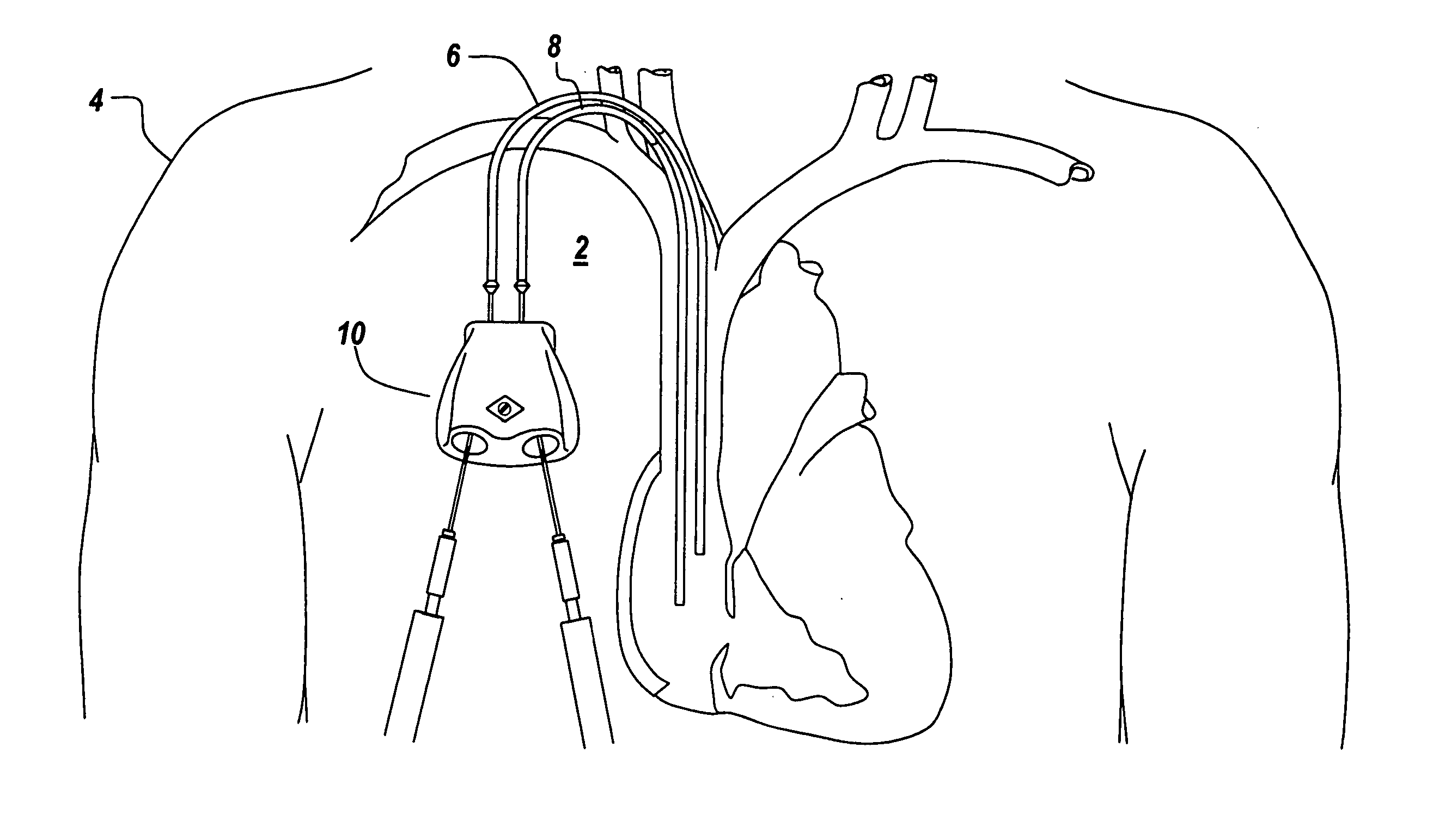
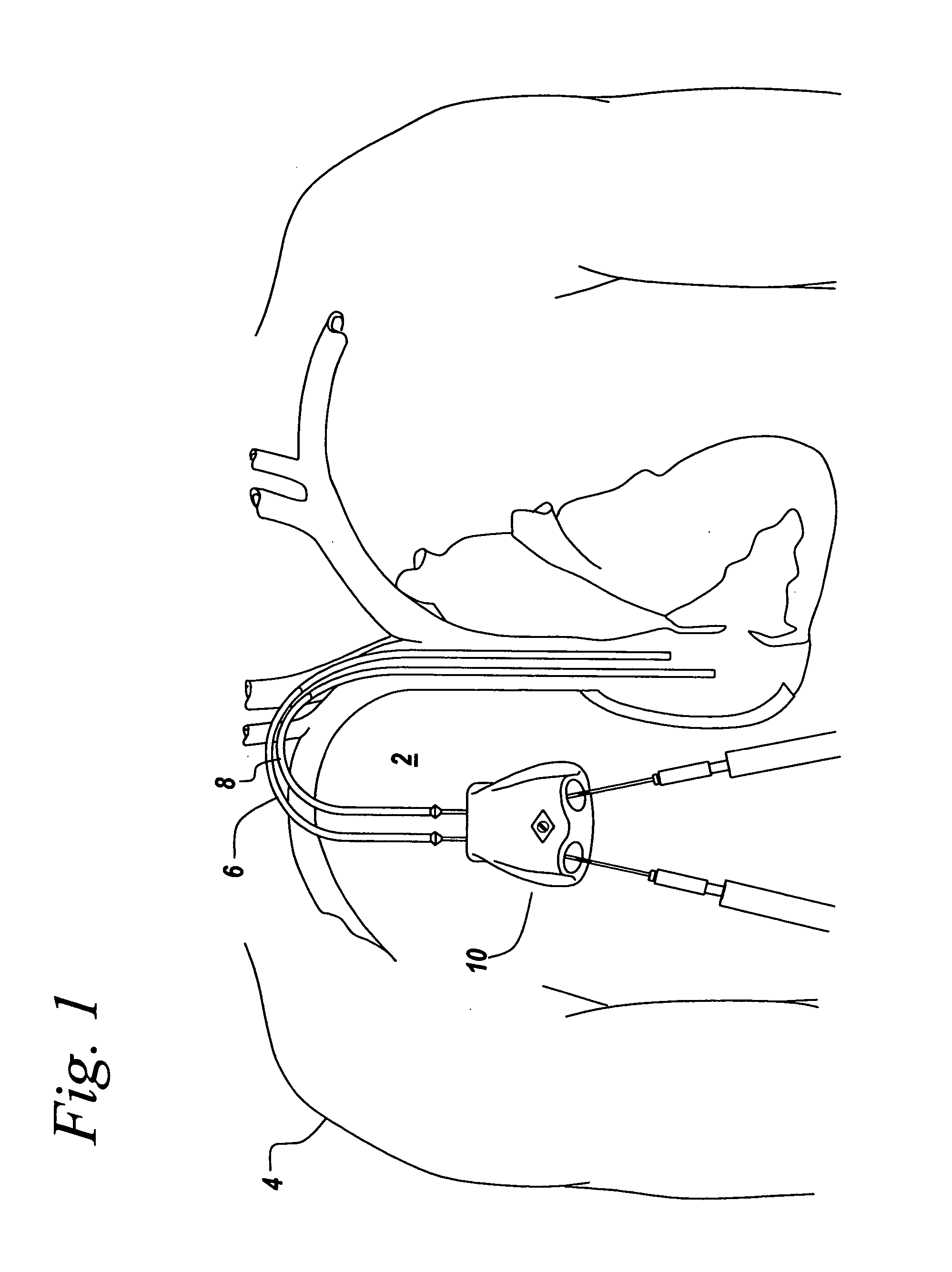
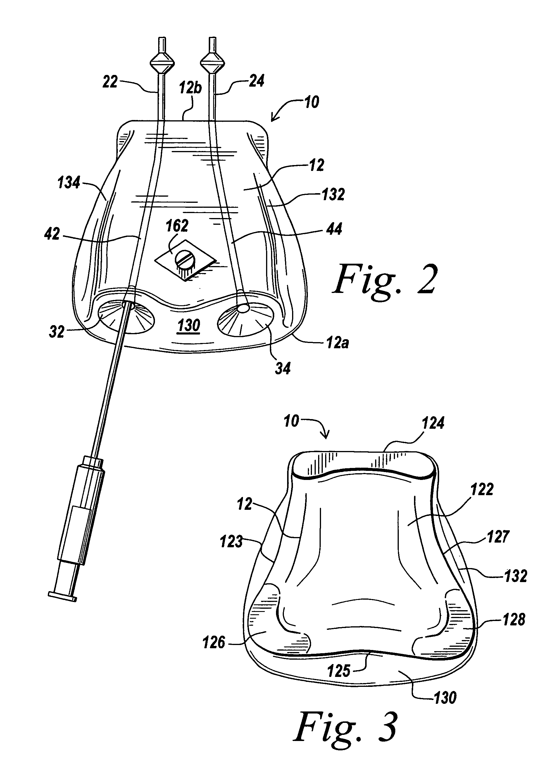
[0026] An illustrative embodiment of the present invention provides a subcutaneous needle connection system for providing access to a vascular system of a patient that exhibits enhanced mechanical stability under the skin, improved fluid flow dynamics, improved needle hole healing potential of the skin, improved needle edge interfacing to minimize flow turbulence and blood cell trauma during use, and other features. The invention will be described below relative to certain illustrative embodiments. Those skilled in the art will appreciate that the present invention may be implemented in a number of different applications and embodiments and is not specifically limited in its application to the particular embodiments depicted herein.

[0027]FIGS. 1-7 illustrates an embodiment of a subcutaneous needle connection system 10 for providing access to a vascular system of a patient according to an illustrative embodiment of the invention. The illustrative subcutaneous needle connection syste...


PUM


Abstract

An apparatus for establishing a re-usable, recurring, mechanical connection to an organ within a patient is provided. A subcutaneous needle connection system for providing bidirectional, straight and turbulent-free fluid flow access to a vascular system of a patient includes a housing defining a needle access opening for receiving a needle, a cannula opening in communication with the vascular system and a passageway connecting the needle access opening to the cannula opening. The housing can additionally have a second needle access opening connected to a second cannula opening via a second passageway. The subcutaneous needle connection system allows for blood to be removed from the vascular system via one cannula opening, passed to a hemodialysis system via a needle inserted in an associated needle access opening, treated using the hemodialysis system, and returned to the vascular system via the other needle access opening and associated cannula opening. The implantable needle connection system provides enhanced mechanical stability under the skin, improved fluid flow dynamics, improved needle hole healing potential of the skin, and improved needle edge interfacing to minimize flow turbulence and blood cell trauma during use.

Description

FIELD OF THE INVENTION [0001] The present invention relates to a surgical implant to provide a subcutaneous connection to a vascular system of a patient. More particularly the present invention relates to a subcutaneous needle connection system for providing repeated access to the vascular system of a patient. BACKGROUND OF THE INVENTION [0002] A number of patients today undergo recurring medical procedures requiring repeated skin penetration to access the patient's vascular system and internal organs, including organs contained within the mediastinal, chest, abdominal and peritoneal cavities. One such recurring medical procedure is hemodialysis, which is used to treat kidney failure by removing harmful wastes and excess salts and fluid from a patient's blood. [0003] Currently, over one million patients worldwide suffer from End State Renal Disease (ESRD) conditions and require some form of daily or thrice weekly dialysis treatment via needle or catheter access. Peritoneal Dialysis ...

Claims


Application Information

Patent Timeline
Patent Type & Authority Applications(United States)
IPC IPC(8): A61M31/00
CPCA61M1/285A61M39/0208A61M2039/0211A61M1/3661
Inventor MUTO, RUDOLPHHERWECK, STEVE A.
Owner ATRIUM MEDICAL
Learn More