
Methods to ameliorate and image angioplasty-induced vascular injury

a vascular injury and angioplasty technology, applied in the field of vascular injury amelioration and image angioplasty, can solve the problems of tissue ischemia or infarction, most of the drug never reaches its intramural target, and the medical device has several significant limitations, so as to reduce the thrombosis and/or restenosis, prevent restenosis, and maximize the effect of distribution into the injured wall

Inactive Publication Date: 2007-06-21
BARNES JEWISH HOSPITAL
View PDF4 Cites 15 Cited by
  • Summary
  • Abstract
  • Description
  • Claims
  • Application Information

AI Technical Summary

Benefits of technology

"The present invention provides a method to prevent restenosis after angioplasty by using targeted emulsions containing anti-restenotic, anti-cell migratory, or anti-cell proliferative agents that allow the intima to heal and prevent restenosis. The emulsions can be administered before or during the interventional procedure and are designed to maximize distribution into the injured wall and minimize downstream losses. The method also includes the use of imaging agents to verify the delivery of the therapeutic agent to the blood vessel wall."

Problems solved by technology

The angioplasty procedure and the stents used have a number of drawbacks, including effecting neointimal proliferation leading to restenosis, and acute or delayed induction of arterial thrombosis, resulting in tissue ischemia or infarction.
Despite the benefits of drug eluting stents, these medical devices have several significant limitations.
Due to size and inflexibility, many sites of stenosis in distal lesions or small branches cannot be treated with DES, resulting in the use of a traditional non-DES or no stent at all.
However, most of the drug never reaches its intramural target.
The majority is washed from the stent and carried downstream, where it can impair the normal vasomotor functions of arterial wall, and the residual drug remaining with the stent, greatly delays intimal repair, a process dependent on the formation of the endothelial lining over the implanted metal struts of the stents.
Because of this delay in healing, the risk of thrombosis is maintained for many months and up to two years.
Unfortunately, in some situations, despite all efforts to avoid these risks, the subject may experience acute episodes of ischemia, infarction and frequently death.
In some cases these adverse events occur despite maximum medical therapy, in other instances treatment is prematurely discontinued by the physician or patient.
In other cases, the cost of these medications exceeds patient financial resources over the multi-month or year(s).

Method used


Image

  • Methods to ameliorate and image angioplasty-induced vascular injury
  • Methods to ameliorate and image angioplasty-induced vascular injury
  • Methods to ameliorate and image angioplasty-induced vascular injury

Examples


example 1

[0083] Rabbits were fed an atherogenic diet for three weeks, and then subjected to balloon stretch injury. A catheter was inserted from the left common carotid artery, and a double balloon expanded into each artery. From the space between the two balloons, in test rabbits, αvβ3 nanoparticles prepared as in preparation A and comprising rapamycin were administered. In a control contralateral vessel, the emulsion prepared in preparation A without rapamycin was administered.

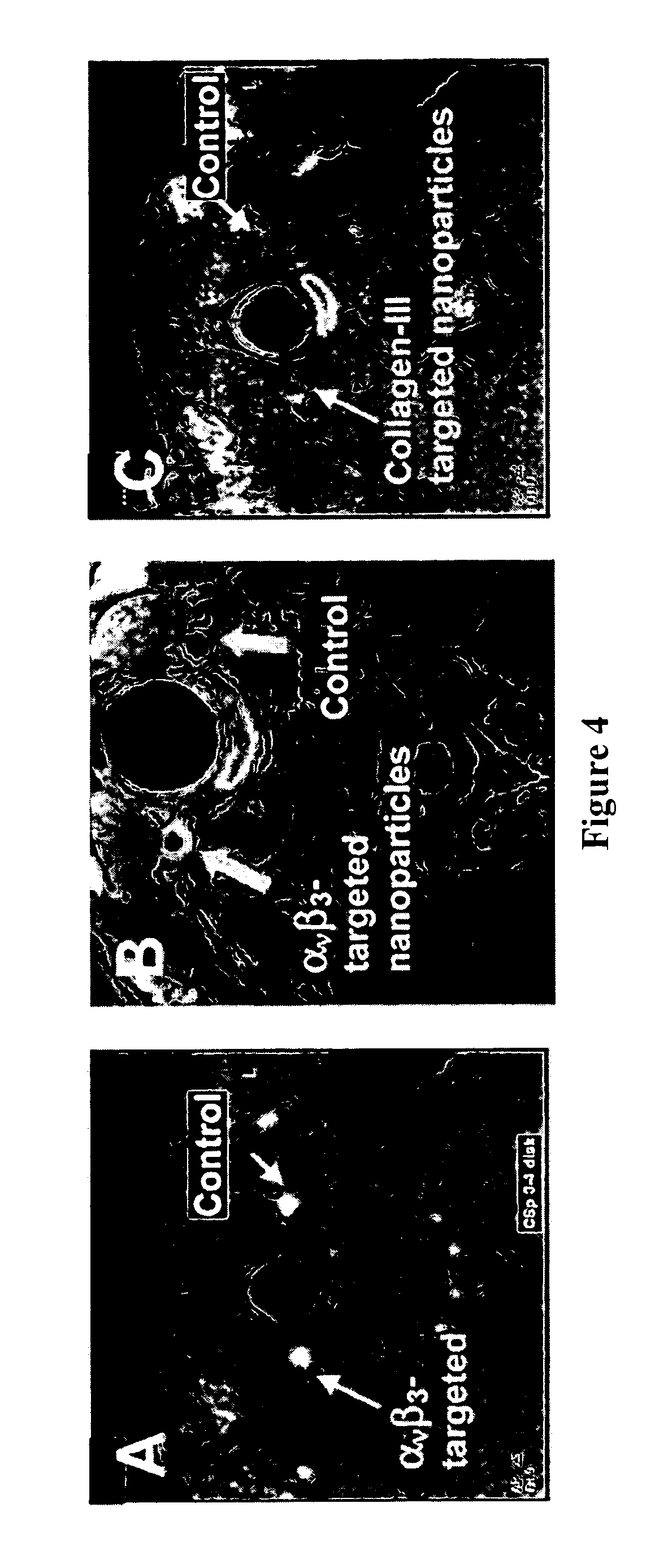
[0084] The test and controlled arteries were imaged by MRI contrast enhancement to detect the injury pattern and the distribution of nanoparticles. Plaque development after treatment was determined by microscopic methods.

[0085] Imaging with the αvβ3 integrin-targeted paramagnetic nanoparticles showed delineation of the stretch injury pattern. Magnetic resonance imaging is performed at 1.5 T, a clinically relevant field strength, using a clinical scanner (NT Intera CV, Philips Medical Systems) and a quadrature birdc...

example 2

[0087] New Zealand White Rabbits were fed 0.25% cholesterol diet for four months which resulted in plaque formation in the femoral artery. The artery was opened using balloon angioplasty using a dual balloon catheter and dispensing 0.4 ml of αvβ3-integrin-targeted perfluorocarbon nanoparticles containing 0.3 mol % rapamycin in 12 of the rabbits, or non-targeted nanoparticles in 6 of the rabbits, or saline in 6 of the rabbits over the course of five minutes. The release of the drug was determined with dissolution studies and after 3 days more than 97% of the rapamycin was still incorporated in the nanoparticle emulsion. The emulsion also contained 99mTc label permitting detection of the local delivery of the targeted emulsion, but not the nontargeted emulsion into the femoral arteries.

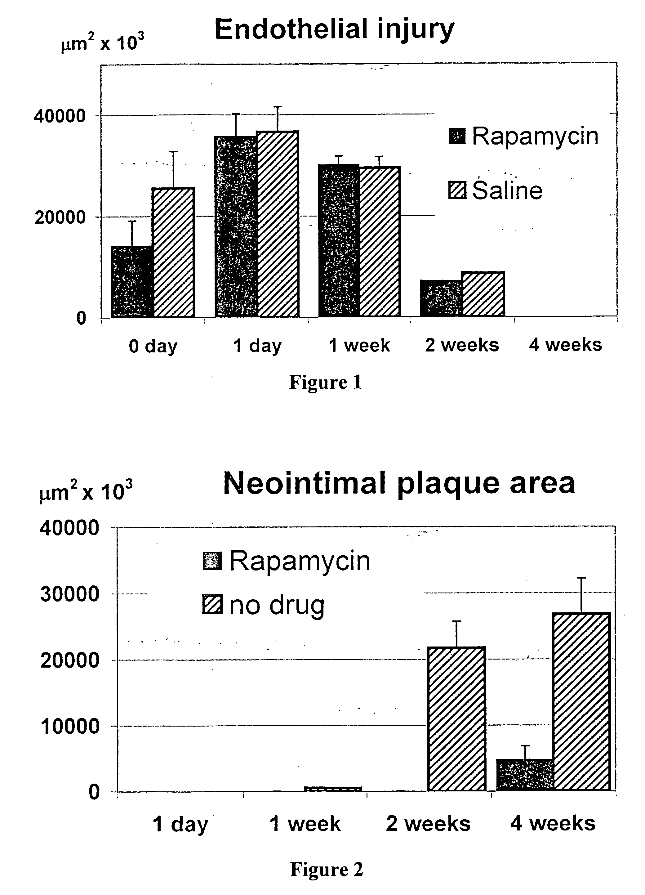
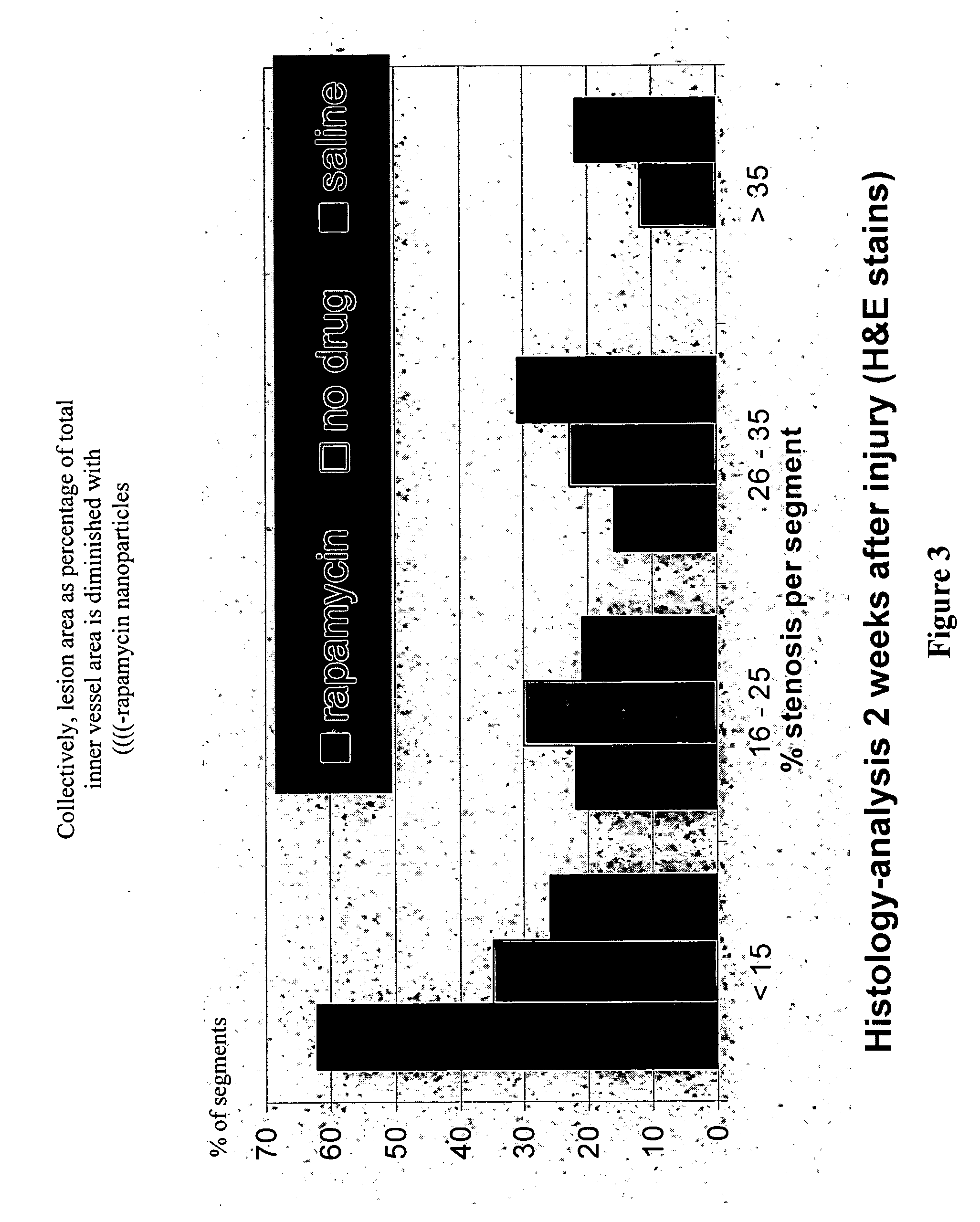
[0088] Stenosis developed in balloon-injured, but untreated femoral arteries, but not in those exposed to the αvβ3-targeted perfluorocarbon nanoparticles with rapamycin A, 4 weeks after injury.

[0089] ...

example 3

[0092] Ligand-targeted paramagnetic nanoparticles were prepared as previously described. Briefly, the nanoparticles comprised 20% (volume / volume) perfluorooctylbromide (PFOB; Exfluor Research, Round Rock, Tex., USA) and 1.5% (weight / volume) of a surfactant co-mixture, 1.7% (w / v) glycerin and water for the balance. The surfactant co-mixture included 69.9 mole % lecithin (Avanti Polar Lipids, Inc., Alabaster, Ala., USA), 0.1 mole % peptidomimetic vitronectin antagonist (Bristol-Myers Squibb Medical Imaging, Billerica, Mass., USA) or anti-collagen III f(ab) (CSIRO, Victoria, Australia) coupled to MPB-PEG2000-phosphatidylethanolamine (Northern Lipids, Inc., Vancouver, British Columbia, Canada), and 30 mole % of gadolinium diethylene-triamine-pentaacetic acid-bis-oleate (Gateway Chemical Technologies, St. Louis, Mo., USA). Nontargeted, paramagnetic particles were prepared by substituting the ligand-lipid conjugate with lecithin. The nominal sizes for each formulation were measured with a...


PUM

PropertyMeasurementUnit
mole %aaaaaaaaaa
concentrationaaaaaaaaaa
areaaaaaaaaaaa

Abstract

Methods for inhibiting restenosis in blood vessels expanded by angioplasty are described. The method comprises administering blood vessel wall-targeted emulsion containing an anti-restenotic agent.

Description

RELATED APPLICATION [0001] This application claims benefit of U.S. provisional application 60 / 741,929 filed 2 Dec. 2005. The contents of this document are incorporated herein by reference in their entirety.TECHNICAL FIELD [0002] This invention relates to methods to prevent restenosis and ameliorate vascular injury induced by angioplasty. More particularly, the invention relates to the use of targeted particulate emulsions comprising a therapeutic agent that may aid in repair of an injured blood vessel, as well as retarding restenosis. Images of the injury may also be obtained. BACKGROUND ART [0003] Clogged or constricted blood vessels are often treated by angioplasty—i.e., insertion of an inflatable device to effect opening of the vessel, usually followed by placement of a stent in the open vessel to address vascular recoil due to spasm or local wall dissections caused by the original procedure. The angioplasty procedure and the stents used have a number of drawbacks, including effe...

Claims


Application Information

Patent Timeline
Patent Type & Authority Applications(United States)
IPC IPC(8): A61K51/00A61K39/395A61K38/17A61K31/704A61K31/4745A61K31/337A61K31/336
CPCA61K9/0024A61K9/10A61K31/336A61K31/337A61K31/4745A61K31/704A61K38/17A61K47/48238A61K47/488A61K47/48815A61K49/0002A61K49/1809A61K49/1812A61K49/1866A61K51/1217B82Y5/00A61K47/62A61K47/6907A61K47/6911
Inventor LANZA, GREGORYWICKLINE, SAMUEL A.
Owner BARNES JEWISH HOSPITAL
Learn More