Method for stimulating angiogenesis and wound healing

a technology of angiogenesis and wound healing, applied in the direction of angiogenin, prosthesis, peptide/protein ingredients, etc., can solve the problems of promiscuous induction of angiogenesis in tissues, short half-life of vegf protein, and significant challenge in the delivery of such angiogenic promoters such as vegf protein, so as to achieve efficient binding of a protein and promote angiogenesis

Inactive Publication Date: 2007-06-21
TRUSTEES OF BOSTON UNIV
View PDF3 Cites 44 Cited by
  • Summary
  • Abstract
  • Description
  • Claims
  • Application Information

AI Technical Summary

Benefits of technology

[0014] The present invention is directed to using a device that will efficiently bind a protein under acidic conditions, while permitting its release under normal physiological conditions. One can use any protein that contains a domain that binds to heparin or fibronectin, preferably heparin, where that binding is pH dependent such as occurs with VEGF. Preferably, the protein contains or is altered to contain a heparin-binding consensus sequence such as XBBBXXBX or XBBXBX, where B is a basic amino acid residue such as Lys, Arg, or His, and X is any amino acid residue. Preferably, the protein contains or is altered to contain a pH sensitive residue. For example, His has a pH sensitive ionizable moiety. The fibroblast growth factor family of heparin-binding proteins have a stretch of about nine amino acids that are believed to play a role in heparin-binding specificity. [Ashikari-Hada, S., et al., J. Biol. Chem (2004); Luo, Y. et al., Biochemistry 37:16506-15(1998)]

Problems solved by technology

The delivery of such angiogenic promoters such as VEGF protein remains a significant challenge.
For example, the half-life of VEGF protein is very short; the administration of high doses of VEGF protein is associated with hypotension, and systemic administration of VEGF protein can cause promiscuous induction of angiogenesis in tissues other than that which has been targeted.
Promiscuous induction of angiogenesis can cause blindness, increase the aggressiveness of tumor cells, and lead to a multitude of other negative side-effects.
If too little VEGF protein is delivered, angiogenesis will not be induced, and a significant therapeutic benefit will not be achieved.
If too much VEGF protein is delivered, the formation of disorganized vasculature beds, loss of function

Method used


Image

  • Method for stimulating angiogenesis and wound healing
  • Method for stimulating angiogenesis and wound healing
  • Method for stimulating angiogenesis and wound healing

Examples


example 1

Affect of Heparin and Heparin-Derived Compounds on Binding

Results

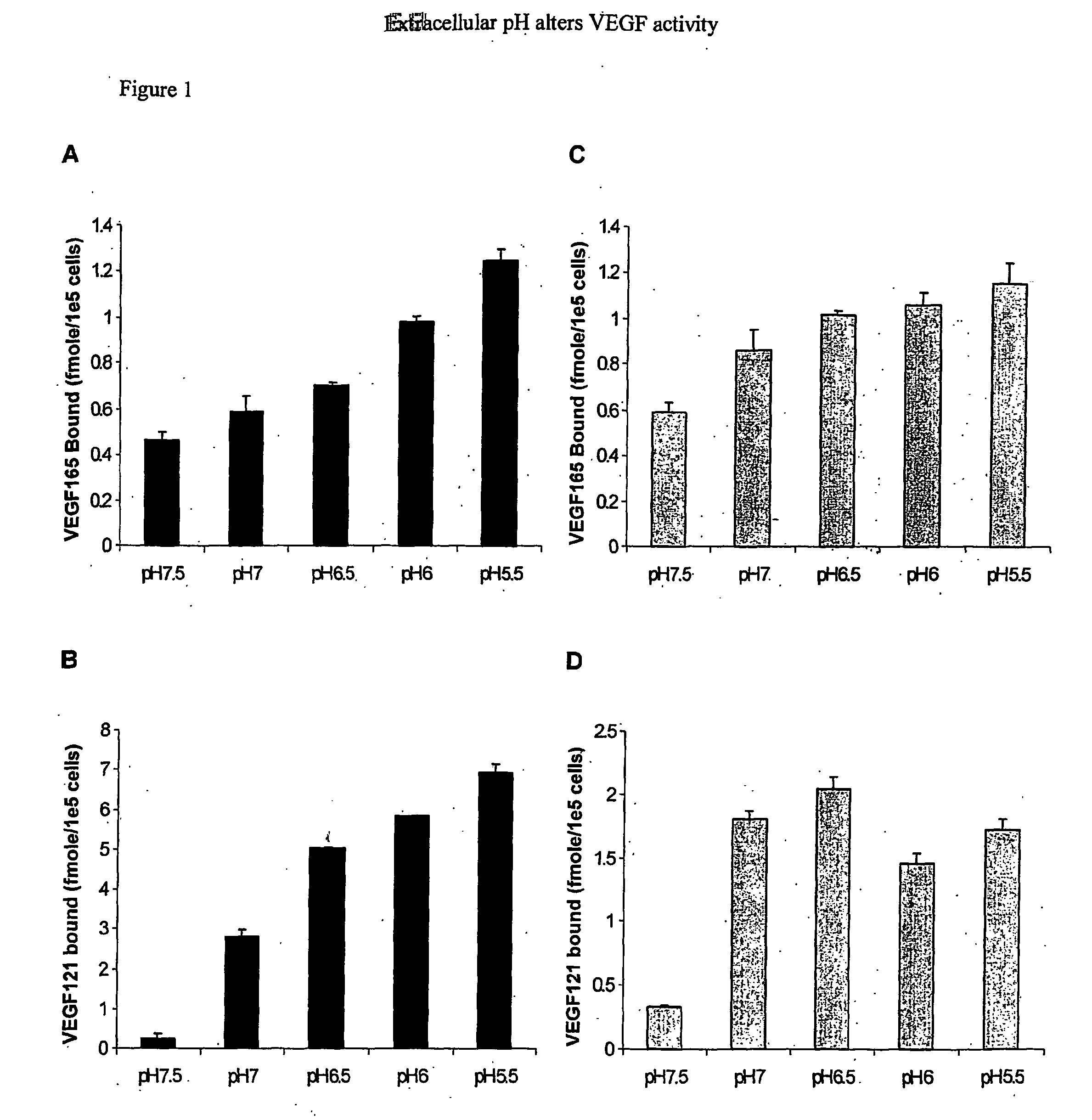
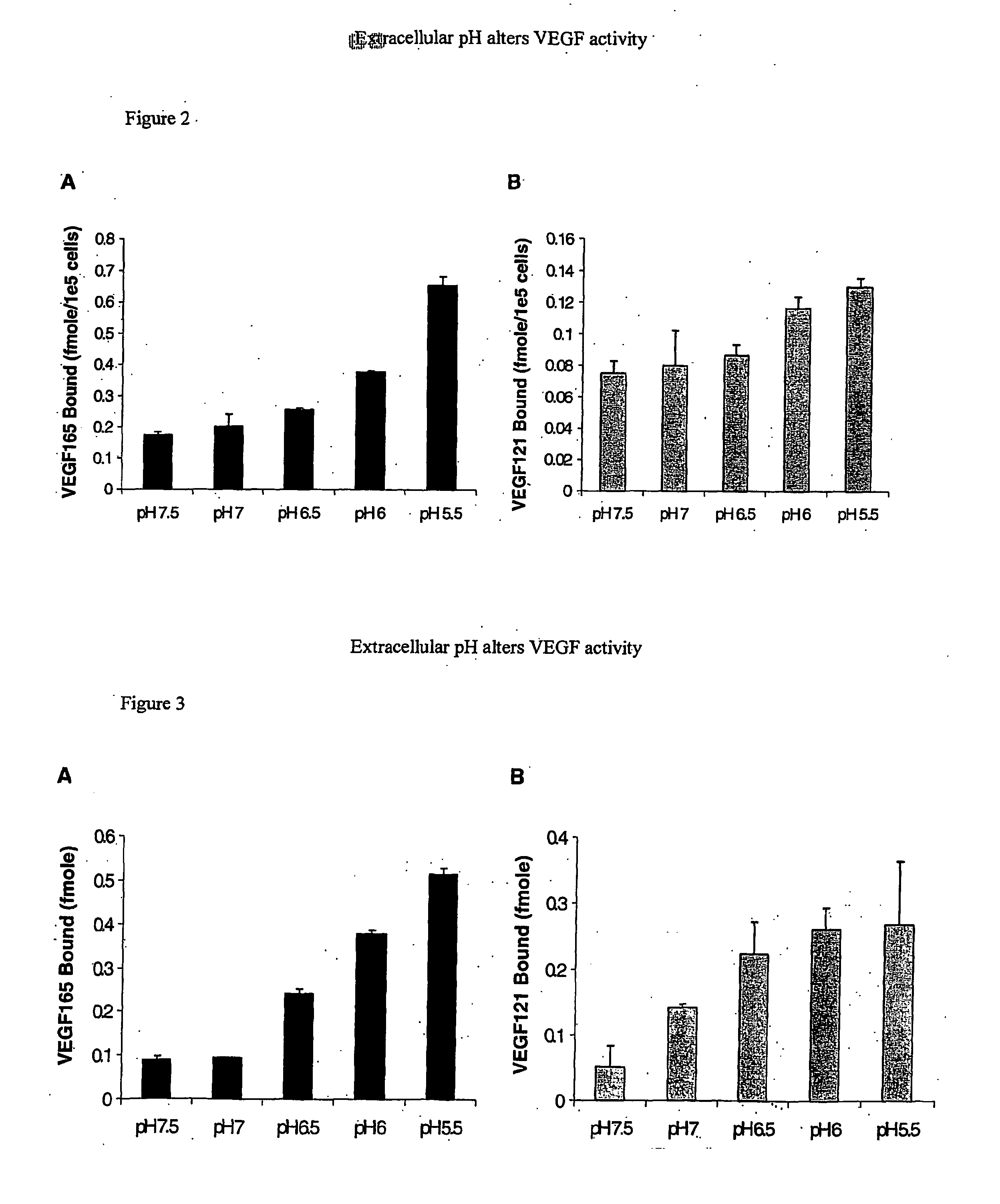
VEGF165 and VEGF121 Binding to Cell Surfaces are Altered by pH

[0095] At sites of angiogenesis, such as tumors and wounds, the environment is rather hypoxic. Hypoxia increases expression of VEGF thereby promoting increased rates of migration and proliferation of endothelial cells. Hypoxic environments also lead to decreases in extracellular pH. While the intracellular signaling events occurring under hypoxic conditions in response to VEGF have been of major focus, there has been little attention to how acidity affects VEGF outside of cells. Therefore, to investigate the role of extracellular pH on VEGF165 and VEGF121 interactions with cell surfaces, binding assays were conducted with confluent BAEC at various pHs ranging from pH 7.5-5.5 (FIG. 1). It was found that as pH decreased, VEGF165 and VEGF121 binding to BAEC increased dramatically. HSPG-mediated VEGF165 binding at pH 5.5 was ˜2.5 fold greater than that at p...

example 2

[0108] Factors in Addition to Heparin and Heparin-Derived Compounds Affecting Binding

Activation of Erk½ in BAEC

[0109] Erk12 / activation in BAEC was evaluated in response to VEGF that had been pre-bound to BAEC at pH 5.5. BAEC were plated at 20,000 cells per well in 6-well dishes. After 24 h, the medium was replaced with DMEM containing 0.5% CS for 24 h, to quiesce the cells. Cells were incubated with binding buffer at pH 5.5 for 10 min at 37° C. VEGF165 (0.6 nM) was added to the cells at pH 5.5 for 60 miin at 37° C. Unbound VEGF165 was removed. Cells were incubated for 10 min in buffer at pH 7.5, 7.0, 6.5, 6.0, or 5.5. Erk½ activation in BAEC was evaluated in response to VEGF that had been dissociated from fibronectin. Quiescent endothelial cells were generated as stated above. Binding buffer (1 mL) was added to cells at pH 7.5 or pH 5.5, for 90 min. VEGF165 (3.0 nM) and VEGF121 (3.5 nM) were allowed to bind to fibronectin-coated dishes in the presence of 1 μg / mL of heparin for 1...

example 3

Preparation of VEGF Encapsulated Spheres

[0122] A 1:1 heparin-Sepharose slurry (45 mg / ml, pH 7.5) was combined with fibronectin (0.30 mg / ml), VEGF165 / 121 (1.25 ug / ml), and binding buffer (10 mM HEPES, 1 mg / ml BSA, pH 5.5). The final pH of the mixture was determined to be 5.5. The mixture was incubated for 2 hours at 4° C. with rotation. After incubation, sodium alginate (1.8% w / v, pH 5.5) was added to the solution to produce a final alginate concentration of 1.2% w / v. The solution was gently mixed to ensure uniform distribution. The alginate mixture was then extruded though a 22 gauge needle into cold calcium chloride solution (1.5% w / v, pH 5.5). Calcium alginate spheres were immediately formed upon entering the hardening solution. The alginate spheres were allowed to further crosslink for 5 minutes with gentle mixing, followed by 15 minutes without mixing. The spheres were then washed three times with cold HEPES buffer (10 mM HEPES, pH 5.5). Each sphere had an average diameter of 3...


PUM


Abstract

A device comprising an extracellular matrix having an internal pH between 4.0 and 6.0 is discussed. This matrix contains heparin or a heparin-related compound such as heparan sulfate. Preferably the matrix also contains fibronectin or a fragment thereof. The matrix will bind to a protein having a pH dependent binding to heparin such as VEGF, preferably VEGF 121 or VEGF 165. The device will release the protein as the pH increases to physiological pH, such as 7.0 to 7.5. The device can be used to deliver a drug to a specific site. For example, with VEGF to a site in need of angiogenesis.

Description

[0001] This invention was supported by National Institutes of Health grant HL56200 and the government of the United States has certain rights thereto.FIELD OF THE INVENTION [0002] The present application is directed to a method of delivering a protein bound to an extracellular matrix under acidic conditions that is released upon exposure to physiological pH and a device comprising such an extracellular matrix. Preferably such an extracellular matrix bound protein can be used in stimulating angiogenesis and for example, enhancing wound healing, where the bound protein is an angiogenic promoter, preferably VEGF, that is released upon exposure to physiological pH. BACKGROUND OF THE INVENTION [0003] Angiogenesis, the growth of new blood vessels, is a complex process involving the disruption of vascular basement membranes, migration and proliferation of endothelial cells, and subsequent blood vessel formation and maturation. Several mediators are known to elicit angiogenic responses, and...

Claims


Application Information

Patent Timeline
IPC IPC(8): A61F2/00A61K38/16A61B
CPCA61K47/4823A61K47/48853A61L15/425A61L15/44A61L2300/414A61K38/1866A61K47/61A61K47/6921
Inventor NUGENT, MATTHEW A.GEORGES, ADRIENNE L.
Owner TRUSTEES OF BOSTON UNIV
Why Eureka
Try Eureka