Artificial cartilage tissue and production method thereof

a technology of cartilage and production method, which is applied in the field of artificial cartilage tissue, can solve the problems of poor self-regeneration ability, acute pain in the joint, and no way to regenerate cartilage, and achieves superior engraftablility, fixed mechanical strength, and high safety.

Active Publication Date: 2007-08-09
PG RES
View PDF7 Cites 11 Cited by
  • Summary
  • Abstract
  • Description
  • Claims
  • Application Information

AI Technical Summary

Benefits of technology

[0019] The present invention takes the above problems into consideration with the object of: establishing a preparation method of an artificial cartilage tissue which is substantially free of exogenous scaffolding material and heterologous contaminant protein, has a high safety, and has a fixed mechanical strength suturable with a suture thread; and providing an artificial cartilage tissue which has a fixed mechanical strength or composition showing a superior engraftablility, and a production method thereof.

Problems solved by technology

Such a structure in which these cells are dispersed throughout a matrix, that is, the fact that a small number of cells exist per unit volume, and the absence of vasculature means a very poor ability of self-regeneration when an injury occurs.
When articular cartilage is damaged due to aging, trauma, or the like, smooth movement of the joint is interferred and an acute pain occurs in the joint.
However, if the cartilage tissue itself is defective, there is no way to regenerate the cartilage other than implantation of external chondrocytes or cartilage tissue, or introduction of myeloid cells from the bone marrow underneath the cartilage so as to form a cartilage.
However, the regenerated cartilage is fibrocartilage which is poor in mechanical strength, and thus it is said that joint function lasts only for several years.
Moreover, it is difficult to introduce myeloid cells to form a cartilage at the cartilage defective site of osteoarthritis, which is a chronic degenerative disease of cartilage.
The current state is that there is no way other than artificial joint replacement as a treatment method for the last stage of osteoarthritis.
Artificial joint replacement is effective for relieving acute pains in the joint, but it is regarded as a “last resort” since the joint function is largely limited and the life span of the artificial joint is about 15 years (the interval between the bone of the patient and the artificial joint is loosened).
The method of using an osteochondral graft collected from a dead body involves problems of donor shortages and risks of infection.
However, there are shortcomings in that the collection quantity is limited, and a large osteochondral defect wound remains at the collection site.
However, in the method of reference 1, if chondrocytes are monolayer cultured, inherent characteristics of the chondrocytes partially disappear (dedifferentiation), and therefore, requirements for the-implant material, such as “redifferentiation”, “biosynthesis of the matrix”, and “repair of the defective site” are increased, thus the time required for recovery is inevitably elongated.
Moreover, a large number of the implanted cells are scattered and lost during that period, and thus many researchers are raising questions about the effect.
However, a drawback is the necessity of the periosteal patch.
Furthermore, because of the paste-like form, the strength is much weaker compared to the actual cartilage.
This means that it takes a long time to form the actual cartilage, and it is difficult for it to be compatible with the cartilage of the weight-bearing region of the joint.
Furthermore, in the method of reference 3, when a synthetic polymer is used for articular cartilage, the engraftment rate after implantation is poor.
The reason is that, since the cartilage is an avascular tissue without a bloodstream, it takes a long time for the synthetic polymer to disappear even if it is a bioabsorbable synthetic polymer, which may interfere with the fusion between the implant material and the surrounding tissue, and the alteration of the matrix composition into that of original articular cartilage during the time until the polymer disappears.
On the other hand, serum or plasma used for culturing involves a very big problem in terms of the safety of the prepared artificial cartilage tissue.
FBS shows a superior growth-stimulative effect and matrix synthesis-stimulative effect for chondrocytes, and thus is very effective for preparing artificial cartilage tissues, but has a risk of bovine spongiform encephalopathy (BSE) and a risk of immunogenicity due to a contaminant protein derived from FBS mixed in the artificial cartilage tissue.
If no serum nor plasma is used, such risks can be completely excluded, but preparation of engraftable artificial cartilage tissues is very difficult in serum-free media or plasma-free media.
However, it is difficult to prepare artificial cartilage tissues which are suturable with a superior engraftablility, that is, having a mechanical strength above a fixed level.

Method used


Image

  • Artificial cartilage tissue and production method thereof
  • Artificial cartilage tissue and production method thereof
  • Artificial cartilage tissue and production method thereof

Examples


example 1

Preparation of Artificial Cartilage Tissue Using Cells Derived from Porcine Articular Cartilage and its Mechanical and Biochemical Properties

1. Purpose

[0099] The purpose is to prepare an artificial cartilage tissue using cells derived from a porcine articular cartilage, to select the main constituents that reflect the mechanical strength, and to understand the contents of the main constituents having the suturable mechanical strength in the artificial cartilage tissue.

2. Cell Isolation

[0100] A hind-leg of an approximately one-year old LWD porcine was purchased from Shimoda Chikusan Limited. The knee joint was opened, and femur articular cartilage was aseptically collected. Hereinafter, according to a partially modified method of Mok S. S. et al. (J. Biol. Chem., 1994, 269, 33021-33027), cells derived from the articular cartilage (hereunder, referred to as chondrocytes) were isolated.

[0101] The collected cartilage was put in a DMEM / F-12 medium (medium containing Dulbecco's Mo...

example 2

[0115] Implantation of Artificial Cartilage Tissue Prepared by Using Cells Derived from Porcine Articular Cartilage into Pig Knee Joint

1. Purpose

[0116] The purpose is to discuss the safety of an artificial cartilage tissue prepared by using serum derived from a heterologous animal or an animal of the same species, and to understand the range of water retention and collagen content required for engraftment.

2. Artificial Cartilage Tissue

[0117] According to the method described in Example 1, six artificial cartilage tissues were prepared by using cells derived from porcine articular cartilage. However, two samples were prepared by using fetal bovine serum (FBS) instead of porcine serum. The diameter of the prepared artificial cartilage tissue was about 14 mm and the thickness thereof was about 1 mm. These samples were punched out in a disc-shape having a diameter of 8 mm, and the obtained samples were used for implantation. After the disc-shaped samples were punched out, the rema...

example 3

Transforming Growth Factor-β1 and Mechanical Strength

1. Purpose

[0123] The purpose is to ensure the effect of the transforming growth factor-β1 on the mechanical strength of an artificial cartilage tissue, and to discuss the concentration to be added into the medium.

2. Preparation of Artificial Cartilage Tissue

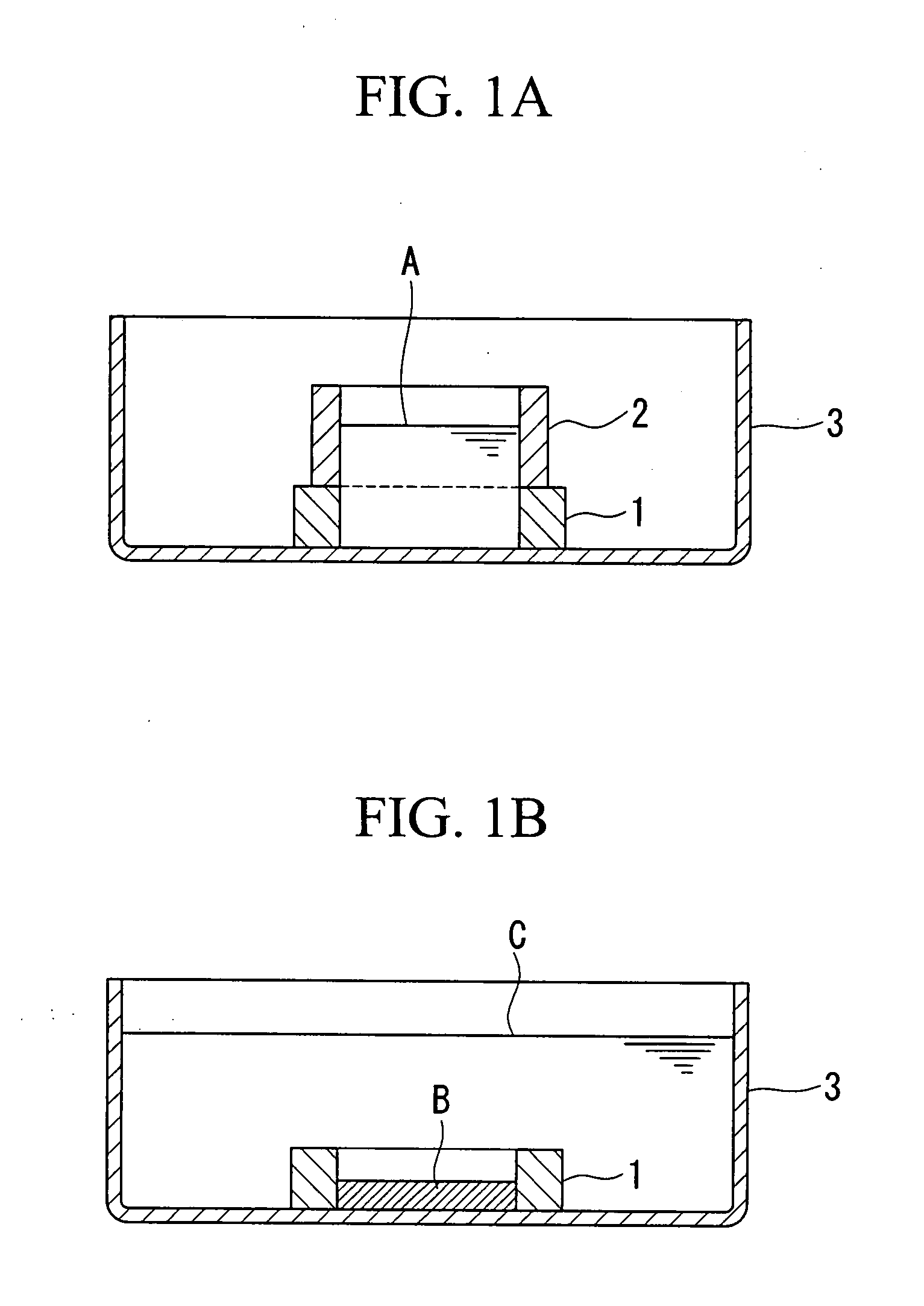
[0124] According to the method described in Example 1, artificial cartilage tissues were prepared using cells derived from porcine articular cartilage. In the preparation step using the silicon rings shown in FIG. 1A and FIG. 1B, media containing different concentrations of the transforming growth factor-β1 (TGF-β1) were used. The TGF-β1 concentration in the medium was 2.5, 5, 10, 20, and 40 ng / mL, each of which was referred to as the 2.5 ng / mL group, the 5 ng / mL group, the 10 ng / mL group, the 20 ng / mL group, and the 40 ng / mL group.

(3) Measurement of Tear Strength

[0125] According to the method described in Measurement Example 1 (1), the tear strength of the prepared p...


PUM

PropertyMeasurementUnit
dry weightaaaaaaaaaa
wet weightaaaaaaaaaa

Abstract

To obtain an artificial cartilage tissue having high strength and biocompatibility. There is provided an artificial cartilage tissue which is substantially free of heterologous protein and is substantially free of scaffold derived from non-cartilage-derived cells, but contains mammalian cartilage-derived cells and an extracellular matrix formed by the cartilage-derived cells, where: (1) the water retention per mg of tissue dry weight is 17.8 mg or less; (2) collagen of 5.57 μg or more per mg of tissue wet weight is contained; (3) proteoglycan of 9.22 μg or more per mg of tissue wet weight is contained.

Description

BACKGROUND OF THE INVENTION [0001] 1. Field of the Invention [0002] The present invention relates to an artificial cartilage tissue prepared using a tissue engineering technique, and a production method thereof. [0003] This application is based on Japanese Patent Application No. 2005-224469, the content of which is incorporated herein by reference. [0004] 2. Description of Related Art [0005] Articular cartilage is an important tissue which covers the osteoepiphysis and is in charge of the smooth movement of joints. The articular cartilage has a structure in which chondrocytes are dispersed throughout a non-cellular tissue called an extracellular matrix (hereunder, referred to as “matrix”) that is neither innervarated nor vascularized. Such a structure in which these cells are dispersed throughout a matrix, that is, the fact that a small number of cells exist per unit volume, and the absence of vasculature means a very poor ability of self-regeneration when an injury occurs. [0006] W...

Claims


Application Information

Patent Timeline
Patent Type & Authority Applications(United States)
IPC IPC(8): C12N5/08C12N5/077
CPCA61F2/30756C12N5/0655C12N2501/15C12N2501/115C12N2501/105
Inventor MIYAUCHI, SATOSHIMIYAZAKI, TATSUYAMATSUZAKA, SATOSHI
Owner PG RES
Why Eureka
Try Eureka