
Device and method for detecting antibiotic inactivating enzymes

Inactive Publication Date: 2007-08-16
CREIGHTON UNIVERSITY
View PDF10 Cites 8 Cited by
  • Summary
  • Abstract
  • Description
  • Claims
  • Application Information

AI Technical Summary

Benefits of technology

[0024] The 3-dimensional test thus allows the laboratory to report to the clinician not only the susceptibility or resistance of a microorganism to antibiotics, but also the ability of the microorganism to inactivate the antibiotics. As a hypothetical example, whereas a conventional antibiotic susceptibility test might indicate that a microorganism was susceptible to the two antibiotics, cefaclor and cefoxitin, the 3-dimensional test might provide additional information to show that the microorganism produced

Problems solved by technology

A problem with currently available antimicrobial susceptibility tests is their failure to reliably predict the in vivo effect and therefore the outcome of clinical therapy.
Sometimes an antibiotic will fail to cure an infection even though the microorganism is susceptible to the antibiotic in the laboratory test.
That is, the current routine laboratory tests can be misleading and give an over-optimistic impression of the therapeutic potential of antibiotics.
These tests can therefore cause patients to be given ineffective treatments.
In serious infections, this inadequacy of current laboratory tests can have fatal consequences.
However one explanation is error arising from a deficiency in the antibiotic susceptibility test itself.
That deficiency is that current routine antibiotic susceptibility tests do not detect the antibiotic-inactivating potential of some microorganisms.
Such enzymes, which are not reliably detected in routine antibiotic susceptibility tests, may cause sufficient antibiotic inactivation at the site of an infection in vivo to cause a treatment failure.
The genes are typically encoded on large plasmids containing other antibiotic encoding resistance genes, leaving few therapeutic options.
All may cause problems of false susceptibility in routine susceptibility tests.
As a result, these B-lactamases often go undetected.
Without detection, correct therapy for infected patients may not be given and can lead to uncontrolled spread of resistant strains.
This deficiency in the tests places clinicians at a disadvantage in selecting the most appropriate antibiotics for their infected patients.
Both disk diffusion and dilution methods are generally deficient in that they do not yield information about the ability of microorganisms to inactivate antibiotics.
These are complex tests to detect enzymatic inactivation of chloramphenicol and require special instruments capable of measuring the absorbance of light at specific wavelengths or of measuring radioactivity.
Such tests are not antibiotic susceptibility tests and their complexity is such that they are unsuitable for routine clinical microbiology laboratories.
They do not predict the potential for any other bacteria to resist these penicillins, and they do not predict the potential for any bacteria to be resistant to any of the other classes of β-lactam antibiotics, such as cephalosporins, cephamycins, monobactams, monocarbams, penems or carbapenems.
The preincubation procedure thereby invalidates the interpretive tables that are necessary to determine antibiotic susceptibility or resistance.
This is a serious deficiency because it would be unethical to base therapy on this procedure which lacks validated interpretive criteria.
This is inconvenient and therefore a disadvantage.
Furthermore there are doubts about the validity of results obtained with this procedure.
It is therefore inconvenient and limited in scope.
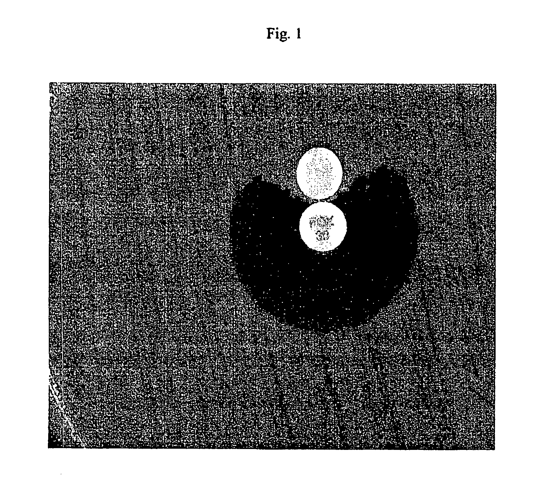
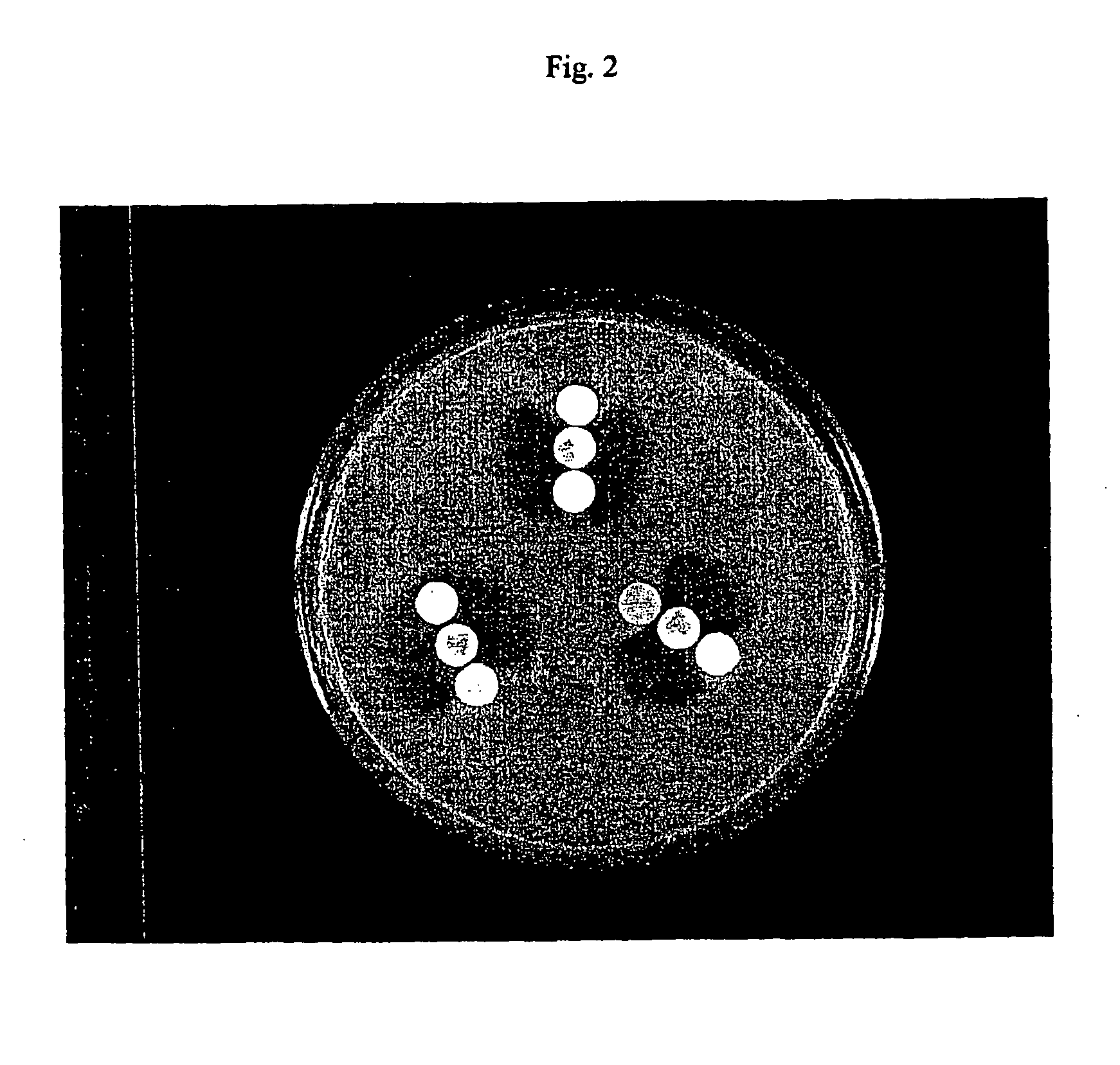
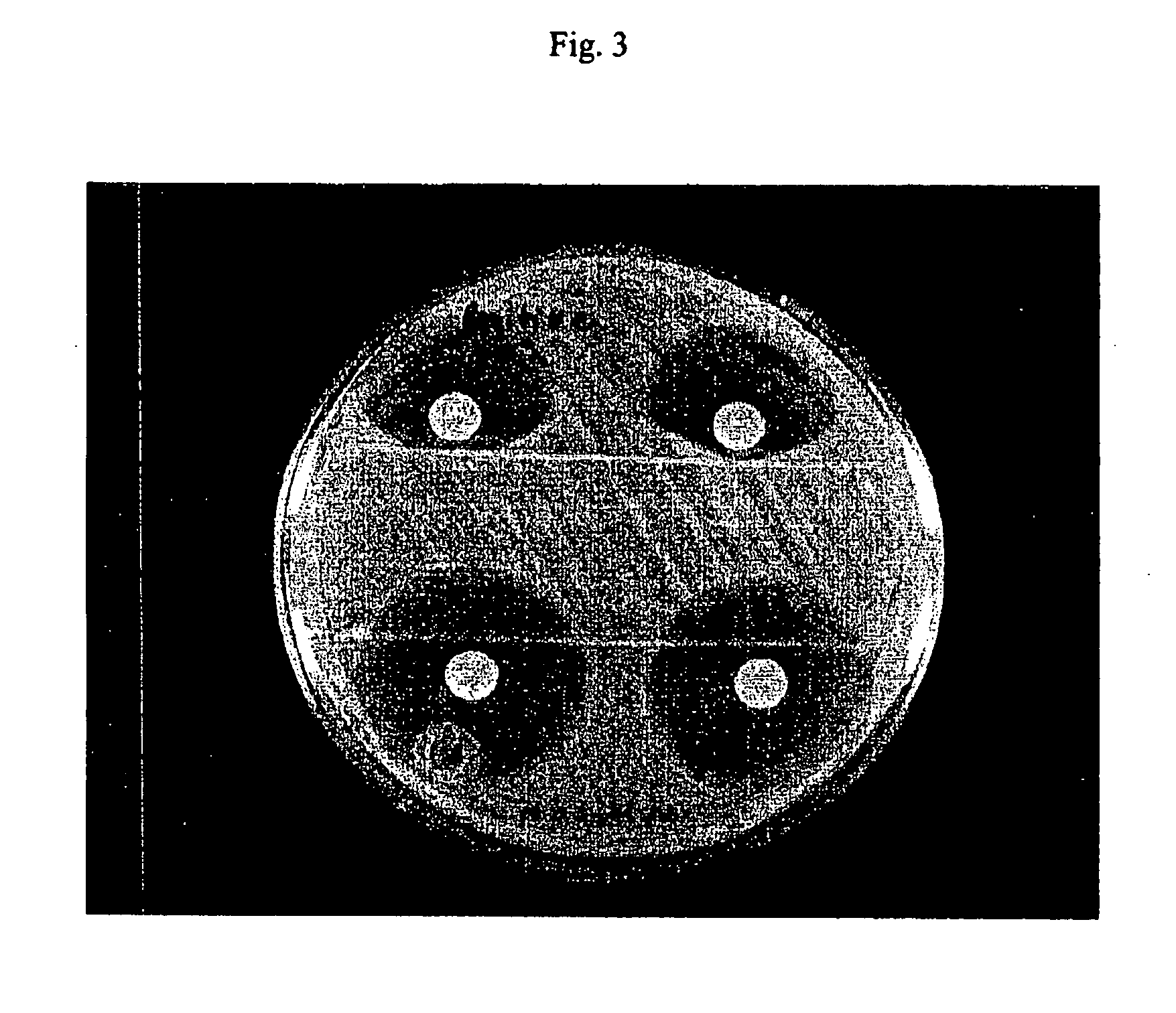
Antibiotic inactivation results in a distortion or discontinuity in the usually circular inhibition zone.
There are several problems with the 3-dimensional test.
a. The procedure for making the slit in the agar for the 3-dimensional test is inconvenient and technically difficult to perform correctly.
B. Making the slit is potentially dangerous to laboratory staff because a scalpel blade contaminated with pathogenic bacteria is an infection hazard.
c. It is also technically difficult to accurately deliver the liquid 3-dimensional inoculum into the slit without overfilling the slit and possibly invalidating the test.
A variety of chemicals have been reported to disrupt or permeabilize microbial membranes, thereby increasing their permeability and causing loss of cellular contents.

Method used


Image

  • Device and method for detecting antibiotic inactivating enzymes
  • Device and method for detecting antibiotic inactivating enzymes
  • Device and method for detecting antibiotic inactivating enzymes

Examples


example 1

Determination of Release of Antibiotic-Inactivating Factors

[0081] In this Example, E. coli MISC 208 (which produces the extended spectrum β-lactamase SHV-2) was used as a test organism and nitrocefin was the substrate in spectrophotometric hydrolysis assays. The E. coli strain was harvested directly into 100 microliters of Mueller Hinton Broth and mixed with 100 microliters of the permeabilizing agent.

[0082] In the hydrolysis assay, units of activity are calculated as: Units=Change⁢ ⁢in⁢ ⁢optical⁢ ⁢density⁢ ⁢(OD) / minute×10Extinction⁢ ⁢coefficient

[0083] The hydrolysis assay was performed essentially as described by O'Callaghan, C. H., et al., A.A.C. 1:283-288 (1972). Briefly, a 100 μM solution of nitrocefin was used as a substrate. The nitrocefin was prewarmed to 37° C. The hydrolysis assay was performed at 37° C. at a wavelength of 389.5 with an extinction coefficient of −0.024. An appropriate cuvette was filled with 0.9 ml of the prewarmed nitrocefin substrate and 100 ul of perm...

example 2

Determination of Release of Antibiotic-Inactivating Factors

[0084] The experiment in Example 1 was repeated using E. coli MISC 208 and cefaclor as substrate in the hydrolysis assays.

Units of β-lactamase ActivityPermeabilizing Agent Preparation(Cefaclor as substrate)Benzalkonium chloride 200 μg / ml16Polymyxin B 10 μg / ml3.8Polymyxin B 30 μg / ml15TE79

example 3

Determination of Release of Antibiotic-Inactivating Factors

[0085] The experiment in Example 1 was repeated using E. coli V1104 to evaluate a variety of permeabilizing agents. Nitrocefan was the hydrolysis substrate.

Units of β-lactamase ActivityPermeabilizing Agent Preparation(Nitrocefin as substrate)Aztreonam210No reagent / 0° C.214No reagent / 45° C.0Ceftazidime196Piperacillin202

[0086] Other exemplary microorganisms that can be used in the determination of release of antibiotic-inactivating factors include those in the following Table:

StrainEnzymeUnits (vs nitrocefin)E. coli V1104202E. coli 165*pI 5.95 TEM ESBL43MG32TEM-1 + TEM-12349K. pneumoniae V1102227


PUM


Abstract

The present invention provides a method of detecting production of antibiotic-inactivating factor and for determining the antibiotic susceptibility of a microorganism comprising the following steps. a culture of the microorganism suspected of producing inactivating factors and / or whose susceptibility is to be determined is admixed with an antibiotic to which susceptibility is to be assayed, and a permeabilizing agent for the microorganism present in a non-growth-inhibiting microorganism-permeabilizing effective amount to form an assay culture. The assay culture is incubated under appropriate culture conditions and for a time sufficient to determine production of antibiotic-inactivating factors and / or the susceptibility of the microorganism to the antibiotic. In another aspect, the present invention provides an improved method for antibiotic susceptibility testing of a microorganism in a culture by admixing the culture with an antibiotic to which susceptibility is to be assayed, and incubating the culture for a time sufficient to determine the susceptibility of the microorganism to the antibiotic, the improvement comprising admixing the culture with a permeabilizing agent for the microorganism present in a non-growth inhibiting microorganism-permeabilizing effective amount.

Description

CROSS-REFERENCE TO RELATED APPLICATIONS [0001] The present application is a continuation-in-part application of U.S. application Ser. No. 11 / 176,420, filed Jul. 7, 2005, which claims priority of U.S. application Ser. No. 10 / 387,788 filed Mar. 13, 2003, which claims priority of U.S. Provisional Application Ser. No. 60 / 364,232 filed Mar. 13, 2002, all of which are incorporated herein by reference in their entirety.STATEMENT REGARDING FEDERALLY SPONSORED RESEARCH OR DEVELOPMENT [0002] None. BACKGROUND OF THE INVENTION [0003] Clinicians and veterinarians often select antibiotic therapies for infections on the basis of laboratory test results. The laboratory tests, known as antimicrobial or antibiotic susceptibility tests, determine the in vitro antimicrobial activity of antibiotics against the microorganisms that cause infections. If the antibiotic susceptibility test indicates that an antibiotic is sufficiently potent to treat an infection, the microorganism causing the infection is re...

Claims


Application Information

Patent Timeline
IPC IPC(8): C12Q1/18
CPCC12Q1/18
Inventor BLACK, JENNIFER A.SMITH MOLAND, ELLEN A.THOMSON, KENNETH S.
Owner CREIGHTON UNIVERSITY
Learn More