Murine Living Tissue Model & UsesTthereof

Inactive Publication Date: 2007-09-20
UNIV OF GLASGOW THE UNIV COURT OF
View PDF0 Cites 0 Cited by
  • Summary
  • Abstract
  • Description
  • Claims
  • Application Information

AI Technical Summary

Benefits of technology

[0004] The present invention provides an in vitro method suitable to allow evaluation of test compounds for oncogenic or anti-cancer properties that can, in part, replace the need to test in live animals.
[0012] The test cell may be a primary cell or a cell line, although cell lines are preferred in order to minimise the number of animals which must be sacrificed in order to prepare the model.
[0020] Mouse cells are used in this model because of the importance of the mouse in studies of carcinogenesis. The model can be constructed of cells from specific genetically modified mice, thereby making use of this important, expanding animal resource, while minimising the numbers of animals needed. Alternatively cells from normal, genetically unmodified mice may be used. As a further alternative, cells may be used which have been genetically modified in vitro. Typically the fibroblasts are primary cells, and are preferably embryonic or neonatal fibroblasts. In preferred embodiments these are combined with normal, benign and / or malignant mouse epithelial cell lines to create epithelial tissue models.
[0030] This protocol has the advantage of providing a matrix which mimics that occurring in vivo, without the use of non-physiological substrates or supports such as nylon mesh, used in other tissue modelling constructs. Such non-physiological substrates typically cannot be degraded by the tumour cells in the same way as a physiological connective tissue matrix. In the present invention, though, the stromal cells are incorporated directly into a contracted gel formed from collagen, which is the major natural component of tissue matrix, and provides a much more physiologically relevant model of the interactions between tumour cells and the underlying tissue.
[0040] Also, by appropriate control of viewing / photographing the model, such as by viewing / photographing several fields at random and thereafter randomly selecting a subset of these, it is possible to minimise any bias which may be introduced by a person analysing the data. It is also possible to observe if the test agent induces or inhibits cellular production of proteins, using suitable techniques known in the art, for example, using immunohistochemisty, immunofluorescence, PCR, microarrays, immunoblotting and zymography.

Problems solved by technology

Conventional animal tests employed to evaluate new therapeutic anti-cancer agents or identify suspect carcinogens are expensive, time consuming, require skilled animal-trained staff and utilise large numbers of animals.
To date in vitro alternatives have relied on the use of conventional cell culture systems which are limited in that they do not allow the three-dimensional interactions that occur between the tumour cells and with their surrounding stromal tissue.
This is a serious disadvantage as such interactions are well documented as having a significant influence on the growth and invasion profiles of tumours.

Method used


Image

  • Murine Living Tissue Model & UsesTthereof
  • Murine Living Tissue Model & UsesTthereof
  • Murine Living Tissue Model & UsesTthereof

Examples


example 1

Preparation of the Living Tissue Model

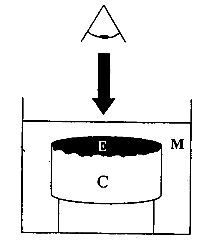
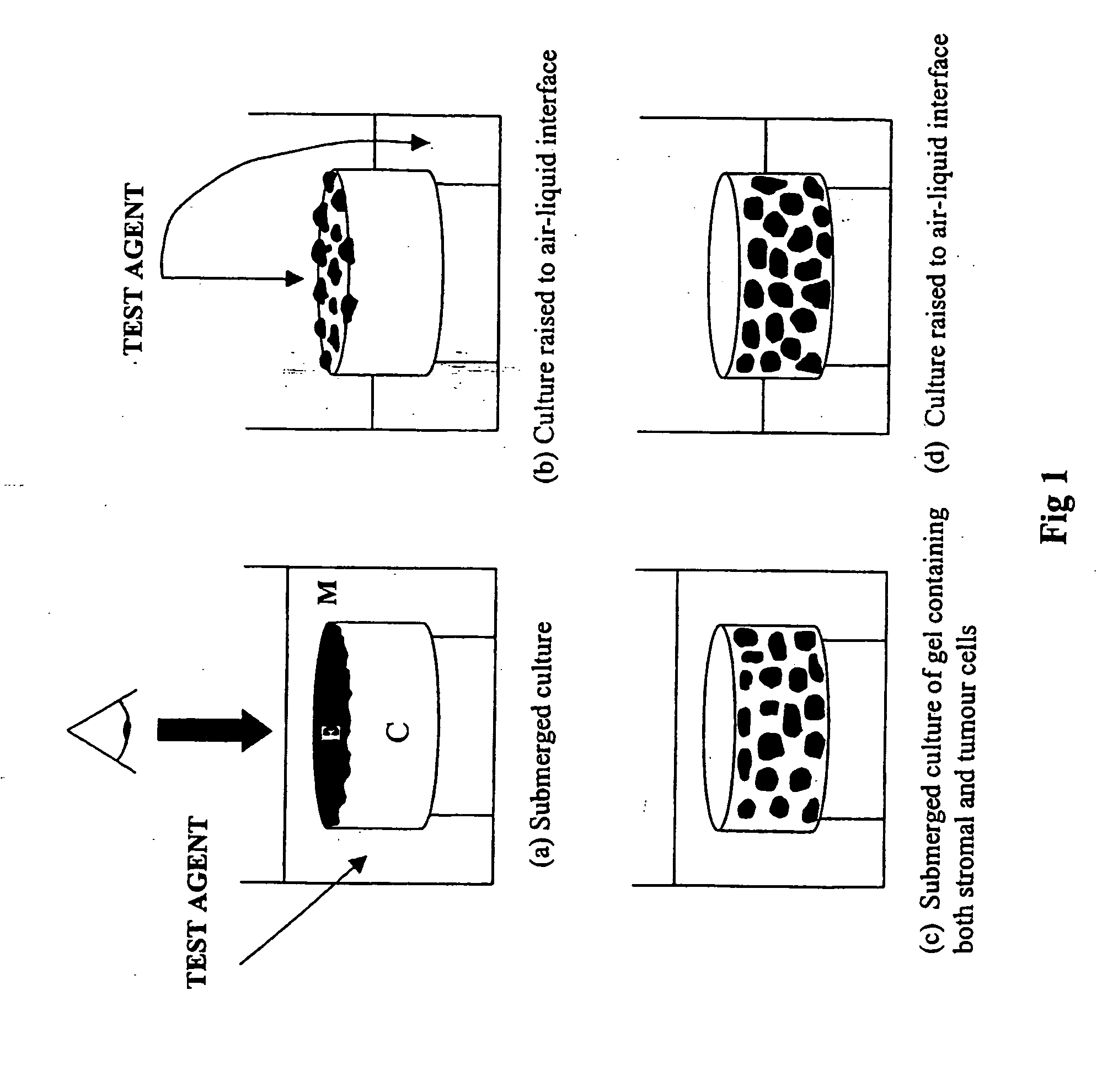
[0052] In summary the in vitro models were developed using contracted collagen gels which supported a layer of mouse tumour cells or corresponding “normal” epithelial cells. The collagen gel was comprised of type I collagen, isolated from rat tail tendon, contracted using primary mouse fibroblasts, which were isolated from the dermis of newborn (or embryonic) mice. The collagen gels contracted to a size approximately 1.5 cm in diameter and were seeded with a single cell suspension of mouse tumour or normal epithelial cells. The models were initially maintained as submerged cultures, which allowed the cells to adhere to the collagen gel and grow. The models may thereafter be raised to the air-liquid interface (semi-submerged culture) to promote cell differentiation and formation of tissue. Benign tumour cells were observed to grow on the surface of the lattice and aggregate to form piles of cells equivalent to wart-like skin papillomas whereas, ...

example 2

Processing of Models and Data Interpretation

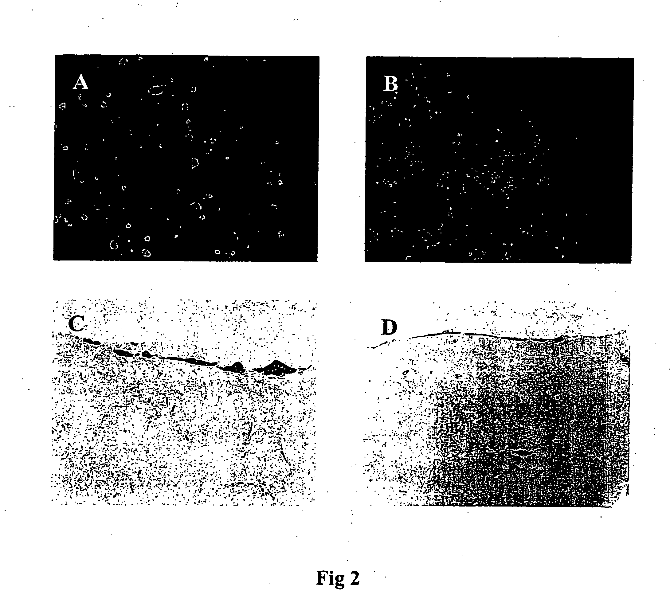
[0057] Cell models were harvested at appropriate time intervals and fixed overnight, at room temperature in a solution of buffered formalin. A small piece of the model was removed and embedded in paraffin wax. Sections were cut and mounted onto glass slides and stained with haematoxylin and eosin (H & E stain). The remainder of the model washed in PBS (phosphate buffered saline), permeabilised with Triton X-100 and stained with propidium iodide (PI). The washed models were stored thereafter in the dark at 4° C. and maintained their fluorescence for several weeks. Whole mounts of the PI stained models were analysed using fluorescent microscopy through coverslips applied directly to the surface of the tissue.

[0058] Propidium iodide staining of the whole mounts allows changes in the cell nuclei to be observed. Condensation and fragmentation of the cell nucleus, indicative of cell death by apoptosis can be clearly identified FIG. 8). Haemato...

example 3

Reproducibility and Viability of the Models

[0064] Reproducibility studies were performed using both BalbMK and SP-1 models grown over a time course and harvested as submerged cultures at day 3 and day 4 and raised to the air interface at day 3, 4, 5 and 6. Models were set up in quadruplicate. Four fields of view were photographed for each of the 48 different models and the area of growth measured in each one. Mean areas and standard errors were calculated for each group. Fields of view compared from within the same gel gave similar areas of growth. Measurements made of different models, which were cultured under the same conditions, gave comparable results. There were instances where areas measured were different from their replicates. This was due primarily to the presence of a large cluster of cells. Replicate models are routinely set up for all treatments studied and several fields of view are studied for each model to minimise errors introduced by natural variations and to act ...


PUM


Abstract

The invention provides an in vitro method for observing an effect of a test agent on a murine tumour model. The model consists a three-dimensional array of murine fibroblasts in a collagen gel which mimics a connective tissue substrate, on or in which are grown benign or malignant murine tumour cells. The model mimics the interactions between the tumour and the underlying tissue substrate, which in turn influence the effect which potential therapeutic or oncogenic agents have on the tumour tissue. Thus the models of the invention provide more physiologically relevant data than monolayer cultures or other available issue models about the effects a particular test agent will have on tumour tissue in vivo. Preferred embodiments provide a model of epithelial tissue and tumours derived therefrom.

Description

FIELD OF THE INVENTION [0001] The present invention relates to methods for testing agents for their effect on murine tissue models, and in particular to methods for assessing the effect of test agents on normal tissue or benign or malignant tumour tissue. The model may be used to represent the progression from normal to benign or malignant tumour tissue and is particularly useful for evaluating any therapeutic or oncogenic properties of a given test agent. The invention also relates to murine living tissue models for use in methods of the invention. BACKGROUND TO THE INVENTION [0002] Conventional animal tests employed to evaluate new therapeutic anti-cancer agents or identify suspect carcinogens are expensive, time consuming, require skilled animal-trained staff and utilise large numbers of animals. To date in vitro alternatives have relied on the use of conventional cell culture systems which are limited in that they do not allow the three-dimensional interactions that occur betwee...

Claims


Application Information

Patent Timeline
IPC IPC(8): G01N33/50
CPCG01N33/5011
Inventor HODGINS, MALCOLM BRIDENGREEN-HALGH, DAVIDEDWARD, MICHAELREITH, ALISON
Owner UNIV OF GLASGOW THE UNIV COURT OF
Why Eureka
Try Eureka