Uniform fluorescent microsphere with hydrophobic surfaces

a fluorescent microsphere and hydrophobic surface technology, applied in the field of fluorescent microspheres, can solve the problems of aggregates, inability to freely flow through the blood stream without being obstructed, etc., and achieve the effects of minimizing interactions with other blood components, maximizing plasma half-life, and improving bioavailability of particles in blood circulation

Inactive Publication Date: 2008-01-24
LIFE TECH CORP
View PDF4 Cites 64 Cited by
  • Summary
  • Abstract
  • Description
  • Claims
  • Application Information

AI Technical Summary

Benefits of technology

[0008] Intravascular tracers of blood circulation can provide a description of the flow field over time and space. Described herein are fluorescent microspheres capable of providing detailed information regarding the intravascular flow field. The microspheres can maximize plasma half-life as well as minimize interactions with other blood components. The bioavailability of the particles in the

Problems solved by technology

One drawback of existing microspheres is their inability to freely flow through the b

Method used


Image

  • Uniform fluorescent microsphere with hydrophobic surfaces
  • Uniform fluorescent microsphere with hydrophobic surfaces
  • Uniform fluorescent microsphere with hydrophobic surfaces

Examples


example 1

Preparation of 0.1 μm Microspheres with NIR Emission (715 ex / 755 em)

[0253] The staining solution is prepared by adding 700 μL of the dye stock (BODIPY® 670 / 735 [difluoro(1-((3-(2-(5-hexyl)thienyl)-2H-isoindol-1-yl) methylene)-3-(2-(5-hexyl) thienyl)-1H-isoindolato-N1,N2)boron] 10 mg / mL stock solution in CH2Cl2) to a 10 mL glass test tube, adding 300 μL of CH2Cl2, and mixing. 2 mL of Ethanol is then added and sonicated for 30 second to ensure complete mixing. The microspheres are loaded with dye by first adding 10 ml of a vortexed microsphere stock (0.11 μm sulfate polystyrene microspheres (0.1 μm), 8.1% solids, with surface charge content of 6 μEq / g (measured from conductometric titration)) to a 250 ml round bottom flask and then slowly adding 14 mL of methanol and stirring for 5 minutes. The staining solution is added dropwise to the stirred microsphere suspension and incubated for 30 minutes with continual stirring. The organic solvents are evaporated in a BUCHI R-124 vacuum eva...

example 2

Preparation of 2 μm Microspheres with NIR Emission (715 ex / 755 em)

[0255] These fluorescent microspheres are prepared essentially as in Example 1, except that 1.1 mL of dye stock is added to the 10 ml tube and the microsphere stock is 2.0 μm sulfate polystyrene microspheres (8.1% solids, with surface charge content with surface charge content of 6 μEq / g (measured from conductometric titration)).

Methods:

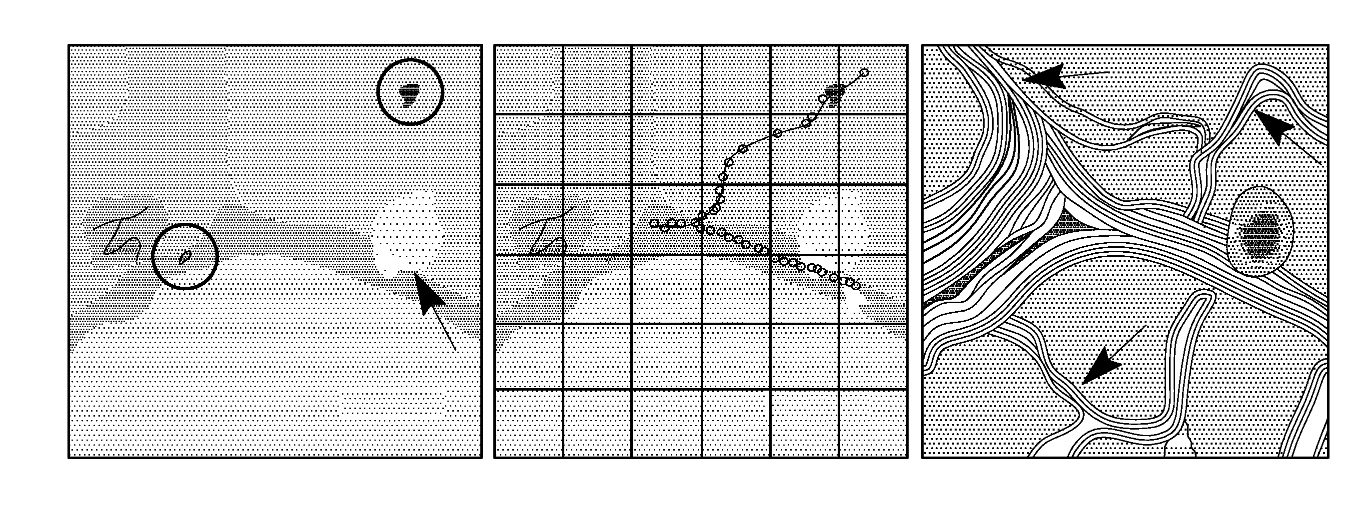
[0256] Studies of blood flow regulation have focused on the accurate description of the velocity field because other flow property calculations follow directly from these measurements. To obtain a detailed description of the velocity field in the microcirculation, conventional velocimetry techniques consider the field to be composed of a very large number of particles (Raffel et al., 2000; Westerweel, 1997). The spatial displacement of these particles in two separate images provides a measurement of the instantaneous in-plane velocity vector field (Adrian, 1984; Adrian, 1991). Thes...


PUM


Abstract

Fluorescent microspheres for the measurement of blood flow are provided. The microspheres are substantially uniform in diameter and have a hydrophobic surface, which allows them to circulate more freely throughout bloodstream, while reducing immunogenicity, particle aggregation and bioaccumulation. The hydrophobic surface on each microsphere is generally comprised of polymeric material having a limited surface charge.

Description

CROSS-REFERENCE TO RELATED APPLICATIONS [0001] This application claims priority of U.S. Ser. No. 60 / 806,382, filed Jun. 30, 2006, which disclosure is herein incorporated by reference.FIELD OF THE INVENTION [0002] This invention relates to fluorescent microspheres having a hydrophobic outer surface, as well as methods for their preparation. The invention also relates to methods for measuring blood flow by administering the fluorescent microspheres to a subject. BACKGROUND OF THE INVENTION [0003] Detectable microspheres are useful tools as signaling tracers for blood flow measurements. After entering the animal's circulatory system, a signal from the microspheres can be detected through a proper imaging instrument and a range of characteristics of the blood flow in live animals can then be monitored and recorded for many different types of studies. [0004] For some time, radioactively labeled microspheres have been used as tracers to estimate regional organ perfusion. In order to minim...

Claims


Application Information

Patent Timeline
IPC IPC(8): A61K49/00
CPCA61K49/0004A61K49/0093A61K49/0021
Inventor ZHANG, YU-ZHONG
Owner LIFE TECH CORP
Why Eureka
Try Eureka