Scaffolds for cell transplantation

a cell transplantation and scaffolding technology, applied in the field of scaffolds for cell transplantation, can solve the problems of limited success of cell transplantation in musculoskeletal regenerative medicine, net loss of muscle mass and resulting strength, and so as to promote cell-based maintenance of tissue structure and function, promote cell-based migration, and promote the effect of tissue destruction

Active Publication Date: 2008-02-21
RGT UNIV OF MICHIGAN +1
View PDF6 Cites 192 Cited by
  • Summary
  • Abstract
  • Description
  • Claims
  • Application Information

AI Technical Summary

Benefits of technology

[0004] The devices and methods of the invention provide a solution to several problems associated with previous cell transplantation protocols. Transplantation systems that enhance the viability of the cells and induce their outward migration to populate injured or defective bodily tissues enhance the success of tissue regeneration, e.g., the regeneration of muscle tissue or other tissues as well as angiogenesis. Such a device that controls cell function and / or behavior, e.g., locomotion, contains a scaffold composition and one or more bioactive compositions. The bioactive composition is incorporated into or coated onto the scaffold composition. The scaffold composition and / or bioactive composition temporally and spatially (directionally) controls egress of a resident cell or progeny thereof.
[0005] The devices mediate active recruitment, modification, and release of host cells from the material in vivo, thereby improving the function of cells that have resided in the scaffold. For example, the device attracts or recruits cells already resident in the body to the scaffold material, and programs or reprograms the resident cells to a desired fate (e.g., immune activation or tissue regeneration).
[0007] Alternatively or in addition, egress is regulated by a bioactive composition. By varying the concentration of growth factors, homing / migration factors, morphogens, differentiation factors, oligonucleotides, hormones, neurotransmitters, neurotransmitter or growth factor receptors, interferons, interleukins, chemokines, cytokines, colony stimulating factors, chemotactic factors, extracellular matrix components, adhesion molecules and other bioactive compounds in different areas of the device. The device controls and directs the migration of cells through its structure. Chemical affinities are used to channel cells towards a specific area of egress. For example, adhesion molecules are used to attract or retard the migration of cells. By varying the density and mixture of those bioactive substances, the device controls the timing of the migration and egress. The density and mixture of these bioactive substances is controlled by initial doping levels or concentration gradient of the substance, by embedding the bioactive substances in scaffold material with a known leaching rate, by release as the scaffold material degrades, by diffusion from an area of concentration, by interaction of precursor chemicals diffusing into an area, or by production / excretion of compositions by resident support cells. The physical or chemical structure of the scaffold also regulates the diffusion of bioactive agents through the device.

Problems solved by technology

When these normal processes are compromised by disease or age, damaged muscle fibers are instead replaced by infiltrating fibrous tissue or fat, leading to a net loss of muscle mass and a resultant loss of strength.
Cell transplantation has been used in regenerative medicine for musculoskeletal disorders as well as degenerative conditions such as diabetes with limited success.
Limitations of earlier approaches include loss of cell viability and function following transplantation.

Method used


Image

  • Scaffolds for cell transplantation
  • Scaffolds for cell transplantation
  • Scaffolds for cell transplantation

Examples


example 2

Niche Scaffolds Promote Muscle Regeneration using Transplanted Cells

[0133] Transplanting myoblasts within synthetic niches that maintain viability, prevent terminal differentiation, and promote outward migration significantly enhances their repopulation and regeneration of damaged host muscle. Myoblasts were expanded in culture, and delivered to tibialis anterior muscle laceration sites in mice by direct injection into muscle, transplantation on a macroporous delivery vehicle releasing factors that induce myoblast activation and migration (HGF and FGF), or transplantation on materials lacking factor release. Controls included the implantation of blank scaffolds, and scaffolds releasing factors without cells. Injected cells in the absence of a scaffold demonstrated a limited repopulation of damaged muscle, and led to a slight improvement in muscle regeneration. Delivery of cells on scaffolds that did not promote migration resulted in no improvement in muscle regeneration. Strikingly...

example 3

Treatment Skin Wounds

[0152] In the context of a skin defect, the goals of the therapy are dictated by the type of wound (e.g., acute bum, revision of scar, or chronic ulcer) and size of the wound. In the case of a small chronic ulcer, the objective is closure of the wound by regeneration of the dermis. The epidermis regenerates via migration of host keratinocytes from the adjacent epidermis. For large wounds, keratinocytes, optimally autologous, are provided by the device to promote regeneration of the epidermis. Cells are loaded into a scaffold material that is placed directly over the wound site, e.g., a scaffold structure in the form of a bandage. The material provides a stream of appropriate cells to promote regeneration. For dermal regeneration, fibroblasts cells are used to seed the device, and these cells are either autologous (biopsy taken from another location and expanded before transplantation) or allogeneic. Advantages of autologous cells include a decreased risk of dis...

example 4

Devices and Systems for Promoting Angiogenesis

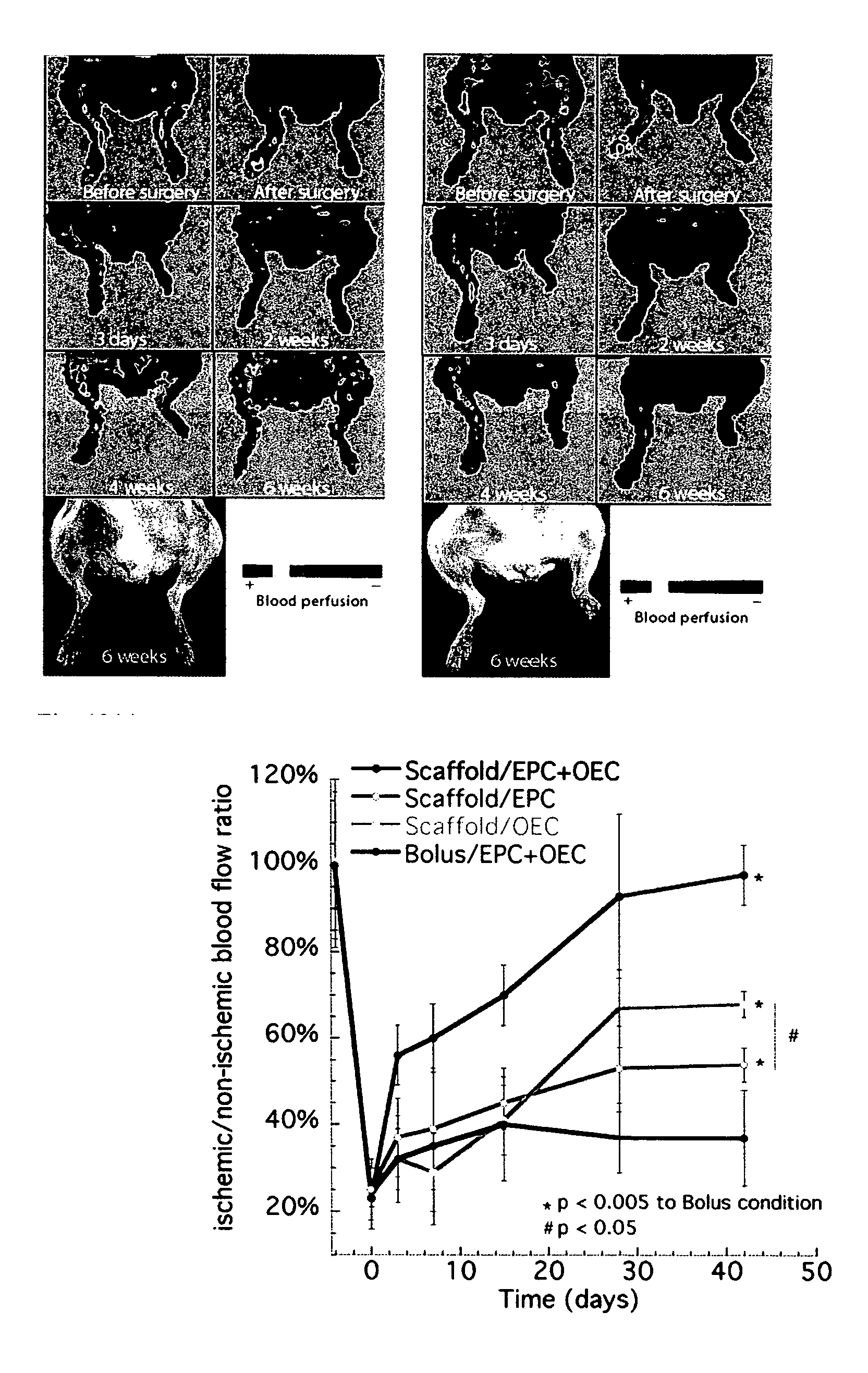
[0155] Angiogenesis is a critical element in any tissue regeneration effort, and the temporally distinct signaling of vascular endothelial growth factor (VEGF) is crucial in this process. Devices that contain compositions which promote angiogenesis together with endothelial cells resulted in a synergistic angiogenic effect. The approach utilizes cells that play a role in the angiogenic process, e.g., endothelial progenitor cells and outgrowth endothelial cells (e.g., derived from cord blood or from peripheral blood samples).

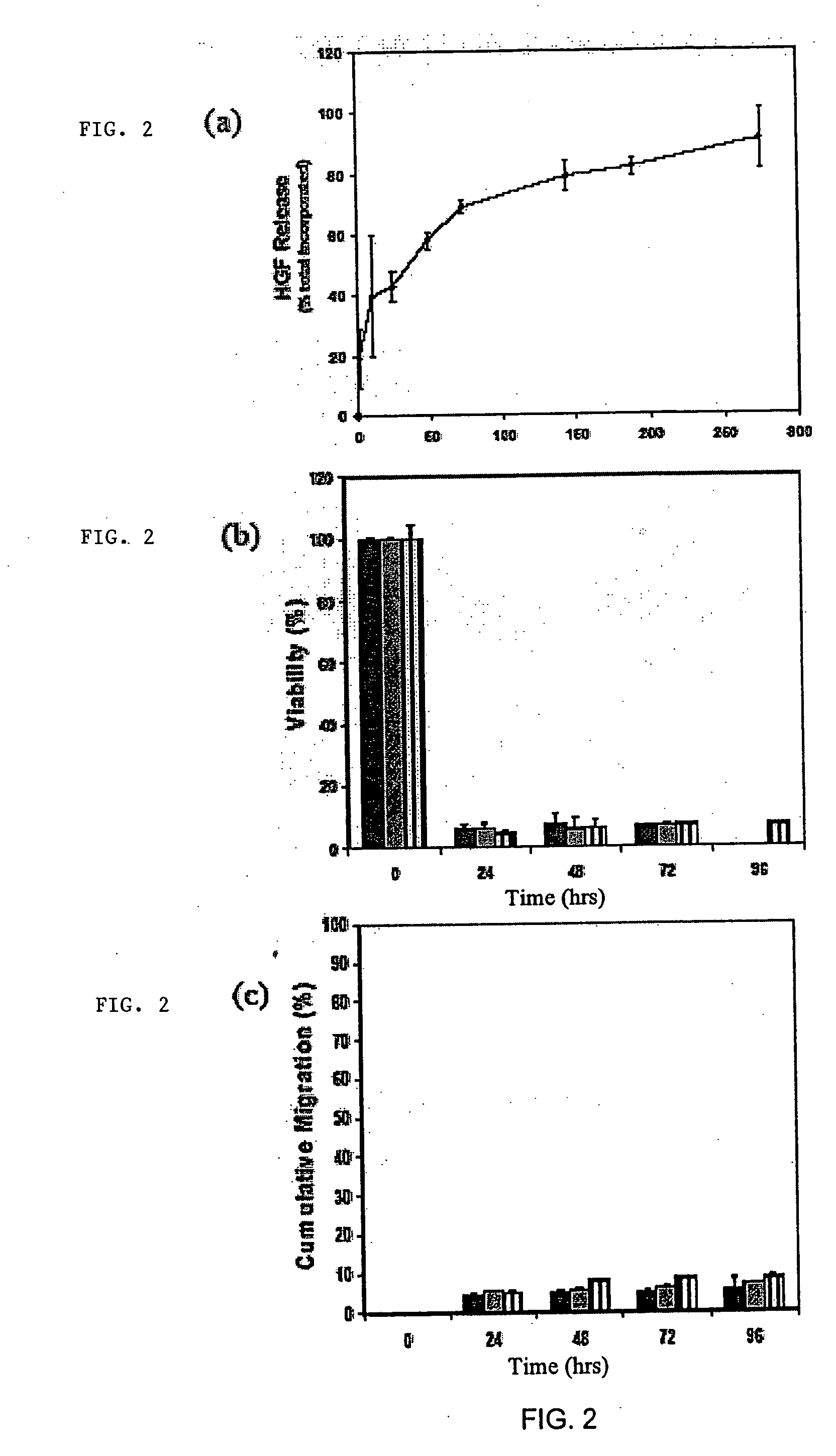
[0156] An injectable alginate hydrogel was developed to provide spatial distribution and temporal control of factors inducing neovascularization of hypoxic tissues. The hindlimbs of C57BL / 6J mice were made ischemic by femoral artery and vein ligation, and the hydrogel containing growth factors was directly injected into the ischemic muscle (bolus delivery of VEGF was used as a control), and the in vivo release kinet...


PUM

PropertyMeasurementUnit
pore sizeaaaaaaaaaa
diameteraaaaaaaaaa
diameteraaaaaaaaaa

Abstract

A device that includes a scaffold composition and a bioactive composition with the bioactive composition being incorporated into or coated onto the scaffold composition such that the scaffold composition and / or a bioactive composition controls egress of a resident cell or progeny thereof. The devices mediate active recruitment, modification, and release of host cells from the material.

Description

GOVERNMENT SUPPORT [0001] The invention was supported, in whole, or in part, by NIH / NICDR grant number RO1 DE13349 and HL069957. The Government has certain rights in the invention.BACKGROUND OF THE INVENTION [0002] The total costs of musculoskeletal disease in the US in 2000 have been estimated at US $ 254 billion and, in developing countries, the figure is estimated at US$ 100 billion. Under normal conditions, muscles can repair themselves by regenerating damaged muscle fibers and restoring muscle strength. Following an initial necrosis of damaged muscle fibers, an inflammatory response is initiated that activates a resident population of quiescent cells termed satellite cells. These myogenic cells proliferate, migrate to the site of injury, differentiate and fuse to form mature myofibers, or fuse with existing myofibers thus regenerating damaged muscle fibers and restoring their function. When these normal processes are compromised by disease or age, damaged muscle fibers are inst...

Claims


Application Information

Patent Timeline
Patent Type & Authority Applications(United States)
IPC IPC(8): C12N5/06C12M1/00
CPCA61L27/3633A61L27/38A61L27/54A61L27/56A61L27/58A61L2300/604A61L2300/438A61L2300/426A61L2300/606A61L2300/252A61K39/0011A61L2300/414B33Y80/00A61K38/1866A61K38/39A61K38/193A61P35/00A61P43/00Y02A50/30A61K39/001184A61K39/001182A61K39/00117A61K39/001191A61K39/001156A61K39/00118A61K39/001192A61K39/001171A61K39/001186A61K39/001195A61K2039/54A61K2039/55561A61K2039/6093A61L27/18A61L2400/18A61L2430/30A61K39/21A61K39/39A61L27/20A61L2300/412C12N7/00C12N15/117C12N2310/17C12N2320/32C12N2740/16034A61K9/0024A61K9/1647A61K2039/55516A61K2039/55522A61K2039/55583A61L27/3826A61L2300/232A61L2430/34
Inventor MOONEY, DAVID J.ALI, OMAR ABDEL-RAHMANBARROS E SILVA, EDUARDO ALEXANDREKONG, HYUN JOONHILL, ELLIOTT EARL JR.BOONTHEEKUL, TANYARUT
Owner RGT UNIV OF MICHIGAN
Why Eureka
Try Eureka