Ion-detecting microspheres and methods of use thereof

a technology of ion-detecting microspheres and microspheres, which is applied in chemical methods analysis, instruments, material analysis, etc., can solve the problems of large sample volumes, high sensing volume of optode films, and long response times (many hours), and achieve high-throughput analysis of samples, high detection efficiency, and high detection efficiency.

Inactive Publication Date: 2008-07-24
AUBURN UNIV +1
View PDF28 Cites 0 Cited by
  • Summary
  • Abstract
  • Description
  • Claims
  • Application Information

AI Technical Summary

Benefits of technology

[0015]Accordingly, this invention provides ion-detecting microspheres for detecting target ions in a sample, and to methods of use thereof in clinical laboratory instrumentation such as flow cytometry for sample analysis. This invention is based on the discovery that ion-detecting microspheres can be used in conjunction with existing clinical laboratory instrumentation such as flow cytometry as a diagnostic tool for rapid, high-throughput analysis of a sample. The ion-detecting microspheres of this invention were shown to possess acceptable sensitivity, selectivity, and precision for the potential clinical determination of ions. This invention further demonstrates that multiplexed measurements of two or more types of ions in a sample are possible using this analytical method.
[0023]In particular, lead ion-detecting microspheres are provided that respond with a high degree of sensitivity (i.e., can detect nanomolar levels of lead ions) and display extremely enhanced equilibrium response times (i.e., on the order of minutes). In addition, the lead ion-detecting microspheres only require sample volumes on the order of milliliters, and demonstrate high response stability for various lead ion concentration changes.

Problems solved by technology

Electrolytes, however, are a class of analytes that have never been assessed with this technique.
However, a significant drawback is the high sensing volume of the optode film, which requires typically on the order of 10 μmoles of ions to be extracted from the sample in order to achieve the desired optical response.
Consequently, massive volumes of sample (many liters) and very long response times (many hours) in a continuous flowing system have so far been required to accurately measure low levels of heavy metals in aqueous samples.

Method used


Image

  • Ion-detecting microspheres and methods of use thereof
  • Ion-detecting microspheres and methods of use thereof
  • Ion-detecting microspheres and methods of use thereof

Examples


example 1

Preparation of Ion-Detecting Microspheres

[0088]A polymer cocktail containing PVC, DOS, ionophore, ETH 5294 and NaTFPB was dissolved in 5 mL of cyclohexanone and diluted with 100 mL of dichloromethane. The addition of 1 mL of xylenes to the solution aided in the aesthetic appearance of the cast particles. Potassium sensing microspheres contained PVC (33 wt %), DOS (66 wt %), ETH 5294 (0.2 mmol / kg), NaTFPB (0.3 mmol / kg), and the potassium ionophore BME-44 (21.1 mmol / kg). Sodium sensing microspheres contained PVC (33 wt %), DOS (66 wt %), ETH 5294 (0.2 mmol / kg), and either sodium ion-ionophore X (10.0 mmol / kg) and NaTFPB (0.3 mmol / kg) (type 1) or sodium ion-ionophore (89.3 mmol / kg) and NaTFPB (27.1 mmol / kg) (type 2). For segregation of particle subsets in the parallel analyses, type 1 sodium sensing particles also contained RLC reference dye (3.9×10⁻³ mmol / kg). Particles were prepared using a particle casting apparatus as described by Tsagkatakis et al. (16).

[0089]The following setting...

example 2

[0090]Instrumentation and Measurement

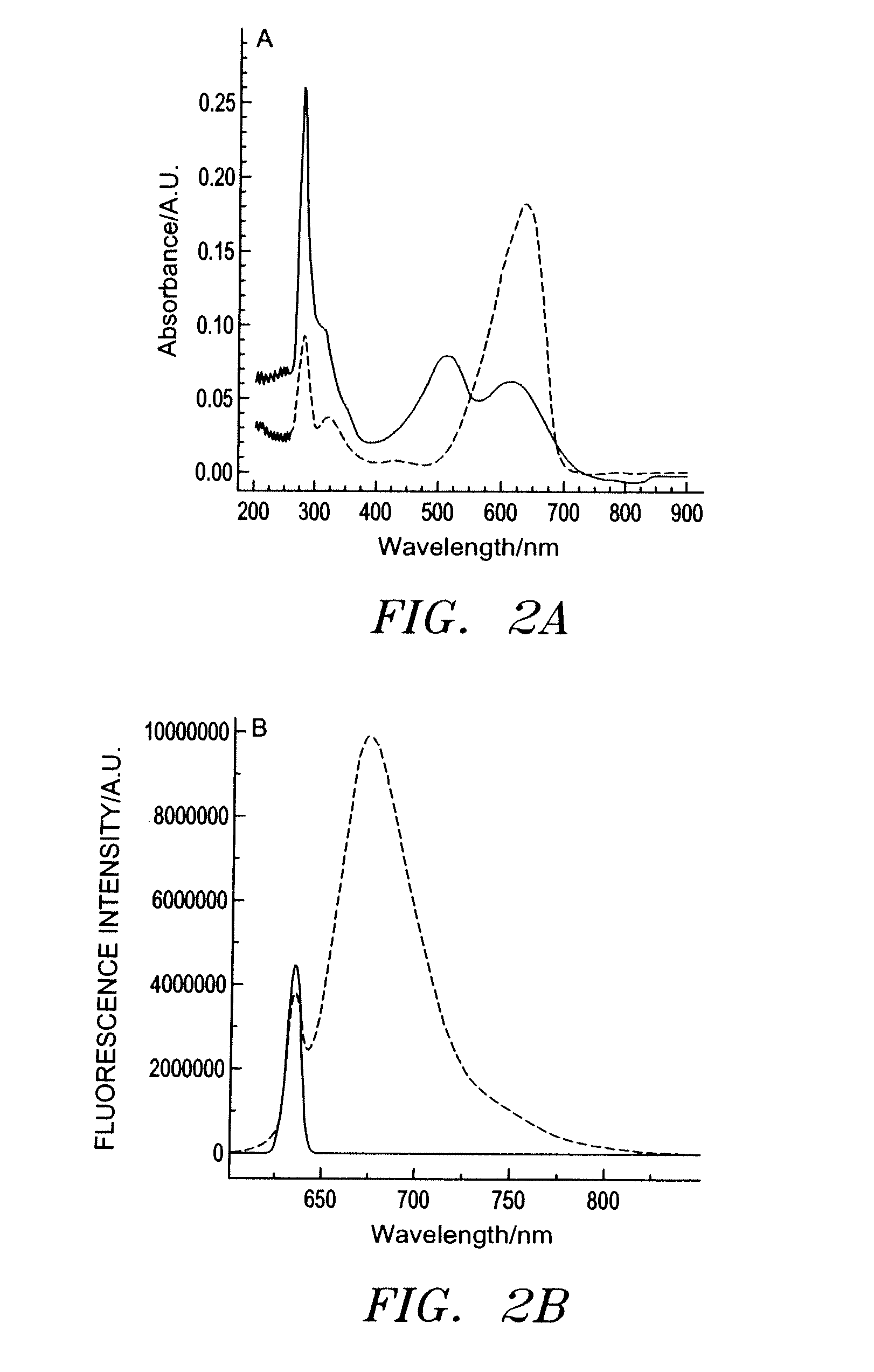
[0091]The absorption behavior of ETH 5294 in both its protonated and deprotonated forms was determined using a DU 70 spectrophotometer (Beckman Coulter, Inc., Fullerton, Calif.). For fluorescence characterization, a 1×10⁻⁵ M solution of ETH 5294 in DMSO was mixed with a 1% (v / v) aqueous solution of either HCl or NaOH and excited at 635 nm using a Fluorolog 3 fluorometer (Instruments SA, Edison, N.J.) to determine the emission behavior of the protonated and deprotonated forms at this wavelength. Optical characterization of RLC in methanol found λex=820 nm and λem=860 nm.

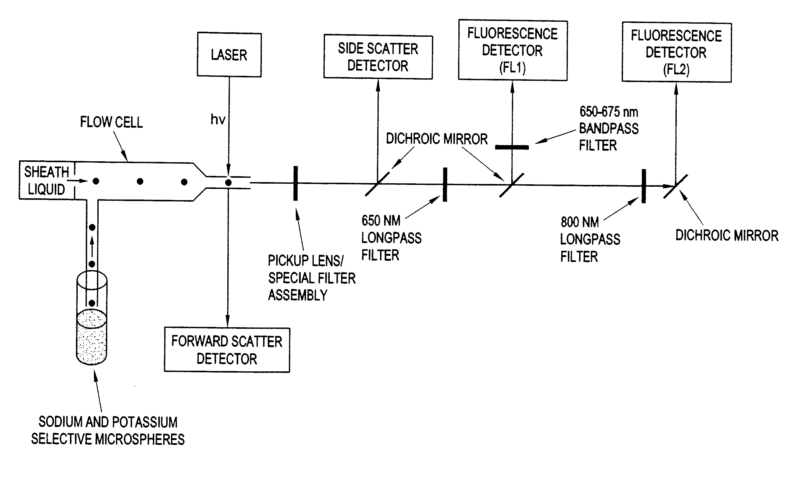
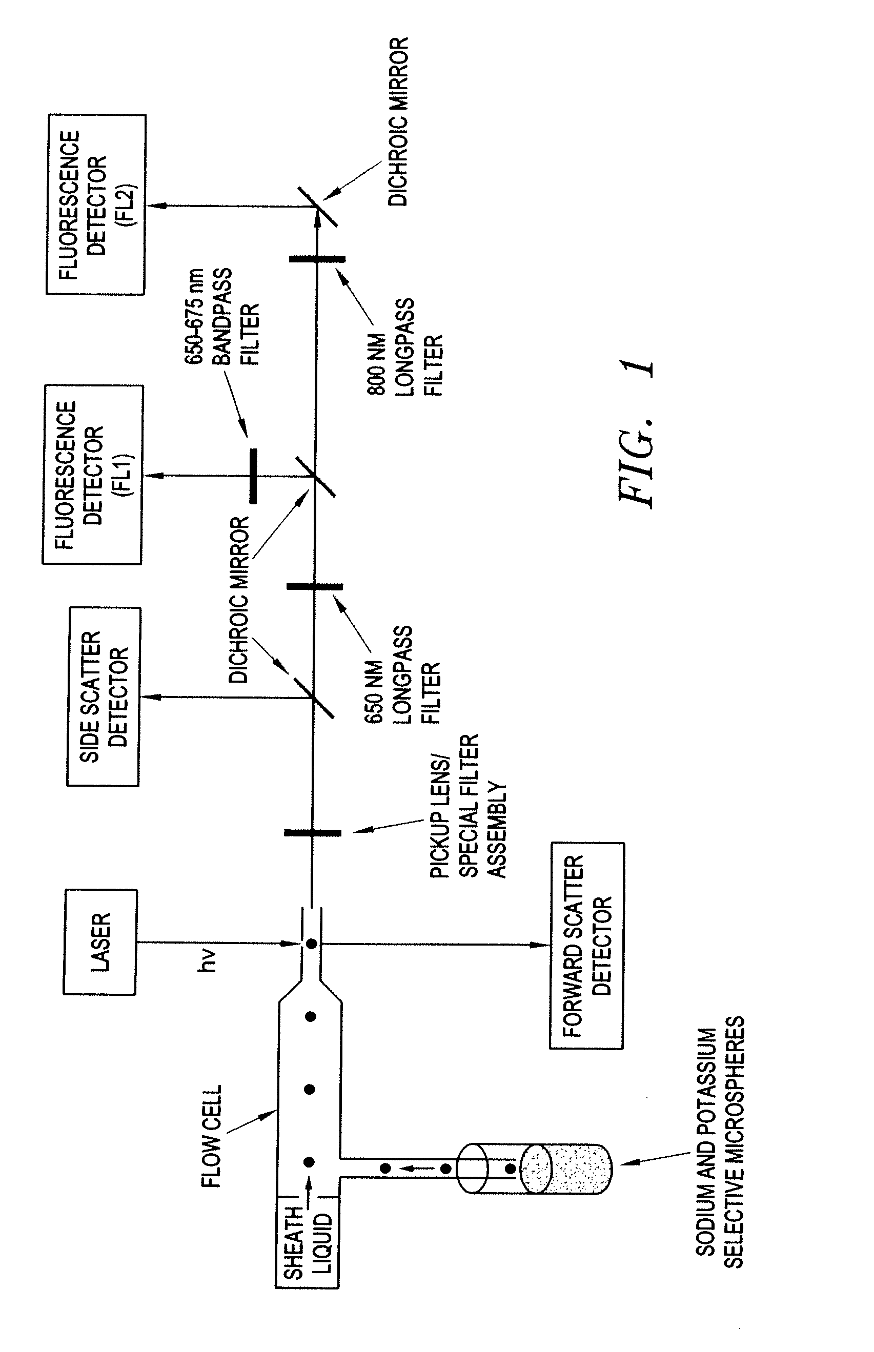
[0092]A Beckman Coulter EPICS XL flow cytometer modified for both 635-nm and 785-nm excitation was used to interrogate the sensing microspheres. A 650-nm long-pass emission filter and a 660 (±15) nm band-pass filter were used to collect fluorescence emitted between 650 and 675 nm, and an 800-nm long-pass emission filter was used to collect fluorescence emitted above 800 nm. A schem...

example 3

Preparation of Lead Ion-Detecting Microspheres

[0093]The schematic set-up and the general protocol for the preparation of sensing particles have been reported recently (17) and it applies here with minor modifications. Unless otherwise indicated a cocktail mixture was prepared by weighing out 58.5 mg PVC, 116 mg DOS, 1.44 mg (10.2 mmol / kg) chromoionophore, 1.27 mg (6.9 mmol / kg) reference dye DiIC18, 4.05 mg (23.8 mmol / kg) ion exchanger NaTFPB, 14.8 mg (87.2 mmol / kg) ionophore and dissolving it in 5 mL of cyclohexanone. The mixture was shaken in a vortex mixer for approximately one hour and then added dropwise to 100 mL dichloromethane under gentle stirring. After adding 1 mL xylenes the solution was filtered through a 0.45 μm Gelman filter and 50 mL transferred to a gas-tight Hamilton syringe. The syringe containing the polymer core solution was mounted on a syringe pump (Stoelting, Wood Date, Ill.) and set to flow at a rate of 0.263 mL min−1. Deionized water used as the sheath liqui...


PUM

PropertyMeasurementUnit
diameteraaaaaaaaaa
diameteraaaaaaaaaa
diameteraaaaaaaaaa

Abstract

This invention provides methods of using ion-detecting microspheres containing an ionphore and a chromoionphore in clinical laboratory instrumentation such as flow cytometry for sample analysis. In one embodiment, the microspheres are contacted with a flowing stream of a sample under conditions that allow the ion-selective ionophores to complex with the ions in the sample, and to cause deprotonation of the chromoionophore. The complexes are then exposed to an excitation wavelength light source suitable for exciting the deprotonated chromoionophore to emit a fluorescence signal pattern. Detection of the fluorescence signal pattern emitted by the deprotonated chromoionophore in microspheres containing the complexes allows for determination of the presence of the target ions in the sample. In one embodiment, lead ion-detecting microspheres are provided that can detect nanomolar levels of lead ions with response times on the order of minutes.

Description

CROSS-REFERENCE TO RELATED APPLICATIONS[0001]This is a divisional of application Ser. No. 10 / 384,082 filed Mar. 7, 2003, the entire contents of which are incorporated by reference. The present invention claims priority to Provisional Application No. 60 / 363,180, filed on Mar. 11, 2002, entitled “Rapid Trace Level Sensing Fluorescent Microspheres.”GOVERNMENT INTERESTS[0002]The invention was made in the course of work supported by grant No. GM59716 from the National Institutes of Health. The United States government has certain rights in this invention.BACKGROUND OF THE INVENTION[0003]1. Field of the Invention[0004]The present invention is related to systems for detecting target ions in a sample, and more specifically, to ion-detecting microspheres and methods of use thereof in clinical laboratory instrumentation such as flow cytometry for sample analysis.[0005]2. Description of the Prior Art[0006]Throughout this application, various references are referred to within parentheses. Discl...

Claims


Application Information

Patent Timeline
Patent Type & Authority Applications(United States)
IPC IPC(8): G01N33/20G01N33/84
CPCG01N33/84
Inventor BAKKER, ERICTELTING-DIAZ, MARTINBELL, MIKE
Owner AUBURN UNIV
Why Eureka
Try Eureka