Systems and methods for the detection and analysis of in vivo circulating cells, entities, and nanobots

a technology nanobots, applied in the field of systems and methods for the detection and analysis of in vivo circulating cells, entities, nanobots, etc., can solve the problems of inability to perform serial testing, high mortality rate of sepsis, and nearly universal restrictions of methods to ex vivo use, so as to improve specificity, eliminate large blood draws, and improve specificity

Inactive Publication Date: 2008-10-02
J FITNESS LLC
View PDF13 Cites 11 Cited by
  • Summary
  • Abstract
  • Description
  • Claims
  • Application Information

AI Technical Summary

Benefits of technology

[0009]A salient feature of the present invention is that cells move in vivo, creating a signal that can be analyzed, such as in capillaries with flowing blood.
[0019]Moreover, embodiments of the present invention provide for a flexible platform for testing of multiple assays, including white blood cell counts, differential cell counts, and the like.

Problems solved by technology

Because of this, these methods are nearly universally restricted to ex vivo uses.
Not all types of cell analyses are amenable to blood sampling.
Such large blood sampling makes this method unacceptable for routine breast cancer diagnosis, or for serial testing to evaluate a response to treatment.
Sepsis has a high mortality rate.
All of the above systems do not perform cell counts or they require blood or tissue sampling in order to perform circulating cell counts, and further are not designed for, and fail to reliably provide real-time analysis in living tissue without such a blood extraction.
None of the above systems suggest or teach a method and system for blood level analysis in vivo.
Such an in vivo analysis has not been successfully commercialized to our knowledge.

Method used


Image

  • Systems and methods for the detection and analysis of in vivo circulating cells, entities, and nanobots
  • Systems and methods for the detection and analysis of in vivo circulating cells, entities, and nanobots
  • Systems and methods for the detection and analysis of in vivo circulating cells, entities, and nanobots

Examples


example 1

Expected Lower Limit of Cell Detection

[0065]We estimated the minimum number of cells detectable.

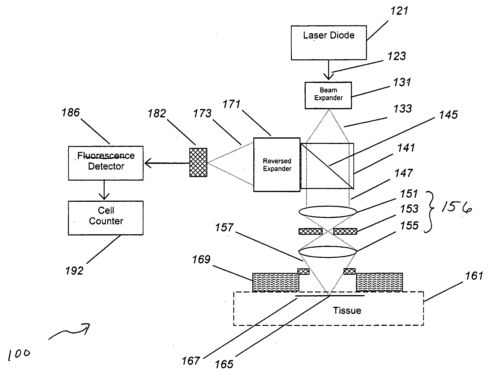
[0066]In many (but not all) cases, the detected cells are in the vascular compartment, which provides the flow needed to generate the cell counting signals. The vascular volume (e.g., the volume of the tissue measured that resides within the vascular compartment) was estimated and then verified by experiment. As an estimate, human tissue has an average blood volume of about 2%, but this can be as high as 10% or more when imaging a capillary-rich bed. With a view of 1 mm and a wide focusing depth of 1 mm, this yields a tissue measurement volume of 1 uL, or a vascular volume of 0.1 uL or less. This level of vascular contact has been confirmed in laboratory tests, with volumes as high as 100 uL for large fiber probes, and volumes under 1 uL for the smallest probes.

[0067]Next, the concentration and count of the target cell was estimated. Cell counts for normal blood elements are known. For wh...

example 2

Detection a Model of Tissue

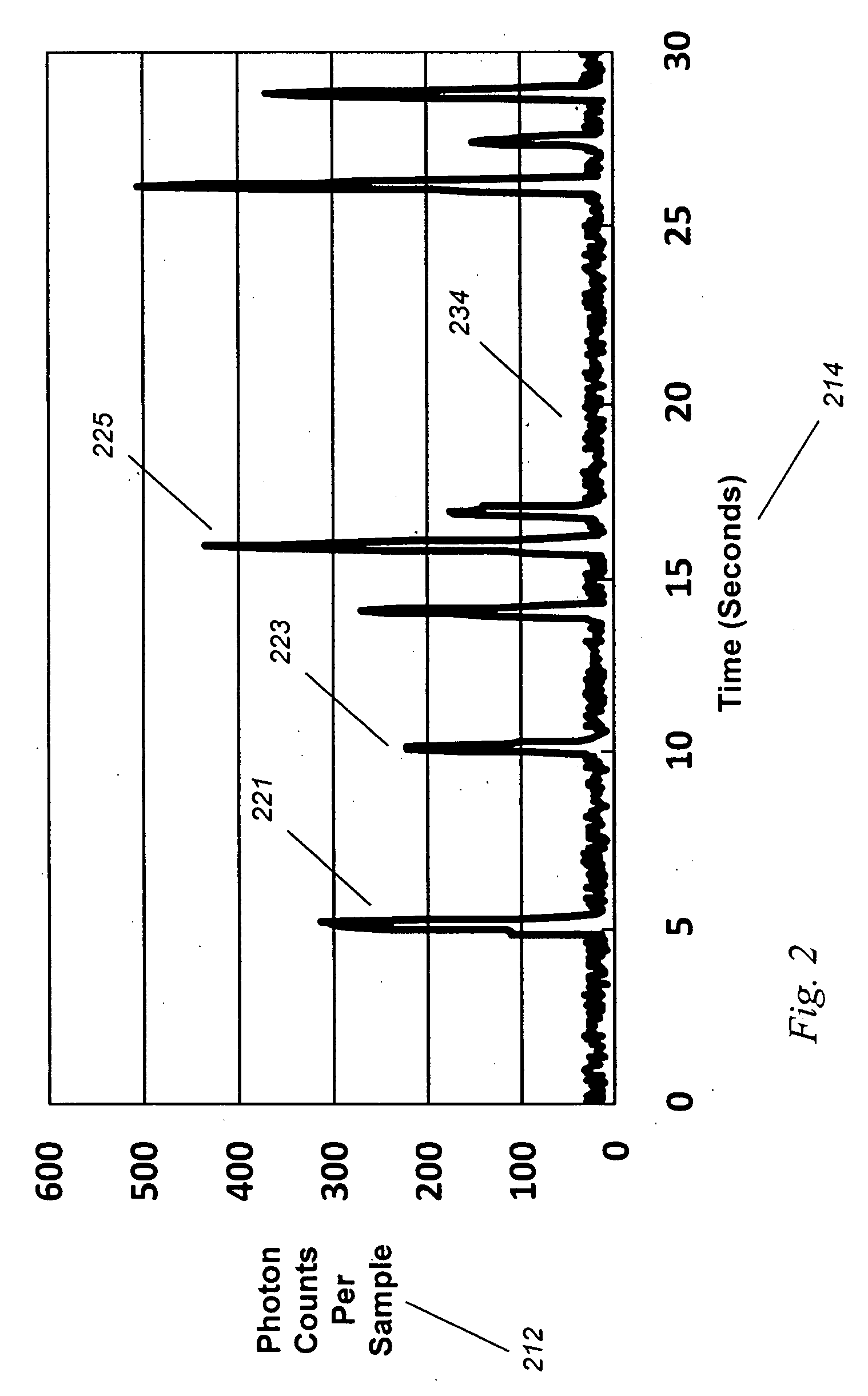
[0074]In order to test the validity of the data generated using the model shown in Example 1, we constructed a working system and tested this in a fluid model of tissue.

[0075]We have shown that in vivo circulating cell counting is feasible. Such improved lens systems may be designed as a standalone device, or embedded into a diagnostic or therapeutic system.

[0076]We have discovered an improved circulating cell counter that operates in vivo. A fiber-based illumination and detection system as been constructed and tested, in which a fiber optic system is used for light collection and collection, and a photodetector has been used to detect and quantify “blips” in returning light. A medical system incorporating the improved device, and medical methods of use, are described. This device has been built and tested in several configurations in models, animals, and planned for humans, and has immediate application to several important problems, both medical and indu...

example 3

Detection of Circulating Prostate Tumor

[0077]By creating a ligand targeted against the extracellular domain of PSMA, a molecule found on the membrane of cells in duct tissue in the prostate gland, one has a binding target that is found only on the surface of prostate cells, and to a lesser extent on new blood vessels (neoangiogenesis). This binding site is also found on circulating tumor cells, such as in prostate cancer.

[0078]We created a ligand using the hj-591 antibody developed by Bander et al. at Cornell University, and coupled this to CyDye (Amersham Health, General Electric, England) using chemistry pathways under the direction of Darryl Bornhop at Vanderbilt University. This work was funded by the US Government (PHS Grant CA107908, David Benaron, Principal Investigator).

[0079]Because the dye binds to prostate cells, circulating tumor cells may be detected using the methods and systems described in Examples 1 and 2.


PUM

PropertyMeasurementUnit
wavelengthsaaaaaaaaaa

Abstract

An improved circulating cell counter for generating light, and for delivering this light to a site in vivo for determining the presence, absence, concentration or count of a target cell, in which a light source such as a laser diode (121) and integrated optics (153) produce a beam transmitted to an in vivo target region (165), such as a capillary bed with flowing cells in a living tissue. Based upon the movement of cells in and out of this region, a circulating cell count (192) is generated, allowing determination of the presence, absence, concentration or count of the target cell. Use with optical, magnetic, or nanobot contrast agents, and methods of use are also described.

Description

U.S. Government Rights[0001]The U.S. government has certain rights in this invention pursuant to Public Health Service contract CA105653 and CA107908, awarded by the National Cancer Institute to the Spectros Corporation.FIELD OF THE INVENTION[0002]The present invention relates to detection systems and methods for providing highly specific cellular analysis of cells, entities, and / or xenograph nanobots in vivo, wherein the traditional ex vivo measurement is replaced by a measurement in living tissue. More particularly the present invention relates to systems and methods employing illuminating optics configured to illuminate and collect light from stained cells in the capillary circulation using a targeted optical dye, thus allowing for cell detection and / or counting in vivo and in real time, and allowing for on-line, real-time analysis of blood components without the need for blood withdrawal and preparation.BACKGROUND OF THE INVENTION[0003]Blood cellular analysis (such as white cell...

Claims


Application Information

Patent Timeline
Patent Type & Authority Applications(United States)
IPC IPC(8): A61B5/145
CPCA61B5/0059A61B5/412A61K47/48776A61K49/0032A61K49/0052A61K49/0058A61K49/0093A61K47/6901
Inventor BENARON, DAVID A.PARACHIKOV, ILLIAN H.
Owner J FITNESS LLC
Why Eureka
Try Eureka