Isolation and Cultivation of Stem/Progenitor Cells From the Amniotic Membrane of Umbilical Cord and Uses of Cells Differentiated Therefrom

a technology of amniotic membrane and stem cells, which is applied in the field of skin equivalent, can solve the problems of many ethical problems, low number of stem cells extracted, and high cost of collection

Active Publication Date: 2008-10-09
CELLRESEARCH CORP PTE LTD
View PDF7 Cites 85 Cited by
  • Summary
  • Abstract
  • Description
  • Claims
  • Application Information

AI Technical Summary

Benefits of technology

[0021]In yet another embodiment of the present invention the UCMC and / or UCEC comprised in the skin equivalent of the present invention are able to proliferate and further differentiate in fibroblasts and keratinocytes, respectively.
[0025]incubating said scaffold in a first medium, which allows said cells to proliferate and further differentiate.

Problems solved by technology

Even though the potential of embryonic stem cells is enormous, their use implies many ethical problems.
In clinical use, however, harvesting of such mesenchymal stem cells causes several problems.
The collection of the cells is a mental and physical burden to the patient as a surgical procedure is required to obtain the cells (for example, the collection of bone marrow is an invasive technique performed with a biopsy needle that requires local or even general anesthesia).
Furthermore, in many cases the number of stem cells extracted is rather low.
However, the existence of mesenchymal stem / progenitor cells is discussed controversially.
On the one hand, such cells could not be isolated or successfully cultured from term umbilical cord blood (Mareschi, K. et al.
However, it is still a matter of debate whether these stem / progenitor cells are indeed derived of umbilical cord tissue.
However, none of these approaches employed thus far has, for example, resulted in the isolation or cultivation of epithelial stem / progenitor cells as a source for epithelial cell-based therapies such as skin resurfacing, liver repair, bladder tissue engineering and other engineered surface tissues.
Depending on the extent of the damage the substitution with patient's own skin (autografts) taken from non-affected parts of the body is sometimes insufficient due to the extensive loss of skin.
However, these mesenchymal stem cells, which are isolated from the bone marrow, are rare and typically 10-20 cc aspirates have to be harvested from a patient in order to obtain enough mesenchymal stem cells.
Furthermore, rapid and efficient methods which are ethically acceptable and do not pose a biomedical burden on the patient for the isolation of epithelial and mesenchymal stem / progenitor cells are still required in order to provide such cells in a sufficient amount for various applications in regenerative medicine and tissue engineering.

Method used


Image

  • Isolation and Cultivation of Stem/Progenitor Cells From the Amniotic Membrane of Umbilical Cord and Uses of Cells Differentiated Therefrom
  • Isolation and Cultivation of Stem/Progenitor Cells From the Amniotic Membrane of Umbilical Cord and Uses of Cells Differentiated Therefrom
  • Isolation and Cultivation of Stem/Progenitor Cells From the Amniotic Membrane of Umbilical Cord and Uses of Cells Differentiated Therefrom

Examples


example 1

Collection of Umbilical Cord Tissue

[0182]Umbilical cord tissue is collected immediately after delivery of the child. The specimen is rinsed clean and immediately transferred into a 500 ml sterile glass bottle containing culture transport medium (L-15 medium supplemented with 50 IU / ml penicillin, 50 μg / ml streptomycin, 250 μg / ml fungizone, 50 μg / ml gentamicin; all reagents purchased from Invitrogen) prior to transport to the laboratory. In the laboratory, stem cell extraction is conducted in a laminar flow hood under sterile conditions. The specimen is first transferred to a sterile stainless steel tray. All remaining blood in the cord vessels is removed by multiple syringing washes using warm phosphate-buffered saline (PBS) supplemented with 5 IU / ml heparin (from Sigma-Aldrich, Missouri, USA). Plain PBS without heparin is used in the final washes. The umbilical cord tissue specimen is then cut into pieces 2 cm in length and transferred into 10 cm diameter cell culture dishes, where ...

example 2

Cell Separation / Cultivation

[0183]Dissection of umbilical cord tissue is first performed to separate the umbilical cord amniotic membrane from Wharton's jelly (i.e. the matrix of umbilical cord) and other internal components. The isolated amniotic membrane is then cut into small pieces (0.5 cm×0.5 cm) for cell isolation. Explant is performed by placing the pieces of umbilical cord amniotic membrane on tissue culture dishes at different cell culture conditions for isolation of either epithelial or mesenchymal stem cells.

[0184]For mesenchymal cell separation / cultivation, the explants were submerged in 5 ml DMEM (Invitrogen Corporation, California, USA) supplemented with 10% fetal bovine serum (Hyclone, Utah, USA) (DMEM / 10% FBS) and maintained in a CO2 cell culture incubator at 37° C. The medium was changed every 2 or 3 days. Cell outgrowth was monitored under light microscopy. Outgrowing cells were harvested by trypsinization (0.125% trypsin / 0.05% EDTA) for further expansion and cryo-p...

example 3

Identification of Stem / Progenitor Cells

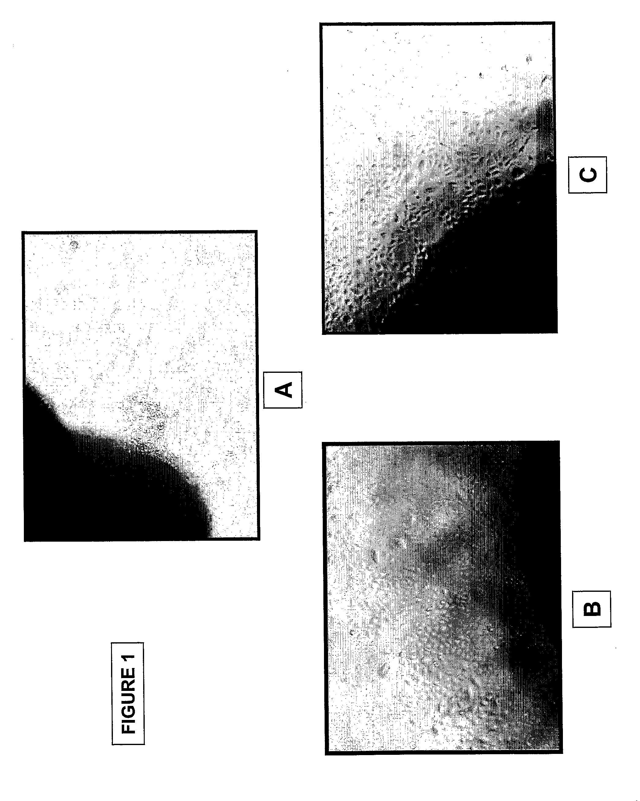
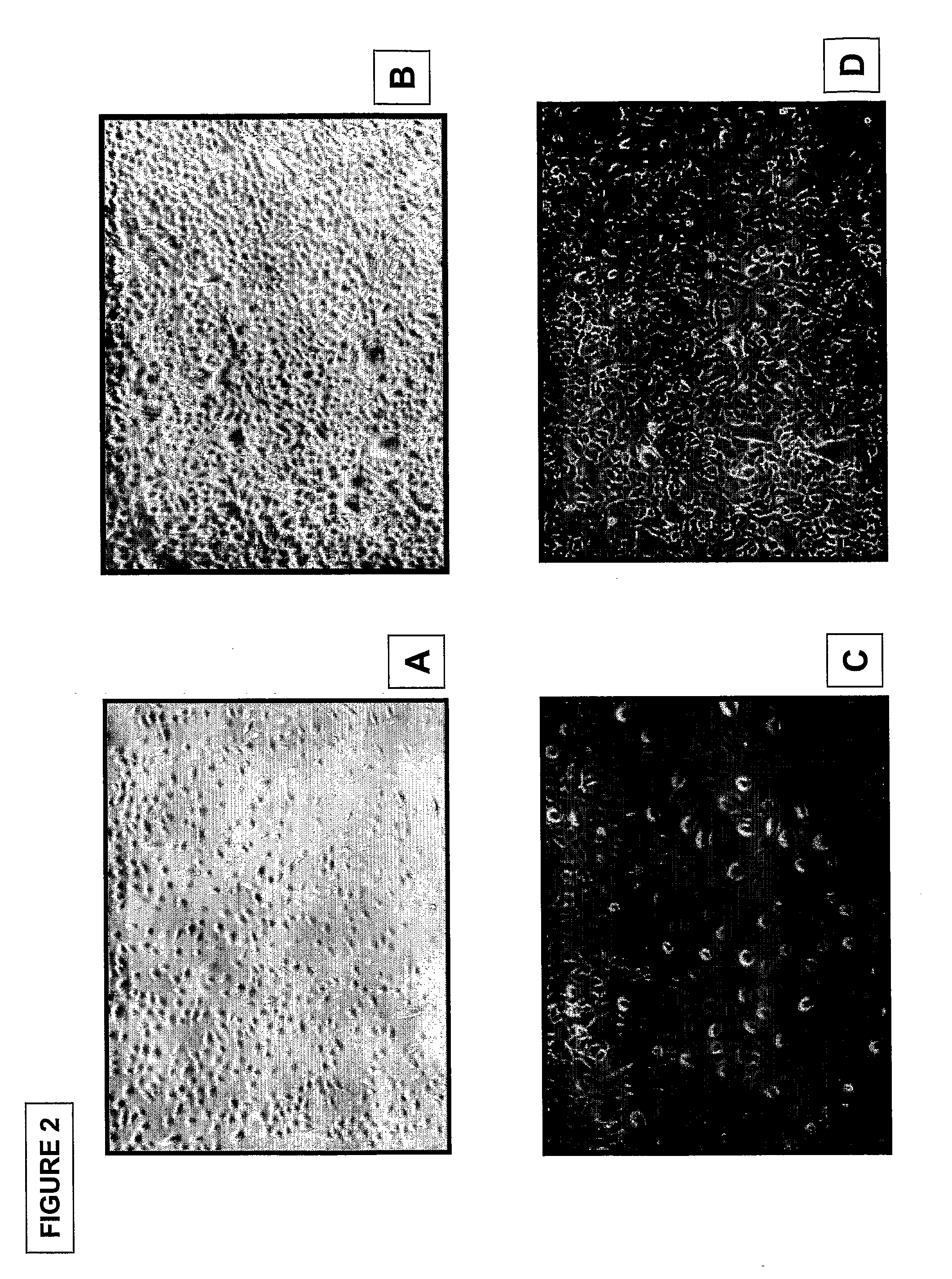
[0190]Epithelial cells: FIG. 1 shows pictures of outgrowing epithelial cells from umbilical cord amniotic membrane prepared by the method using tissue explant (40× magnification). Pictures were taken at day 2 (FIG. 1A) and day 5 (FIG. 1B, C) of tissue culture. Cell morphology analysis demonstrated polyhedral shaped epithelial-like cells. Enzymatic digestion of the umbilical cord segments yielded similar (FIG. 2), epithelial cells at day 2 (FIG. A, C) and day 5 (FIG. B, D) (40× magnification). FIG. 7 shows pictures of colony formation of epithelial stem cells from umbilical cord amniotic membrane cultured on feeder layer using Green's method (40× magnification). A colony of polyhedral shaped epithelial-like cells expanded rapidly from day 3 to day 7.

[0191]Mesenchymal cells: Outgrowth of mesenchymal cells explanted from umbilical cord amniotic membrane was observed as early as 48 hours after placement in tissue culture dishes using CMRL-1066 supp...


PUM

PropertyMeasurementUnit
v/vaaaaaaaaaa
surface areaaaaaaaaaaa
thickaaaaaaaaaa

Abstract

The present invention relates to a skin equivalent and a method for producing the same, wherein the skin equivalent comprises a scaffold and stem / progenitor cells isolated from the amniotic membrane of umbilical cord. These stem / progenitor cells may be mesenchymal (UCMC) and / or epithelial (UCEC) stem cells, which may then be further differentiated to fibroblast and keratinocytes. Further described is a method for isolating stem / progenitor cells from the amniotic membrane of umbilical cord, wherein the method comprises separating the amniotic membrane from the other components of the umbilical cord in vitro, culturing the amniotic membrane tissue under conditions allowing cell proliferation, and isolating the stem / progenitor cells from the tissue cultures. The invention also refers to therapeutic uses of these skin equivalents. Another aspect of the invention relates to the generation of a mucin-producing cell using stem / progenitor cells obtained from the amniotic membrane of umbilical cord and therapeutic uses of such mucin-producing cells. In yet another aspect, the invention relates to a method for generating an insulin-producing cell using stem / progenitor cells isolated from the amniotic membrane of umbilical cord and therapeutic uses thereof. The invention further refers to a method of treating a bone or cartilage disorder using UCMC. Furthermore, the invention refers to a method of generating a dopamin and tyrosin hydroxylase as well as a HLA-G and hepatocytes using UCMC and / or UCEC. The present invention also refers to a method of inducing proliferation of aged keratinocytes using UCMC.

Description

CROSS-REFERENCE TO RELATED APPLICATIONS[0001]This application claims the benefit of priority of U.S. provisional application No. 60 / 729,172, filed Oct. 21, 2005, the contents of which being hereby incorporated by reference it its entirety for all purposes.FIELD OF THE INVENTION[0002]The present invention relates to a skin equivalent and a method for producing the same, wherein the skin equivalent comprises a scaffold and stem / progenitor cells isolated from the amniotic membrane of umbilical cord. These stem / progenitor cells may be mesenchymal (UCMC) and / or epithelial (UCEC) stem cells, which may then be further differentiated to fibroblast and keratinocytes. Further described is a method for isolating stem / progenitor cells from the amniotic membrane of umbilical cord, wherein the method comprises separating the amniotic membrane from the other components of the umbilical cord in vitro, culturing the amniotic membrane tissue under conditions allowing cell proliferation, and isolating...

Claims


Application Information

Patent Timeline
Patent Type & Authority Applications(United States)
IPC IPC(8): A61K35/12A01N1/02A61P3/10A61P11/00A61P19/00C12N5/06C12N5/071C12N5/073C12N5/077
CPCA61L27/18A61L27/3633A61L27/3804A61L27/3839A61L27/56A61L27/60C12N5/0605C12N5/0629C12N5/0668C12N5/0676C12N5/0698C12N2501/11C12N2501/115C12N2502/094C12N2502/1323C12N2506/02C12N2533/54C08L69/00A61P11/00A61P17/02A61P19/00A61P19/08A61P3/10A61K35/32C12N5/0655C12N2506/1369
Inventor PHAN, TOAN-THANG
Owner CELLRESEARCH CORP PTE LTD
Why Eureka
Try Eureka