Method and apparatus for high resolution coherent optical imaging

a coherent optical imaging and high-resolution technology, applied in the field of high-resolution optical imaging, can solve the problems of difficult to obtain high-lateral resolution in vivo conditions, limit the numerical aperture of the imaging system, and may be technically unnecessary, so as to facilitate high-speed operation and simplify the design of the device

Inactive Publication Date: 2008-11-20
HEALTH NETWORK UNIV OF
View PDF9 Cites 16 Cited by
  • Summary
  • Abstract
  • Description
  • Claims
  • Application Information

AI Technical Summary

Benefits of technology

[0017]The present invention provides an endomicroscope with multiple fibers (i.e. channels) employing OCT to allow for different parts of the image to be scanned in parallel, instead of in series as is currently implemented in existing in vivo or ex vivo OCT / OCM systems. Using multiple parallel channels focused to different depths in the tissue, with each channel collecting high-resolution OCT data only across a very small axial range allows for a series of tight focal points to be achieved throughout the entire field of view without dynamic focusing or dynamic compensation. This greatly simplifies the design of the device which may now use mostly fixed optical components, and may facilitate high-speed operation. In addition, the intrinsically miniature dimension of the fiber optic based OCT technique makes the multichannel concept possible to implement in a flexible endoscopic device while the proximal part of the endoscope, which is outside of the patient, allows room for sources, detectors, and other equipment.
[0021]a focusing means in the first optical path for focusing optical radiation from the optical radiation sources into a plurality of respective focal points located on a surface within the first optical path to provide substantially continuous coverage of a selected portion of the first optical path, whereby, in use, a sample can be located at least partially within the selected portion, thereby permitting simultaneous scanning of a plurality of points within the sample.

Problems solved by technology

After reviewing possible clinical applications of the endomicroscope, the following parameters and features were identified since it is difficult to achieve these parameters and features simultaneously with current in vivo OCT systems.
As a result, the outer diameter of these systems is restricted to approximately 2 to 3 mm which limits the numerical aperture of the imaging system.
This makes it difficult to obtain high lateral resolution under in vivo conditions.
These constraints may be technically unnecessary and limit one from fully exploiting the benefit of OCT / OCM in the clinical applications under consideration.

Method used


Image

  • Method and apparatus for high resolution coherent optical imaging
  • Method and apparatus for high resolution coherent optical imaging
  • Method and apparatus for high resolution coherent optical imaging

Examples


Embodiment Construction

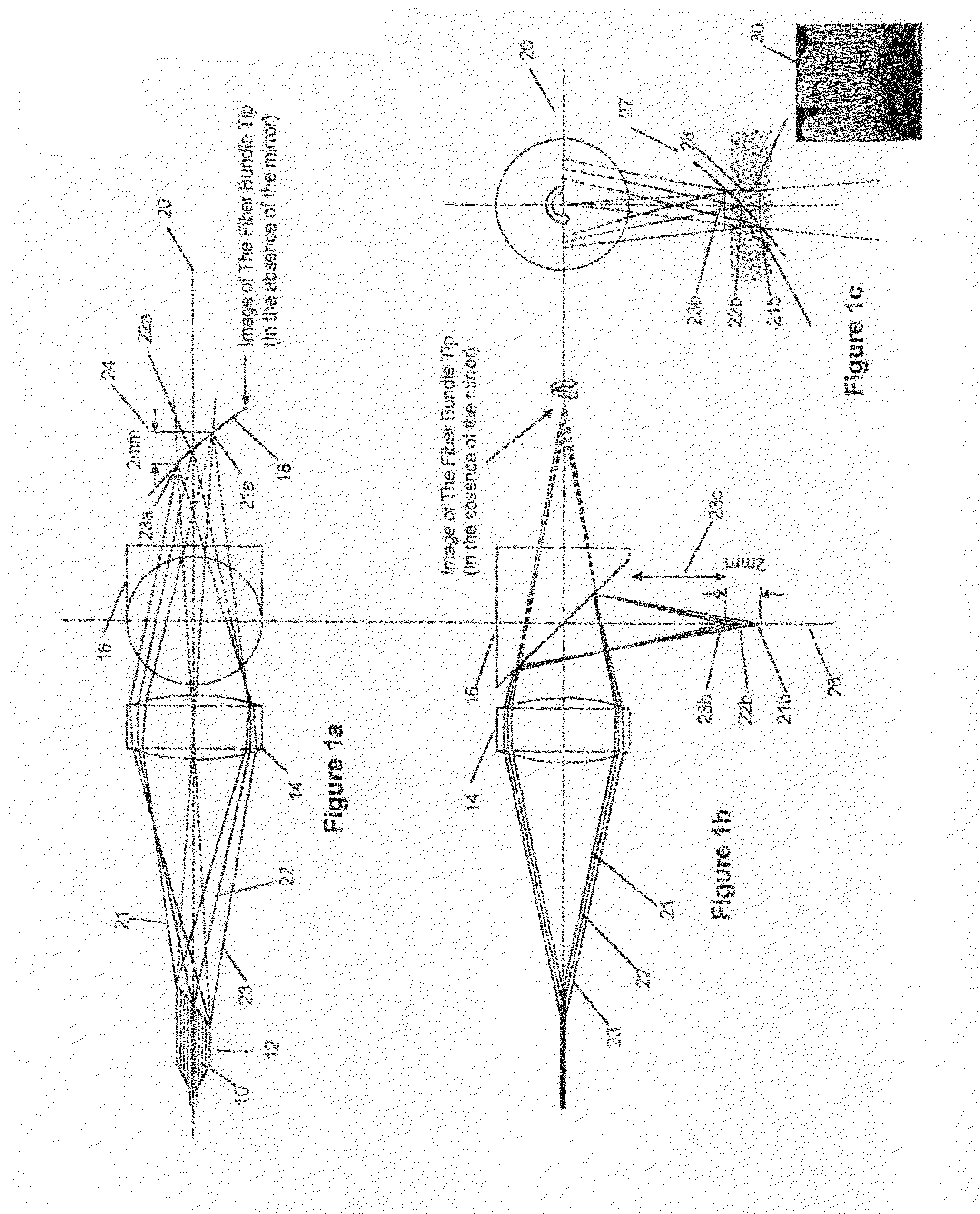
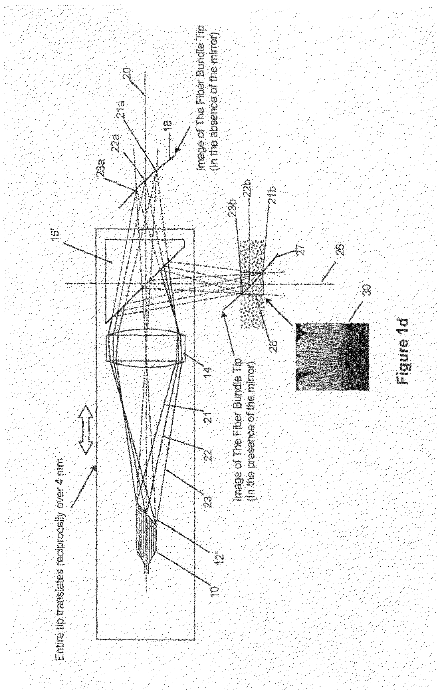
[0070]In the following description, various specific dimensions and other parameters are mentioned such as the wavelength used by the imaging source and the physical dimensions of the optical components used in the apparatus. It is to be appreciated that this is for exemplary purposes only and does not limit this invention. Specific parameters, dimensions and the like may be chosen depending on an intended application of the invention.

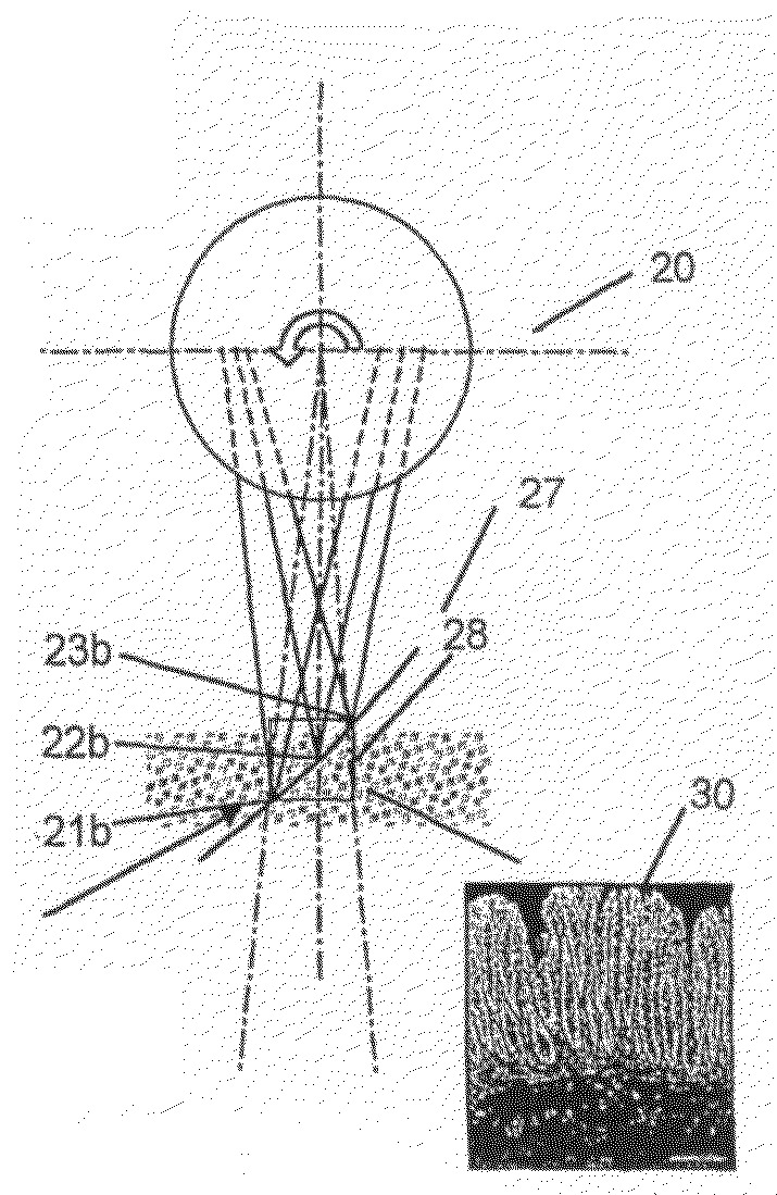
[0071]Referring to FIG. 1, the present invention provides an apparatus comprising an endoscopic coherent optical microscope having multiple single mode fibers 10, a fiber bundle tip 12, a focusing lens 14 and a mirror 16. The multiple single mode fibers 10 and the focusing lens 14 are stationary while the mirror 16 is rotatable. Accordingly, the mirror 16 is mounted for rotation in known manner. Details of the rotating mechanism for the mirror 16 are not described further and can be conventional. Multiple single mode fibers 10 (of which there may be ap...


PUM

PropertyMeasurementUnit
depthsaaaaaaaaaa
distanceaaaaaaaaaa
depthaaaaaaaaaa

Abstract

A method and an apparatus for examining the sub-surface microstructure of a sample are provided. Radiation from a plurality of optical radiation sources travels along a first optical path. In the first optical path, a device focuses the optical radiation from each of the optical sources into a plurality of respective focal points along the first optical path to provide substantially continuous coverage of a selected portion of the first optical path. Then, a sample on the first optical path within the selected length extending into the sample is scanned along said selected portion of the first optical path.

Description

FIELD OF THE INVENTION[0001]This invention relates to both a method and an apparatus for high-resolution optical imaging. More particularly, this invention is concerned with providing high-resolution imaging suitable for incorporation into an endoscope.BACKGROUND OF THE INVENTION[0002]Modern medical imaging techniques have important applications in health care. Modalities such as X-ray computed tomography (CT), magnetic resonance imaging (MRI) and ultrasound imaging are the main tomographic techniques available in most modern medical centers. Visible-light endoscopy is another major imaging modality which is used extensively in procedures like bronchoscopy or colonoscopy. Each of these techniques employs different physical principles and measures different properties of the biological tissue under study with different resolution. Further, they can commonly be performed in-vivo. A third type of imaging, optical microscopy, is still utilized widely in clinical medicine. However, optic...

Claims


Application Information

Patent Timeline
Patent Type & Authority Applications(United States)
IPC IPC(8): G02B6/28G01N21/17A61B1/00A61B1/05A61B5/00G01N21/47G02B6/04G02B23/26
CPCA61B1/00096A61B1/00172G01N21/4795A61B5/0066A61B5/6852A61B1/05
Inventor YANG, VICTOR XIAO DONGVITKIN, I. ALEXWONGKEESONG, LOUIE M.KATZ, SHARONGORDON, MARGARET LESLIEWILSON, BRIAN C.MOK, ALVIN HO KWAN
Owner HEALTH NETWORK UNIV OF
Why Eureka
Try Eureka