Muscle derived cells for the treatment of cardiac pathologies and methods of making and using the same

a technology of muscle derived cells and cardiac pathologies, applied in the field of muscle derived progenitor cells, to achieve the effects of reducing the risk of cardiac disease, and increasing the function of the patient's hear

Active Publication Date: 2009-01-08
UNIVERSITY OF PITTSBURGH +1
View PDF9 Cites 13 Cited by
  • Summary
  • Abstract
  • Description
  • Claims
  • Application Information

AI Technical Summary

Benefits of technology

[0027]MDCs, whether frozen or preserved as a pharmaceutical composition, or used fresh, may be used to treat a number of cardiac conditions. These conditions include myocardial infarction, heart failure, Adams-Stokes disease, congenital heart disease, angina pectoris, arrhythmias, atrial fibrillation, bacterial endocarditis, cardiomyopathy, congestive heart failure, diastolic dysfunction, heart murmurs, and premature ventricular contractions. The MDCs may be used to cure heart defects caus...

Problems solved by technology

These methods result in the enrichment of MDCs that have long-term sur...

Method used


Image

  • Muscle derived cells for the treatment of cardiac pathologies and methods of making and using the same
  • Muscle derived cells for the treatment of cardiac pathologies and methods of making and using the same
  • Muscle derived cells for the treatment of cardiac pathologies and methods of making and using the same

Examples


example 1

MDC Enrichment, Isolation and Analysis

[0094]Enrichment and isolation of MDC: MDCs were prepared as described (U.S. Pat. No. 6,866,842 of Chancellor et al.). Muscle explants were obtained from the hind limbs of a number of sources, namely from 3-week-old mdx (dystrophic) mice (C57BL / 10ScSn mdx / mdx, Jackson Laboratories), 4 6 week-old normal female SD (Sprague Dawley) rats, or SCID (severe combined immunodeficiency) mice. The muscle tissue from each of the animal sources was dissected to remove any bones and minced into a slurry. The slurry was then digested by 1 hour serial incubations with 0.2% type XI collagenase, dispase (grade II, 240 unit), and 0.1% trypsin at 37° C. The resulting cell suspension was passed through 18, 20, and 22 gauge needles and centrifuged at 3000 rpm for 5 minutes. Subsequently, cells were suspended in growth medium (DMEM supplemented with 10% fetal bovine serum, 10% horse serum, 0.5% chick embryo extract, and 2% penicillin / streptomycin). Cells were then pre...

example 2

MDC Vectors and Transfection

[0099]Retrovirus and adenovirus vectors: The MFG-NB (N. Ferry et al., 1991, Proc. Natl. Acad. Sci. USA 88:8377 81) retroviral vector was used for the MDCs experiments. This vector contains a modified LacZ gene (NLS-LacZ) that includes a nuclear-localization sequence cloned from the simian virus (SV40) large T antigen transcribed from the long terminal repeat (LTR). The retroviral stock was grown and prepared as previously described (J. C. van Deutekom et al., 1998, Neuromuscul. Disord. 8:135 48). The retrovirus was titered to 1×10⁷−1×10⁹ cfu / ml.

[0100]An adenovirus vector was also used. This vector contained the LacZ gene under the control of the human cytomegalovirus (HuCMV) promoter (J. Huard et al., 1994, Hum Gene Ther 5:949 58). The E1 E3 deleted recombinant adenovirus was obtained through Dr. I. Kovesdi (Gene Vec Inc., Rockville, Md.).

[0101]Viraltransduction of MDC: For viral transduction, MDCs were plated at a density of 1−1.5×10⁶ in T 75 flasks. PP6...

example 3

Soft Tissue Augmentation of the Skin

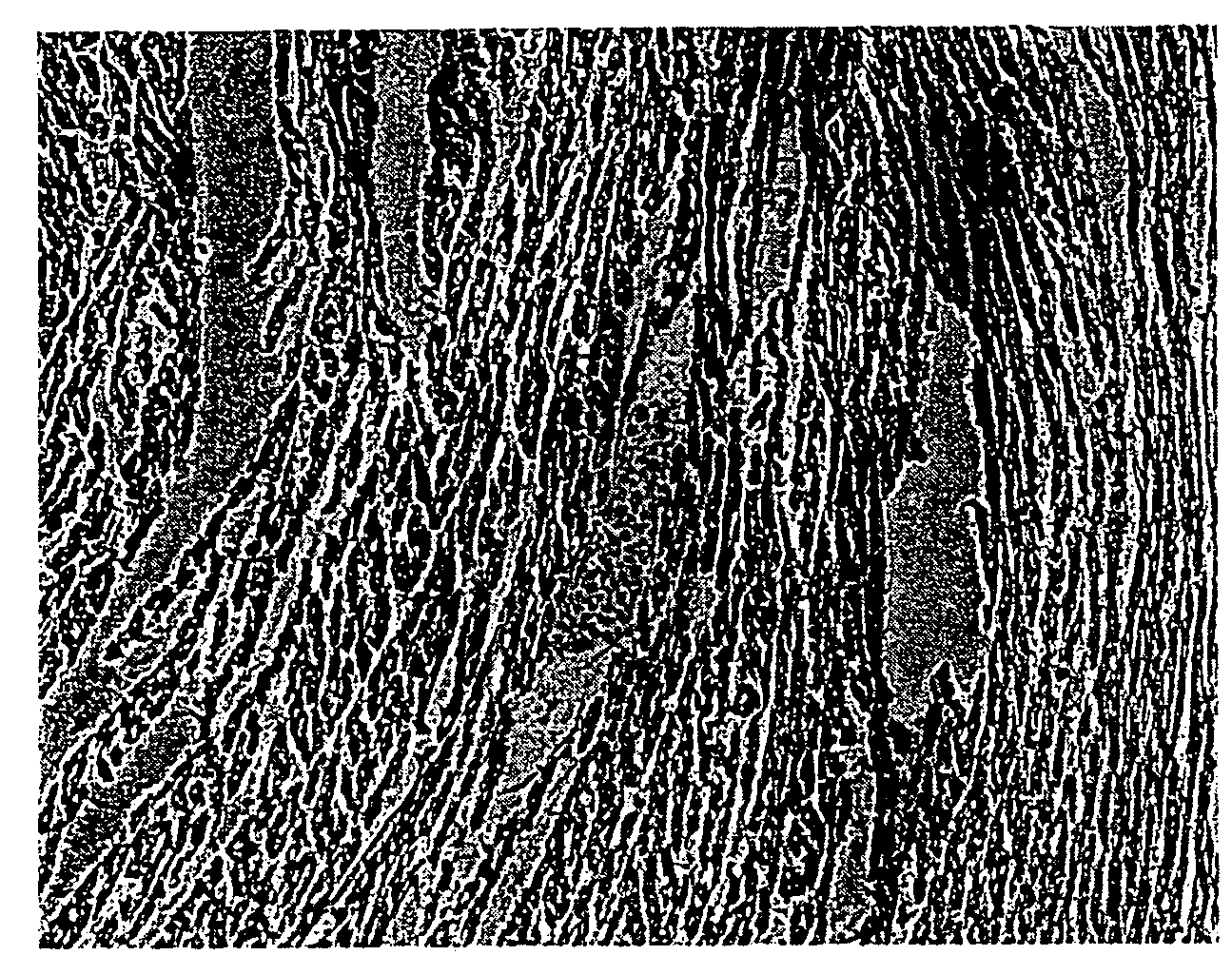
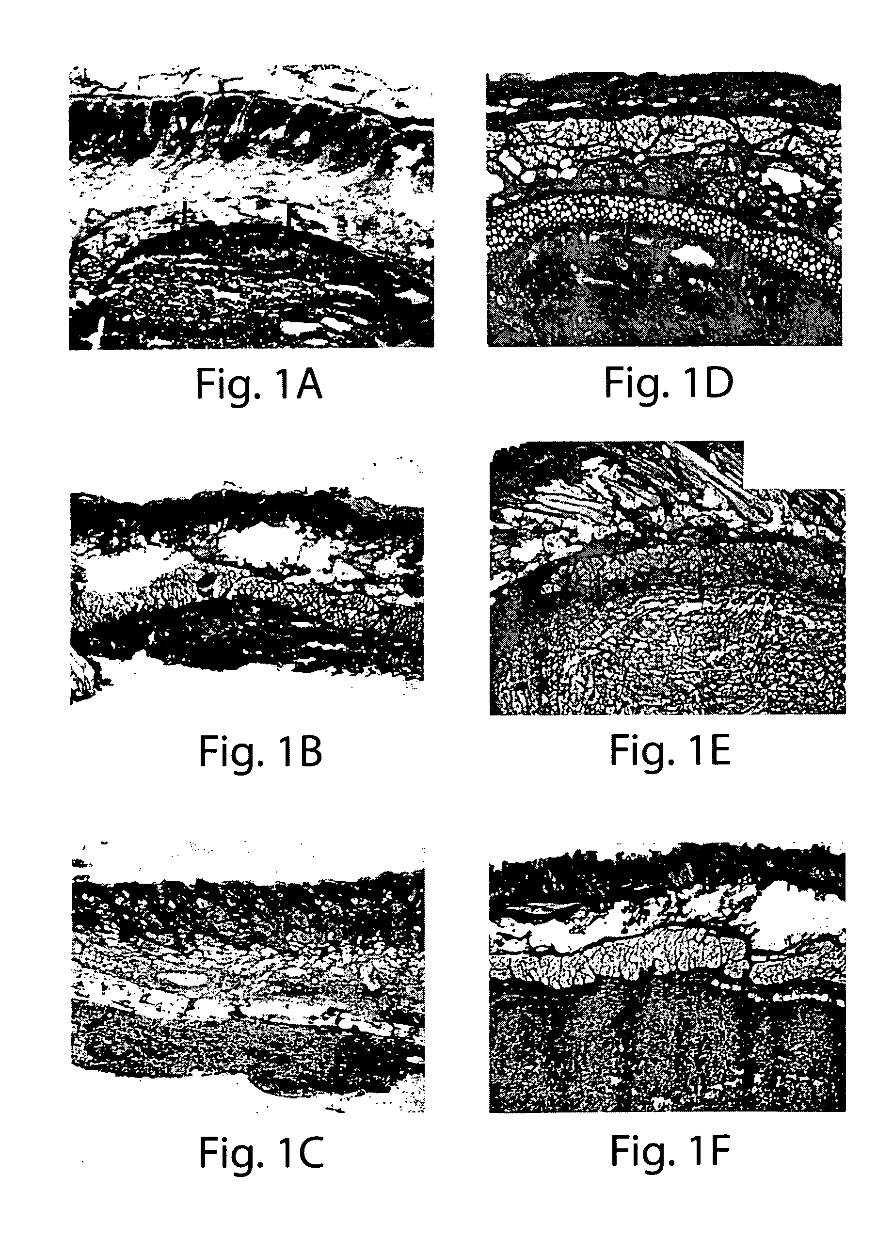
[0102]MDCs and collagen injection: SD rats were prepared for surgery by anesthetizing with halothane using standard methods, and washing the surgical site with Betadine® solution. The skin of the lower abdomen was injected with either 10 microliters (μl) of a MDC suspension in HBSS (approximately 1−1.5×10⁶ cells), 10 μl of commercially available bovine collagen (Contigen™; C. R. Bard, Covington, Ga.), or 10 μl of sterile saline using a Hamilton microsyringe. At 5 days, 2 weeks and 4 weeks post-injection, the area surrounding each injection site was excised, prepared for histochemical analysis, examined microscopically, and photographed. Histochemical analysis included hematoxylin, eosin, or trichrome staining.

[0103]The results demonstrate that MDCs were viable for up to at least 4 weeks following injection into skin tissue, with no evidence of inflammation of the tissue at the injection site (FIGS. 1D-1F). In contrast, collagen was not visible at ...


PUM


Abstract

The present invention provides muscle-derived progenitor cells that show long-term survival following transplantation into body tissues and which can augment soft tissue following introduction (e.g. via injection, transplantation, or implantation) into a site of soft tissue. Also provided are methods of isolating muscle-derived progenitor cells, and methods of genetically modifying the cells for gene transfer therapy. The invention further provides methods of using compositions comprising muscle-derived progenitor cells for the augmentation and bulking of mammalian, including human, soft tissues in the treatment of various cosmetic or functional conditions, including malformation, injury, weakness, disease, or dysfunction. In particular, the present invention provides treatments and amelioration for dermatological conditions, gastroesophageal reflux, vesico-ureteral reflux, urinary incontinence, fecal incontinence, heart failure, and myocardial infarction.

Description

CROSS-REFERENCE TO RELATED APPLICATIONS[0001]This application claims priority from U.S. Provisional Application Ser. No. 60 / 867,405, filed on Nov. 28, 2006, the disclosures of which are incorporated herein by reference in their entireties.[0002]Each of the applications and patents cited in this text, as well as each document or reference cited in each of the applications and patents (including during the prosecution of each issued patent; “application cited documents”), and each of the U.S. and foreign applications or patents corresponding to and / or claiming priority from any of these applications and patents, and each of the documents cited or referenced in each of the application cited documents, are hereby expressly incorporated herein by reference. More generally, documents or references are cited in this text, either in a Reference List before the claims, or in the text itself; and, each of these documents or references (“herein-cited references”), as well as each document or r...

Claims


Application Information

Patent Timeline
IPC IPC(8): A61K35/12C12N5/00C12N5/08A61P9/00A61K35/34C12N5/077
CPCA61K35/12C12N5/0658A61K35/34A61P9/00A61P9/04A61P9/06A61P9/10
Inventor CHANCELLOR, MICHAEL B.PAYNE, THOMASJANKOWSKI, RONALDPRUCHNIC, RYAN
Owner UNIVERSITY OF PITTSBURGH
Why Eureka
Try Eureka