
Device and methods for detecting and quantifying one or more target agents

a technology of target agents and devices, applied in the field of devices and methods for detecting and quantifying one or more target agents, can solve the problems of difficult detection of more than a single agent in a sample in a short time period, pcr can suffer from non-specific amplification of non-targeted nucleic acid sequences, and none of the above problems have been as widely accepted as pcr

Inactive Publication Date: 2009-02-05
ANTARA BIOSCI
View PDF37 Cites 36 Cited by
  • Summary
  • Abstract
  • Description
  • Claims
  • Application Information

AI Technical Summary

Benefits of technology

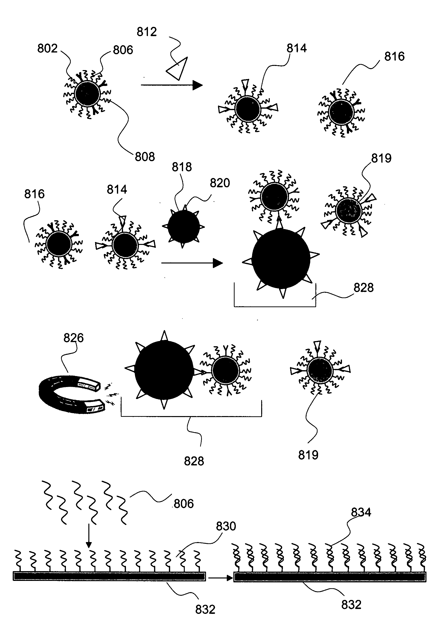
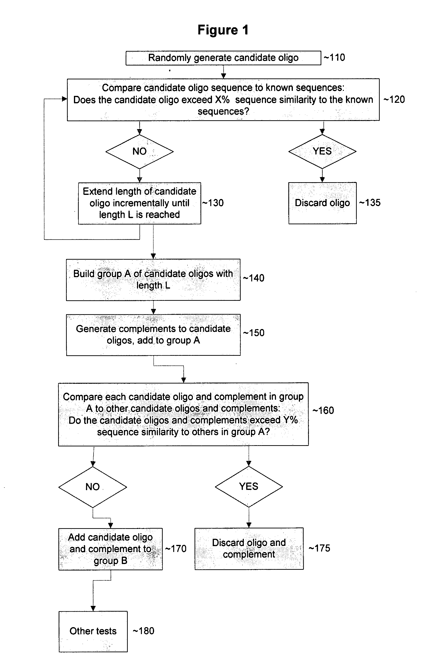
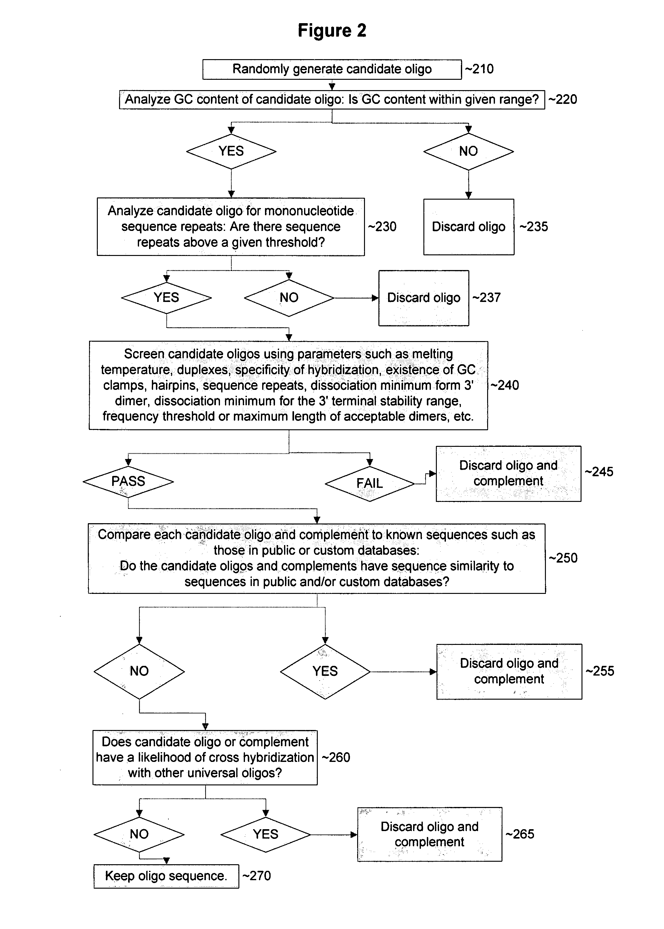
[0016]Certain methods of the present invention solve the problem of multiplex detection for a wide range of target agents by combining the versatility of antibody recognition with the multiplexing capability, speed, and sensitivity of controlled electrochemical detection of nucleic acid hybridization, yet generally minimizing or eliminating the need for nucleic acid isolation / amplification procedures and the problems associated with non-specific nucleic acid hybridization in many embodiments. The non-specific hybridization observed in other detection methods currently known in the art is overcome in these methods by nucleic acid sequences that are rationally designed to minimize the risk of non-specific hybridization, ensuring that sequence-specific hybridization is optimized.
[0028]Another assay embodiment of the invention comprises in varied orders or combinations: (1) exposing a plurality of capture moieties to a sample, each capture moiety comprising a target agent binding domain and a capture-associated oligo having a polymerase recognition sequence; (2) allowing any target agent in the sample to bind to the capture moieties; (3) isolating the capture moiety:target agent complexes; (4) binding an oligonucleotide complementary to the polymerase recognition sequence on each capture-associated oligo to the capture moiety:target agent complexes; (5) reacting the capture-associated oligo with nucleotides and polymerase under conditions to allow linear amplification; and (6) introducing the isolated capture-associated oligo to an electrode having an electrode-associated oligo, each electrode-associated nucleic comprising a detection moiety conjugated to a hairpin loop structure at the unattached end of the electrode-associated oligo, where the electrode-associated oligo is complementary to a specific capture-associated oligo. Binding of the capture-associated oligo to its corresponding electrode-associated oligo will disrupt the hairpin loop structure and position the detection moiety in proximity to the electrode, rendering a redox reaction possible. When the target agent is a nucleic acid duplex in certain embodiment, a single stranded nucleic acid molecule can be conjugated to one strand of the target agent sequence and a different sequence single stranded nucleic acid molecule can be conjugated to the other strand of the target sequence. Because both strands of the target nucleic acid duplex should be present in equal amounts in a sample embodiments' testing for the presence of each strand sequentially or in different aliquots of the same sample can be used as an internal control of the accuracy of the testing.

Problems solved by technology

The need for multiple antibodies, which do not non-specifically cross-react with other antigens, and the incubation steps involved mean that it is difficult to detect more than a single agent in a sample in a short time period.
PCR can suffer from non-specific amplification of non-targeted nucleic acid sequences.
Other variants of methods for the amplification of target nucleic acids exist, but none have been as widely accepted as PCR.
However, the Invader Assay suffers from serious deficiencies including a lack of sensitivity making it unsuitable for various diagnostic applications including infectious disease applications.
Many of these hybridization techniques, while overcoming the problem of non-specific nucleic acid amplification associated with PCR, lack the sensitivity required for many applications, including infectious disease diagnostics.
In particular, hybridization detection techniques such as the cycling probe reaction and the Invader™ Assay that produce a linear amplification of the signaling molecule, rather than the exponential target amplification of PCR, lack the ability to be used for the detection of some infectious disease agents that are typically present in low concentrations.
Additionally, linear amplification techniques may require comparatively substantially longer periods of time to accumulate a detectable signal.
This manipulation makes it possible to conduct tests simultaneously for many different sequences of nucleic acid that may be present in a sample without any substantial cross reactivity (also known as multiplex analysis); however, the possibility of a particular nucleic acid molecule hybridizing to a non-target nucleic acid that may be present cannot be precluded.
Additionally, ascertaining the presence of organisms by detecting specific nucleic acid sequences can involve the extraction and isolation of nucleic acids, which can lead to cross-contamination between samples.
Accordingly, even under the most stringent conditions there may be non-specific hybridization and cross-contamination that can give a false positive result when several nucleic acids of unknown sequence are present in a sample.
Such false indications frequently arise due to factors including faulty isolation techniques.
As discussed above however, whether based on fluorescence or electrochemistry, these hybridization detection methods can be subject to false positive signals due to non-specific hybridization.
Additionally, nucleic acid detection techniques requiring steps of nucleic acid extraction, isolation and purification may lengthen the time taken to achieve a result and also decrease the detection level of the test through the loss of nucleic acid molecules in the many washing steps involved in these isolation steps.
The nucleic acid detection techniques, while overcoming the potential problem of multiplexing associated with ELISA (e.g., the limited number of discriminatory signals), are restricted in use to only detecting nucleic acid molecules.
Therefore, agents such as proteins, chemical species, drugs, hormones, toxins, and prions, which do not contain nucleic acids, cannot be detected by nucleic acid hybridization techniques.

Method used


Image

  • Device and methods for detecting and quantifying one or more target agents
  • Device and methods for detecting and quantifying one or more target agents
  • Device and methods for detecting and quantifying one or more target agents

Examples


example i

Preparation of Monoclonal Antibodies

[0277]A peptide corresponding to amino acid residues in a desired antigen is synthesized with a peptide synthesizer (Applied Biosystems) according to methods known in the art. The peptide emulsified with Freund's complete adjuvant is used as an immunogen and administered to mice by footpad injection for primary immunization (day 0). The booster immunization is performed four times or more in total. The final immunization is carried out by the same procedure two days before the collection of lymph node cells. The lymph node cells collected from each immunized mouse and mouse myeloma cells are mixed at a ratio of 5:1. Hybridomas are prepared by cell fusion using polyethylene glycol 4000 or polyethylene glycol 1500 (GIBCO) as a fusing agent. The lymph node cells of the mouse are fused with mouse myeloma PAI cells (JCR No. B0113; Res. Disclosure Vol. 217, p. 155, 1982), and the resulting hybridomas are selected by culturing the fused cells in an ASF10...

example ii

Preparation of DNA-Antibody Conjugates

[0280]Oligonucleotide #109745 (5′ amino-modified, 88 nucleotides in length) was synthesized using standard phosphoramidite chemistry (Biosearch Technologies, Inc., Novato, Calif.) having the following nucleotide sequence:

5′-ATCTGCAGGGAGTCAACCTTGTCCGTCCATTCTAAACCGTTGTGCGTCCGTCCCGATTAGACCAACCCCCCTATAGTGAGTCGTATTA-3′.

The oligonucleotide was purified using a NAP-5 column (0.1 M / 0.15 M buffer of NaHCO3 / NaCl, pH 8.3).

[0281]0.2 mL of a 100 μM aqueous solution of oligonucleotide #109745 was loaded onto a column. After 0.3 mL buffer was added, 0.8 mL of eluant was collected and quantified. Based on A260 reading, more than 90% of recovery was observed.

[0282]The purified oligonucleotide was chemically modified using Succinimidyl 4-formylbenzoate (C6-SFB). 790 μL of purified oligonucleotide and 36 μL of C6-SFB (20 mM in DMF (dimethylformamide)) were mixed (1:40 ratio) and incubated at room temperature for 2 hours. The reaction product was cleaned up using a...

example iii

Immobilization of an Electrode-Associated Oligo to a Gold Electrode Surface

[0287]The gold electrodes on the chip (Nanostructures, Inc., Santa Clara, Calif.) were cleaned immediately prior to use in UV / ozone cleaner (UVOCS, model T16X16 / OES) for 10 minutes. Cleaned chips were stored in container under inert gas (argon).

[0288]5′-thiolated oligonucleotides with a C6 linker were synthesized using standard phosphoramidite chemistry (Biosearch Technologies, Inc. Novato, Calif.).

[0289]The spotting solution was prepared by mixing 5′-thiolated C6 oligonucleotides with mercaptohexanol (MCH) and KHPO4. Typically, the probe spotting solution consists of a 100 μM thiolated oligo, 1 mM MCH, and 400 mM KHPO4 (pH 3.8) buffer in aqueous solution.

[0290]Chips were printed (30 nl / spot) using BioJet Plus™ series AD3200 non-contact spotter (BioDot, Irvine, Calif.). The relative humidity during the printing was 85%. After incubation of the slides in a humidity chamber for 4 hrs, they were rinsed with an e...


PUM

PropertyMeasurementUnit
Angleaaaaaaaaaa
Contact angleaaaaaaaaaa

Abstract

The present invention provides a device and methods for the detection and quantification of one or more target agents in a sample by rapid and specific electrochemical detection. The present invention includes kits, devices and compositions capable of performing rapid, specific and accurate detection of one or more target agents in a sample.

Description

I. CROSS REFERENCE TO RELATED APPLICATIONS[0001]This application claims the benefit of U.S. Provisional Patent Application Ser. No. 60 / 765,740, filed Feb. 7, 2006, currently pending; U.S. Provisional Patent Application Ser. No. 60 / 801,703, filed May 19, 2006, currently pending; U.S. Provisional Patent Application Ser. No. 60 / 801,950, filed May 19, 2006, currently pending; U.S. Provisional Patent Application Ser. No. 60 / 802,002, filed May 19, 2006, currently pending; U.S. Provisional Patent Application Ser. No. 60 / 802,039, filed May 19, 2006, currently pending; U.S. Provisional Patent Application Ser. No. 60 / 802,049, filed May 19, 2006, currently pending; U.S. Provisional Patent Application Ser. No. 60 / 808,862, filed May 26, 2006, currently pending; U.S. Provisional Patent Application Ser. No. 60 / 812,826, filed Jun. 12, 2006, currently pending; U.S. Provisional Patent Application Ser. No. 60 / 814,566, filed Jun. 16, 2006, currently pending; U.S. Provisional Patent Application Ser. No....

Claims


Application Information

Patent Timeline
IPC IPC(8): C40B10/00G01N33/566C07H21/04G01N27/26C40B30/04C12Q1/68
CPCG01N33/54306
Inventor LABGOLD, MARC R.JOKHADZE, GEORGE G.JEN, I-MIN M.SHEN, NAIPINGKOZLOWSKI, MARK T.AMMINI, CHANDRAMOHAN V.SUHY, DAVID A.NORRIS, MICHAEL C.LOBBAN, PETER E.
Owner ANTARA BIOSCI
Learn More