
Proteomic analysis of biological fluids

a biological fluid and proteome technology, applied in the field of proteome identification of biological fluids, can solve the problems of not providing information on the regulation of protein function through post-translational modifications, proteolysis or compartmentalization, and compromising the well-being of the mother and/or the fetus or newborn,

Inactive Publication Date: 2009-02-26
HOLOGIC INC
View PDF33 Cites 16 Cited by
  • Summary
  • Abstract
  • Description
  • Claims
  • Application Information

AI Technical Summary

Benefits of technology

[0014]The present invention further provides proteomic profiles of biological fluids, such as amniotic fluid and maternal serum, which enable the diagnosis, prognosis, and monitoring of various pathologic fetal / maternal conditions, including, without limitation, intra-amniotic infection (IAI), chromosomal aneuploidies, and fetal diseases associated with fetal growth and maturation. In particular, the present invention provides normal and pathologic proteomic profiles for IAI and chromosomal aneuploidies. The determination of the normal proteomic profile is of great importance, since it enables the elimination of the fetal / maternal condition in question (negative diagnosis), which eliminates the need to subject the patient to unnecessary and potentially dangerous treatment or intervention.

Problems solved by technology

DNA sequence information is helpful in deducing some structural and potential protein modifications based on homology methods, but it does not provide information on regulation of protein function through post-translational modifications, proteolysis or compartmentalization.
There are numerous pathologic maternal and fetal conditions, such as intra-amniotic infection (IAI), preeclampsia, preterm delivery and labor, and chromosomal aneuploidies, that may develop during pregnancy and compromise the well-being or, in some instances, threaten the life of the mother and / or the fetus or newborn.
Unfortunately, early diagnosis for most of these conditions is difficult because the clinical signs and symptoms occur late, and are often non-specific and inconsistent.
For example, the clinical symptoms of IAI typically include maternal fever and leukocytosis, but these symptoms often occur later and are neither sensitive nor specific.
Measurement of cytokines or other biochemical factors is expensive, generally not clinically available, and is primarily a research tool.
Further, the testing efficiency of these tests has not been consistently better than more readily available traditional tests such as amniotic fluid Gram stain and culture, amniotic fluid glucose concentrations, and detection of amniotic fluid leukocyte esterase.
Although all have reasonable sensitivity, specificity, and predictive value none are sufficiently sensitive or specific to be utilized independently of clinical features in the diagnosis of IAI.
This is an invasive procedure associated with a 0.5% risk of loss of the pregnancy.
Further, chromosomal analysis of amniotic fluid cells is a labor-intensive and time-consuming procedure, taking up to 2 weeks.

Method used


Image

  • Proteomic analysis of biological fluids
  • Proteomic analysis of biological fluids
  • Proteomic analysis of biological fluids

Examples


example 1

General Protocols

[0127]Primate Model of Intra-Amniotic Infection

[0128]This protocol was approved by the Institutional Animal Care Utilization Committee of the Oregon National Primate Research Center, and guidelines for humane care were followed. Three pregnant rhesus monkeys (Macaca mulatta) with timed gestations were chronically catheterized as previously described (Haluska G J, et al., Temporal changes in uterine activity and prostaglandin response to RU 486 in rhesus macaques in late gestation, Am J Obstet Gynecol 157: 1487-95 (1987); and Gravett M G, et al., An experimental model for intramniotic infection and preterm labor in rhesus monkeys. Am J Obstet Gynecol 171: 1660-7 (1994)). Briefly, at approximately day 110 of gestation (term is 167 days) pregnant animals were conditioned to a jacket and tether system (Ducssay C A, et al., Simplified vest and tether system for maintenance of chronically catheterized pregnant rhesus monkeys. Lab. Anim Sci 38:343-4 (1988)). After conditio...

example 2

Identification of Proteins and Polypeptides Expressed in the Amniotic Fluid

[0164]Using the materials and methods described in Example 1, proteins and polypeptides expressed in normal and infected amniotic fluid were identified. Human and primate amniotic fluid samples (pooled and individual) were subjected to protein separation techniques (1-D, 2-D and HPLC fractionation) as described in Example 1. The separated proteins (gel bands, spots and fractions) were digested with trypsin to generate peptide pools. The peptide pools were analyzed using tandem MS to decipher their amino acid sequence and composition.

[0165]Five thousand MS spectra were selected using spectral verification programs. These spectral files were analyzed using de novo sequencing programs (Lutefisk, Peaks) to generate the amino acid sequence corresponding to each peptide. The de novo sequences generated from the peptide pool were used to search protein and DNA databases as described in Example 1.

[0166]Using homology...

example 3

Protein Expression Profiles of Primate Amniotic Fluid Following Intrauterine Infection

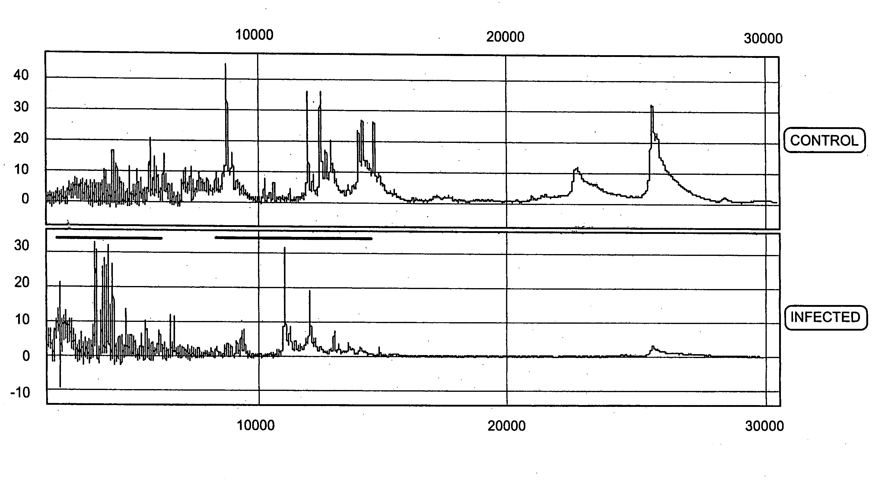
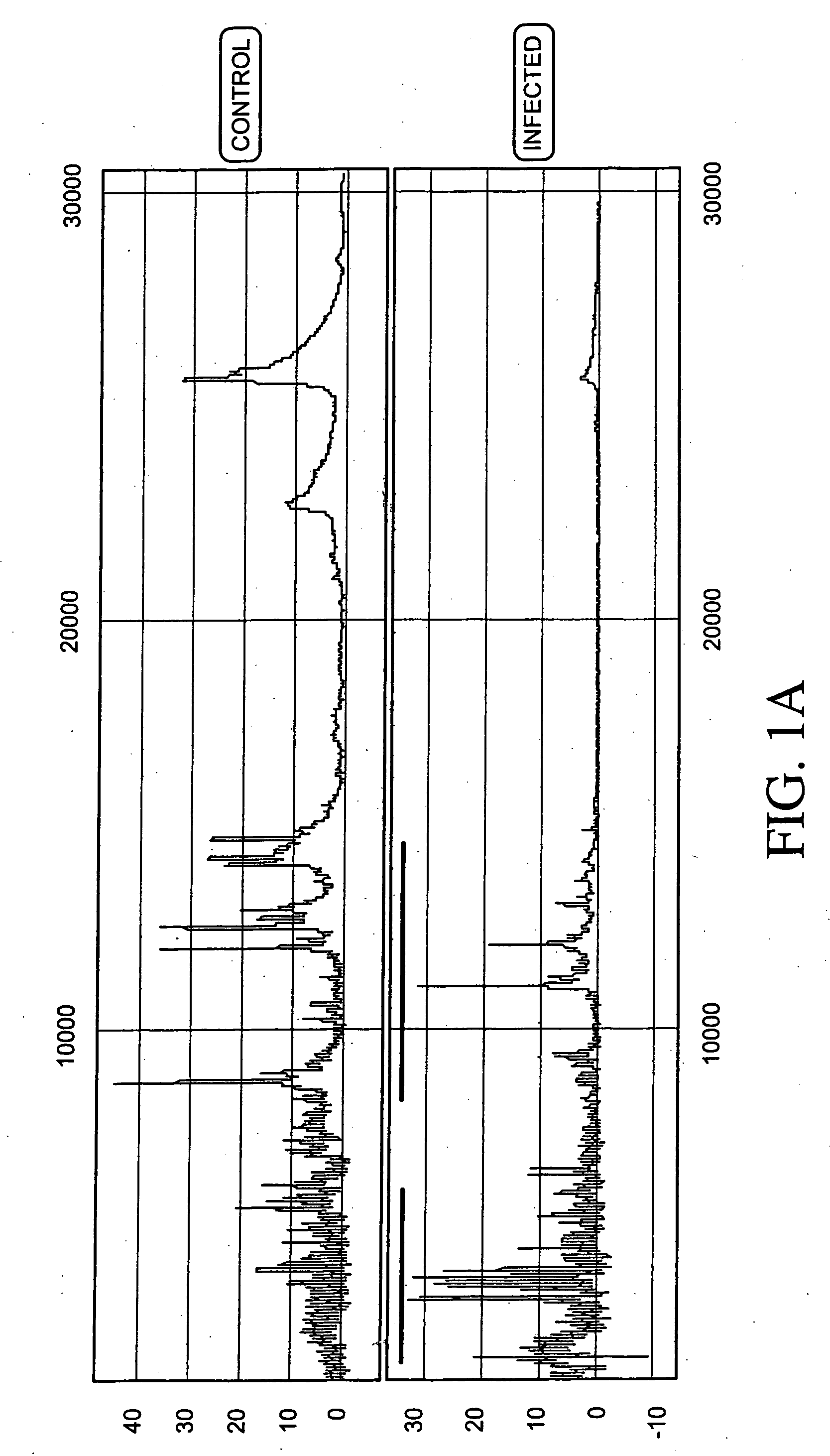
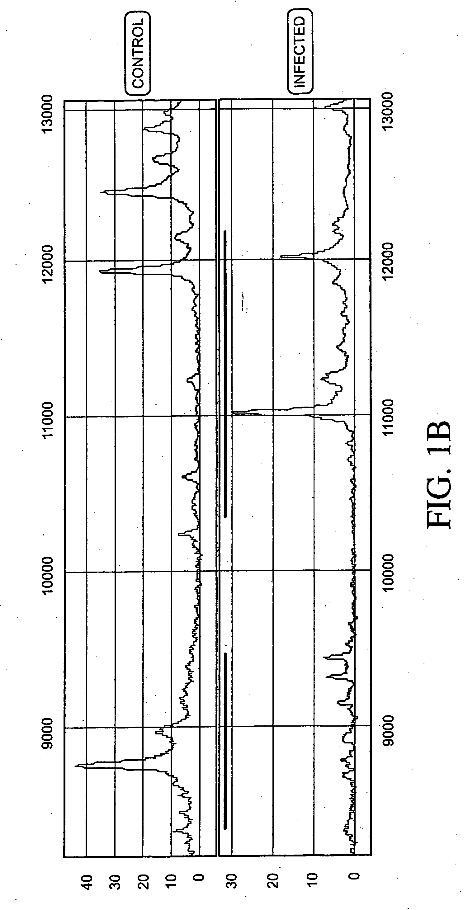
[0187]Protein expression profiles of primate amniotic fluid following intrauterine infection, compared with the corresponding normal expression profiles, are shown in FIGS. 1A-C.

[0188]As illustrated in FIGS. 1A-C, the global protein expression profiles of control and infected amniotic fluid are distinct. A detailed spectra of amniotic fluid profiles in a smaller mass range (FIGS. 1B and 1C), shows distinct and characteristic differences between the protein expression profiles of control and infected samples approximately in the 3-5 KDa and 10-12 KDa range. This illustrates global regulation of protein expression in response to intrauterine infection and the ability to detect a unique expression signature diagnostic of intrauterine infection.


PUM


Abstract

The invention concerns the identification of proteomes of biological fluids and their use in determining the state of maternal / fetal conditions, including maternal conditions of fetal origin, chromosomal aneuploidies, and fetal diseases associated with fetal growth and maturation. In particular, the invention concerns the identification of the proteome of amniotic fluid (multiple proteins representing the composition of amniotic fluid) and the correlation of characteristic changes in the normal proteome with various pathologic maternal / fetal conditions, such as intra-amniotic infection, or chromosomal defects.

Description

[0001]This application is a continuation of U.S. application Ser. No. 10 / 400,005, filed Mar. 25, 2003.BACKGROUND OF THE INVENTION[0002]1. Field of the Invention[0003]The invention concerns the identification of proteomes of biological fluids and their use in determining the state of maternal / fetal conditions, including maternal conditions of fetal origin, chromosomal aneuploidies, and fetal diseases associated with fetal growth and maturation. In particular, the invention concerns the identification of the proteome of amniotic fluid (multiple proteins representing the composition of amniotic fluid) and the correlation of characteristic changes in the normal proteome with various pathologic maternal / fetal conditions, such as intra-amniotic infection, or chromosomal defects.[0004]2. Description of the Related Art[0005]Proteomics[0006]The large-scale analysis of protein expression patterns is emerging as an important and necessary complement to current DNA cloning and gene profiling ap...

Claims


Application Information

Patent Timeline
Patent Type & Authority Applications(United States)
IPC IPC(8): G06F19/00G01N33/68
CPCG01N33/6893G01N33/6803
Inventor ROSENFELD, RONNAGALLA, SRIGRAVETT, MIKE
Owner HOLOGIC INC
Learn More