
Methods for isolating long fragment RNA from fixed samples

Inactive Publication Date: 2009-04-09
RESPONSE GENETICS
View PDF1 Cites 7 Cited by
  • Summary
  • Abstract
  • Description
  • Claims
  • Application Information

AI Technical Summary

Benefits of technology

[0042]One aspect of the present invention is to provide a method for the extraction of RNA from fixed tissue specimens. The in

Problems solved by technology

Problems arise, especially in the application of RNA quantification to clinical studies, in obtaining RNA of sufficient quality for optimal results in most quantification methods.
However, except for small studies in which special efforts are made to collect fresh-frozen tissue samples, biopsy tissue samples taken from patients are typically subjected to formalin fixation and embedding in paraffin.
Morphological examination of fresh-frozen tissue that has been cryostat-sectioned is suboptimal as it makes both molecular histopathological correlations difficult and purification of tumor or other tissues by micro-dissection more difficult.
The secondary reason FFPE is commonly used is the difficulty and expense of storing fresh-frozen tissue samples.
The logistical problems involved in the acquisition and securing of sufficient tissue samples for diagnostic analysis and molecular assays seem insurmountable.
The result is that few, if any, tissue banks worldwide contain enough frozen tissue samples suitable for a wide range of genetic analyses, or which have sufficiently long-term patient follow-up and outcome data.
Unfortunately, the formalin fixation process, while better preserving tissue morphology, has adverse consequences for the RNA in the tissues.
Both of these processes greatly increase the difficulty of using RNA from FFPE specimens in RNA quantitation procedures such as the generation of gene expression profiles.
Short length RNA makes it more difficult to obtain optimal primer-probe sets for quantitative real-time RT-PCR, whereas cross-linking prevents the advancement of the RNA or DNA polymerase enzymes that synthesize the new strands of RNA or DNA that are necessary to carry out successful amplification of the isolated RNA material.
Using randomly-fragmented RNA from FFPE tissue also drastically reduces the yield of amplified RNA compared to that of fresh-frozen tissues, while short fragment length specifically decreases the efficiency and specificity of subsequent hybridization steps.
Extraction of intact high molecular weight (long fragment) RNA from FFPE tissue has been a difficult and inconsistent process.
The studies to date have shown that while it is possible to extract RNA from FFPE that can be successfully subjected to PCR, there are still problems with consistency of isolation yield and quality (length) of the extracted RNA.
These two goals are not always compatible: obtaining the maximal yield of RNA may require conditions that further degrade the length of the RNA, resulting in more RNA but of shorter lengths.
Another factor that is less often recognized and not generally addressed in most previous studies is DNA contamination of the RNA preparations.
Thus, in RNA analysis entailing hybridization technologies, a large amount of DNA contamination can lead to spurious results because DNA may compete with the RNA molecules for binding to the hybridization sites.
Currently accepted models of excision repair suggest that the damage recognition / excision step is rate-limiting to the excision repair process.
However, these studies did not measure quantitative gene expression, but used a semi-quantitative immunohistochemical staining method to measure protein levels.
Studies using binding assays correlated increased EGFR expression with advanced stage NSCLC and shortened overall survival, whereas studies using semi-quantitative techniques for measuring EGFR mRNA or protein expression failed to show a consistent correlation with clinical outcome.
However, these studies reported no consistent correlation of EGFR over expression with lung cancer patient survival.
However, contradictory studies reported no correlation of HER2-neu protein over expression with inferior overall survival in pulmonary adenocarcinomas (AC).
Inconsistent methodologies for the determination of EGFR and HER2-neu expression levels has been at the root of the problem in determining to what extent expression of these genes may be used to prognosticate cancer patient survivability.
In addition, single agent 5-FU therapy continues to be used for patients in whom combination therapy with CPT-11 or oxaliplatin is likely to be excessively toxic.
Nevertheless, the majority of treated patients derive no tangible benefit from having received 5-FU based chemotherapy, and are subjected to significant risk, discomfort, and expense.
Unfortunately, most pre-treatment tumor biopsies are available only as fixed paraffin embedded (FPE) tissues, particularly formalin-fixed paraffin embedded tissues which do not contain active enzyme.
Moreover, biopsies generally contain only a very small amount of heterogeneous tissue.
However, those primers are unsuitable for the quantification of DPD mRNA from fixed tissue by RT-PCR.
As a result, other investigators have made a concerted, yet unsuccessful efforts to obtain oligonucleotide primer sets allowing for such a quantification of DPD expression in paraffinized tissue.
As the most effective single agent for the treatment of colon, head and neck and breast cancers, the primary action of 5-FU is to inhibit TS activity, resulting in depletion of intracellular thymine levels and subsequently leading to cell death.

Method used


Image

  • Methods for isolating long fragment RNA from fixed samples
  • Methods for isolating long fragment RNA from fixed samples
  • Methods for isolating long fragment RNA from fixed samples

Examples


example 1

Long-Fragment RNA Extraction Procedure

[0199]I. Tissue Preparation Standard laboratory procedures are used to mount a 10 micron section of a paraffin block containing a FFPE tissue on a glass slide without a cover slip. For deparaffinization and nuclear fast red (NFR) staining the slides are treated as follows:

[0200]The slides are washed twice in Xylene for 5 minutes, followed by ethanol (“EtoH”) washes. The slide is stained with NFR using standard laboratory procedures.

[0201]Areas of interest (e.g., tumor tissue or stromal tissue) are excised either manually or with a laser capture microdissector (depending on the size of the area to be excised).

[0202]II. RNA Extraction

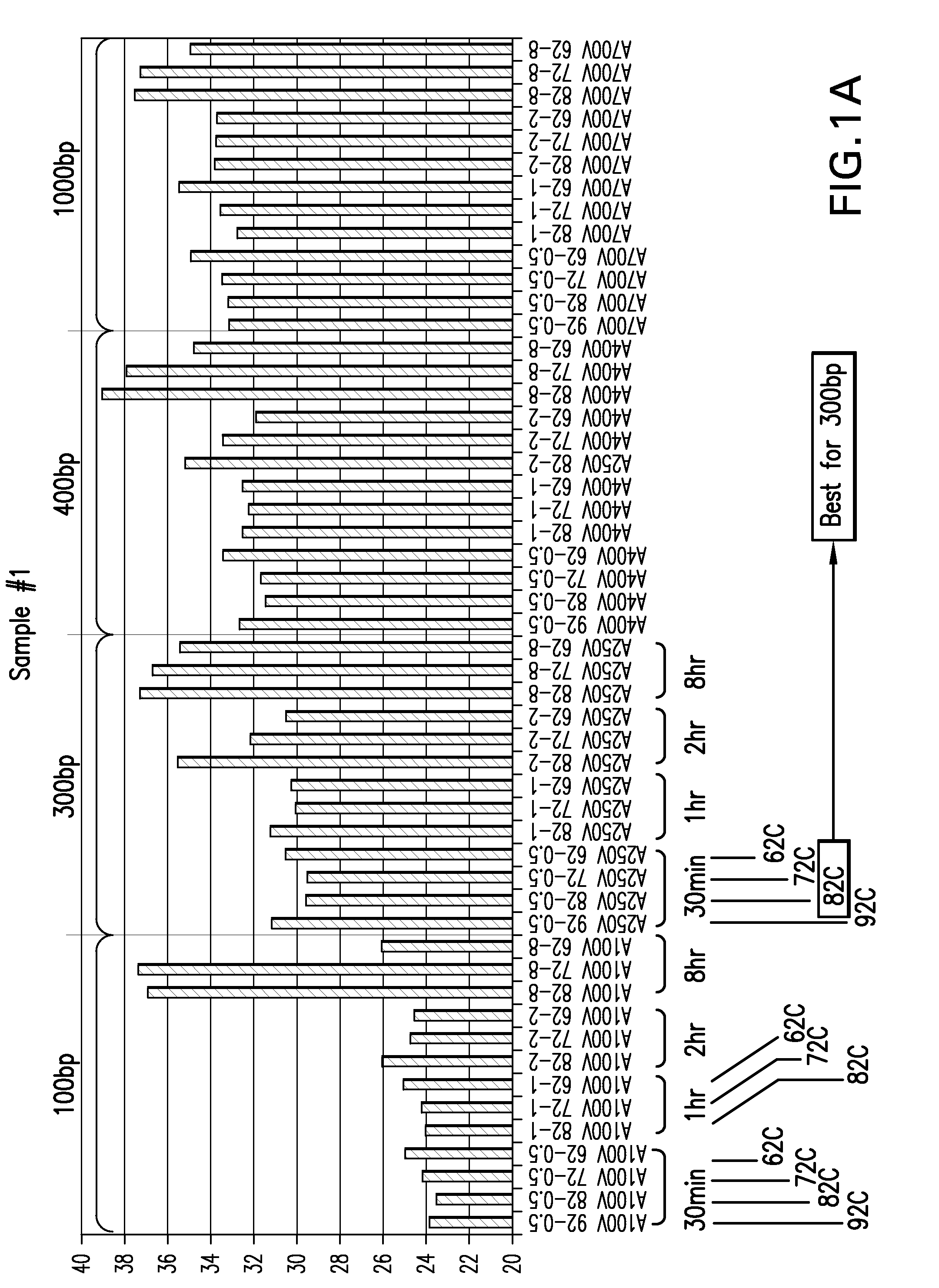
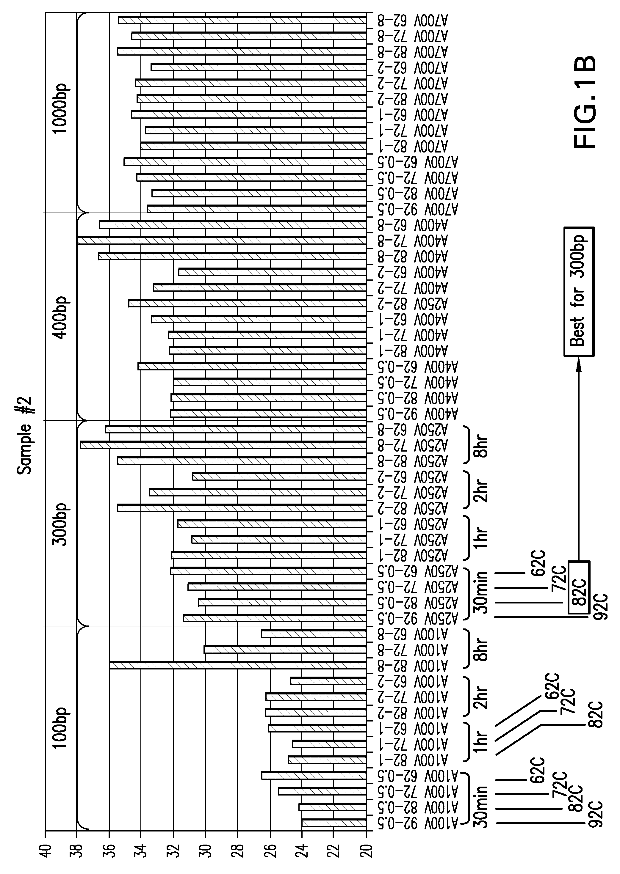
[0203]An extraction solution is prepared containing Tris / HCL, EDTA, SDS and water. A tumor tissue is added to the extraction solution in a centrifuge tube and proteinase K. The sample is then heated at the appropriate temperature and time for the maximal yield of long fragment RNA. For example, the sample is heated at...

example 2

Method of Determining Length Distribution of Isolated RNA

[0206]To determine the relative amounts of various RNA fragment lengths isolated from FFPE tissues, the following strategy was used. RNA isolated from the FFPE specimens using the present invention and other known extraction methods was converted to cDNA using oligo dT primers. This means that only mRNA fragments containing a 3′-oligo A tail would be extended and converted to cDNA, thus providing a starting point from which to measure fragment length. PCR amplification of β-actin mRNA was used to represent the total population of mRNA. Primers were chosen to amplify approximately 100-120 bp segments of the β-actin gene representing locations 100, 300, 400 and 1000 bp from the 3′-end of the mRNA (FIG. 2). With this strategy, any difference in length-dependent efficiency of amplification would be minimized, as opposed to actually trying to amplify 100, 300, 400 and 1000 bp fragments. Thus the Ct of the PCR products of each of th...

example 3

Effects of Proteinase K

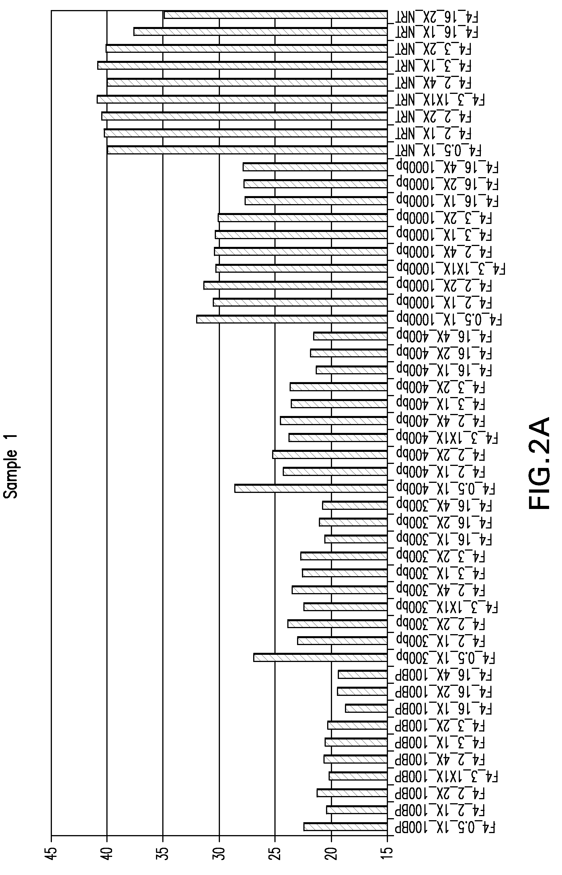
[0207]This example illustrates the effect of proteinase K concentration on RNA yield and DNA contamination. The proteinase K concentration was varied over a 4-fold range (5-20 μg, designated as 1×-4× in the figure) at incubation times of 0.5, 2, 3 and 16 hr. at 50° C. As seen in FIG. 4, 1× (5 μg) of proteinase K gives about a 2-fold (1 Ct) better RNA yield than higher amounts but more importantly, amounts of proteinase K greater than 1× give appreciably higher DNA contamination (2-3 Ct cycles). This experiment also illustrates the influence of incubation time on the amount of DNA extracted, which is from 3 to 7 Ct cycles greater at the 16 hr incubation time than at the shorter incubation times. DNA is detected in the extracts by performing the PCR without first doing a reverse transcription reaction to convert RNA to cDNA (the “no reverse transcription or NRT control”). This way, the only PCR amplification that occurs is of the co-extracted DNA, which if too h...


PUM

PropertyMeasurementUnit
Temperatureaaaaaaaaaa

Abstract

The present invention relates to methods for the extraction of long fragment RNA from fixed tissue specimens. In particular, the present invention relates to methods for the extraction of RNA from formalin-fixed paraffin-embedded tissue specimens for use in biologic applications, including assays based on oligonucleotide hybridization.

Description

[0001]This application claims priority to provisional application 60 / 945,785 filed on Jun. 22, 2007, which is herein incorporated by reference in its entirety.FIELD OF THE INVENTION[0002]The present invention relates to the field of extraction and isolation of high yield and high quality (long fragment) RNA from fixed tissue samples. The present invention relates to the use of these novel extraction methods to provide a method for assessing gene expression levels for genes such as cancer biomarkers in fixed or fixed and paraffin embedded tissues. The present invention also provides a method for determining a chemotherapy based regimen by measuring mRNA levels of a certain biomaker in a patient's tumor cells and comparing it to a predetermined threshold expression levels.BACKGROUND[0003]The quantitative measurement of RNA species is central to the pursuit of modern research in molecular biology. RNA species also has very important clinical significance, for example, in the preparatio...

Claims


Application Information

Patent Timeline
IPC IPC(8): C12Q1/68C07H21/02
CPCC12N15/1003C12Q1/6806C12Q1/6886
Inventor DANENBERG, KATHLEEN
Owner RESPONSE GENETICS
Learn More