Device for the regeneration and prevention of degeneration of the cartilaginous tissue and subchrondral bone, the proliferation of chondrocytes and for blocking or reducing the fibroblastic evolution of chondrocytes and mesenchymal cells by means of a pulsed electromagnetic field

a pulsed electromagnetic field and chondrocyte technology, applied in magnetotherapy, medical science, surgery, etc., can solve the problems of limited cartilagenous damage repair capacity, inability to achieve therapeutic effects, and inability to produce devices, so as to prevent or reduce the differentiation into the fibroblastic phenotype, block the inflammatory phenomenon, and promote the recovery of cartilagenous lesion

Inactive Publication Date: 2009-06-25
IGEA
View PDF3 Cites 12 Cited by
  • Summary
  • Abstract
  • Description
  • Claims
  • Application Information

AI Technical Summary

Benefits of technology

[0028]A further object of the present invention is to provide a method that allows the control of the differentiation of the chondrocytes and of the mesenchymal cells avoiding their fibroblastic differentiation and evolution.
[0029]The preceding object is achieved by the present invention since it relates to a use of a device comprising means for generating a periodic signal / periodic signals u(t) and power amplifying means adapted to apply said signal u(t) to at least one solenoid for the generation of a pulsed electromagnetic field M(t) addressed towards a portion of human / animal tissue containing cartilage, to block or reduce the fibroblastic evolution of chondrocytes and mesenchymal cells.
[0030]According to a second aspect of the present invention, it is provided a method for blocking or reducing the fibroblastic evolution of chondrocytes and mesenchymal cells by means of electromagnetic waves comprising the steps of generating a periodic signal / periodic signals u(t) and applying said signal u(t) to at least one solenoid for the generation of a pulsed electromagnetic field M(t) addressed towards a portion of human / animal tissue containing cartilage.
[0032]Advantageously the use according to the present invention allows to prevent or reduce the differentiation into the fibroblastic phenotype, blocking inflammatory phenomena and thus promoting recovery from a cartilagenous lesion that produces a functional reparative tissue consisting in hyaline cartilage following the activity of the cells that contain a chondrocytal phenotype. The present invention allows moreover to preserve the cartilagenous tissue which is not directly compromised by the lesion, which being exposed to the negative effects of the inflamatory environment could undergo degeneration. Moreover the inhibition of the prostaglandin synthesis according to the present invention protects against and prevents chondrocytes apoptosis. The device moreover allows to increase the proliferation and then the expansion of the number of transplanted cells, chondrocytes and mesenchymal staminal cells, making them more sensible to the activity of growth factors.

Problems solved by technology

Said techniques have not succeeded, however, in producing devices that can be used for a real therapeutic cycle of the cartilaginous tissues / subchondral bone for application in humans, i.e. able to act and modify in a sensitive, specific and significant way the structure of the damaged or inflamed cartilage and subchondral bone.
The lack of adequate preclinical experimentation permitting identification and combination of the optimal, i.e. effective, electromagnetic field parameters (amplitude, waveform frequency and duration of exposure) can result in incorrect choice of the parameters that characterise the electromagnetic field, therefore not obtaining any therapeutic effect, as also highlighted in literature (Fini M et al, Effects of pulsed electromagnetic fields on articular hyaline cartilage: review of experimental and clinical studies.
As compared with other connective tissues, the cartilage is a non vascularized tissue; chondrocytes are fed by diffusion through the extracellular matrix and therefore the cartilage is a tissue that grows and regenerates itself much more slowly than other tissues, in particular it shows limited capacity to repair a cartilagenous damage.
As is known, the degeneration of articular cartilage manifests itself very frequently and it progressively worsens with age.
However, cartilage degeneration is not just a consequence of ageing, but also the end result of a complex series factors related to problems of a biological nature and / or mechanical problems.
These techniques, although they give good results, only allow to achieve the formation of a new fibro-cartilagenous tissue with morphological and structural properties which are quite different, worse than those that identify the cartilage of the articular surface achieving only a slowing down of the arthrosic process.

Method used


Image

  • Device for the regeneration and prevention of degeneration of the cartilaginous tissue and subchrondral bone, the proliferation of chondrocytes and for blocking or reducing the fibroblastic evolution of chondrocytes and mesenchymal cells by means of a pulsed electromagnetic field
  • Device for the regeneration and prevention of degeneration of the cartilaginous tissue and subchrondral bone, the proliferation of chondrocytes and for blocking or reducing the fibroblastic evolution of chondrocytes and mesenchymal cells by means of a pulsed electromagnetic field
  • Device for the regeneration and prevention of degeneration of the cartilaginous tissue and subchrondral bone, the proliferation of chondrocytes and for blocking or reducing the fibroblastic evolution of chondrocytes and mesenchymal cells by means of a pulsed electromagnetic field

Examples


example 1

Method for the Treatment and / or Prevention of Pathologies Affecting the Cartilage and / or Subchondral Bone and for the Proliferation of Chondrocytes

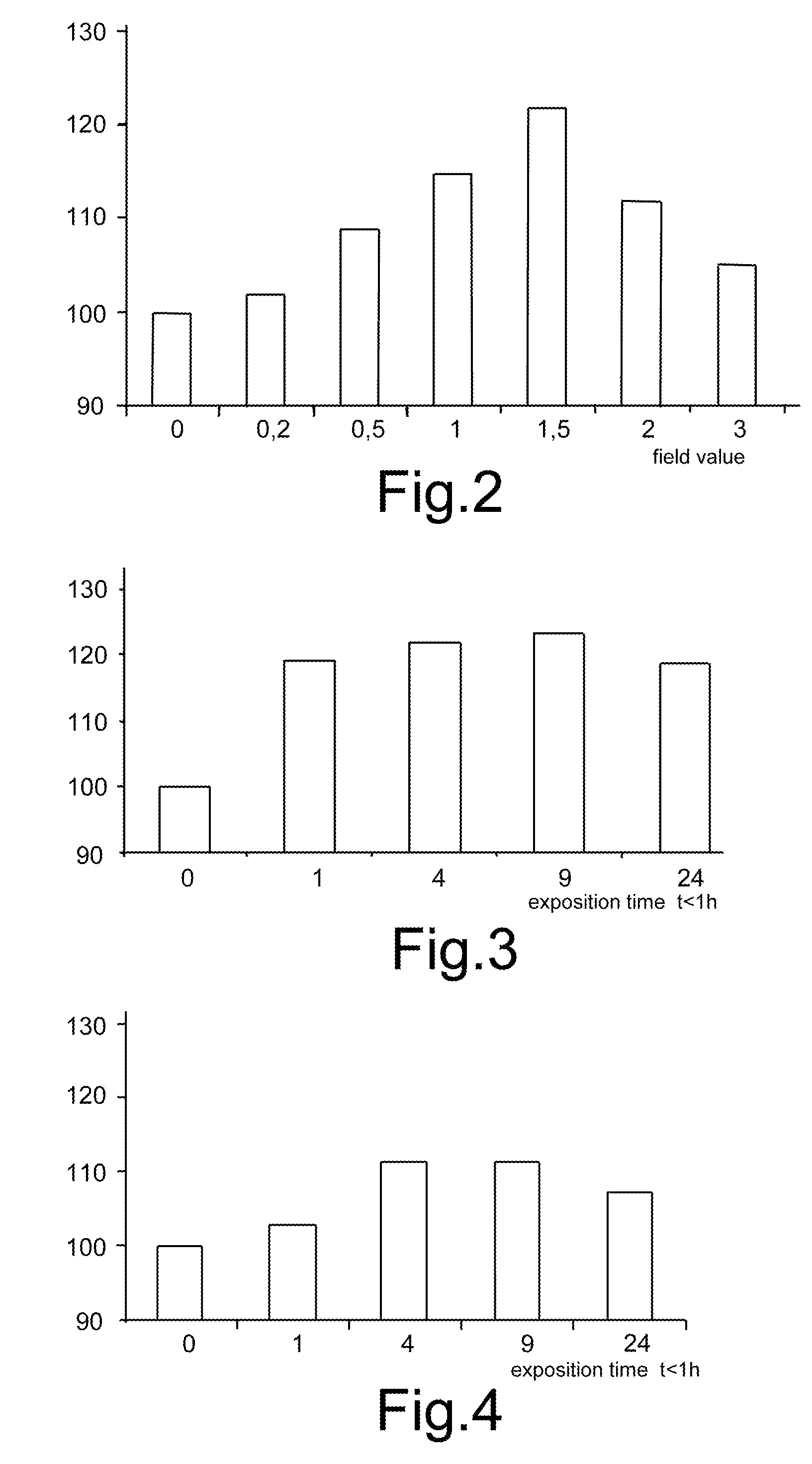
[0094]The method was tested in vitro on bovine cartilage which has a high affinity with human cartilage.

[0095]Explants of articular cartilage in the form of discs were performed from five different animals aged between 14 and 18 months.

[0096]In particular, four explants were performed on each donor animal taken from areas near the same joint thus obtaining twenty discs.

[0097]Each group of explants was divided at random into a first subgroup of explants with test function (therefore subject to the electromagnetic field) and a second group of explants with control function (therefore not subject to the electromagnetic field).

[0098]The explants underwent pre-treatment by placing them for 48 hours in a culture of DMEM / F12 to which 10% of FBS (Fetal Bovine Serum) and antibiotics (penicillin 100 units / ml, streptomycin 0.1 mg / ml) (Life Technolog...

example 2

Method for Blocking or Reducing the Fibroblastic Evolution of Chondrocytes and Mesenchymal Cells

[0148]A sample of bone marrow was collected from the acetabulum and femoral head of patients undergoing total hip arthroplasty. Bone marrow mononuclear cells were obtained by ficoll (Sigma, Italy) density gradient centrifugation and depleted of CD45+ and glycophorin-A (GlyA)+ cells using micromagnetic beads (Miltenyi Biotec, Italy). CD45− / GlyA− cells were plated in 75 cm2 culture flasks (Corning Inc, NY, USA) in mesencult+stimulatory supplement (both from StemCell, Technologies Inc, BC, Canada) and 1% penicillin-streptomycin (Gibco, Italy) for 14 days. Near-confluence cultures were then trypsinized and expanded through six sequential passages to confluence. At each passage, cells were characterised with the FACSCalibur flow cytometry system (Becton Dickinson, CA, USA) using antibodies against the following surface antigens: CD3, CD34, CD14, CD45, CD90 and CD105 (Becton Dickinson).

[0149]Ce...

example 3

In Vitro Effects

Inflammation Control

[0153]Anatomic Biophysical Chondroprotection acts in a specific manner on the adenosinic receptors A2A of the cellular membrane of pro-inflammatory cells, neutrophils, rendering then available to binding with adenosine. Within the sphere of adenosinic receptors, the receptors A2A are those of greater anti-inflammatory effect.

[0154]The bonding with adenosine causes: inhibition of the production of pro-inflammatory cytokines, reduction in the synthesis of free radicals, increase in the production of ATP and cytokines with anti-inflammatory action, TGF□, and the inhibition of cycloxygenase 2 activity.

[0155]The kinetic studies carried out by the applicant have shown how the stimulator device implemented in accordance with this invention permits an anti-inflammatory effect to be achieved.

[0156]In cases of inflammation, by using the device 201 it is in fact possible to activate the adenosinic receptors on the cell membrane via the generated biophysical ...


PUM


Abstract

Device for the regeneration and prevention of degeneration of the cartilage and subchondral bone, the proliferation of chondrocytes and for blocking or reducing the fibroblastic evolution of chondrocytes and mesenchymal cells by means of electromagnetic waves comprising a device for generating a periodic signal u(t) and a power amplifier suitable for applying the signal u(t) to a pair of solenoids for the generation of an electromagnetic field M(t) addressed towards a portion of human / animal body containing cartilage. Setting means are provided for the generation of an electromagnetic field having intensity between 0.5 and 2 milliTesla, frequency between 37 and 75 Hz and period of application between 1 hour and 9 hours.

Description

[0001]This is a continuation-in-part application of U.S. patent application Ser. No. 12 / 300,233, filed Nov. 10, 2008, by Stefania Setti and Ruggero Cadossi, entitled DEVICE FOR THE REGENERATION AND PREVENTION OF DEGENERATION OF THE CARTILAGINOUS TISSUE AND SUBCHRONDRAL BONE AND THE PROLIFERATION OF CHONDROCYTES BY MEANS OF A PULSED ELECTROMAGNETIC FIELD, the content of which is incorporated herein by reference.TECHNICAL FIELD[0002]The present invention relates to a device for the regeneration and prevention of degeneration of the cartilaginous tissue and subchrondral bone and the proliferation of chondrocytes by means of a pulsed electromagnetic field. Further the present invention refers to a method for blocking, preventing or reducing the fibroblastic evolution and the expression of its chondrocyte phenotype and its mesenchymal cells used to repair the cartilaginous tissue by means of the application of a pulsed electromagnetic field. Lastly this invention concerns an electromagne...

Claims


Application Information

Patent Timeline
Patent Type & Authority Applications(United States)
IPC IPC(8): A61N2/04
CPCA61N1/326A61N2/02A61N2/004
Inventor SETTI, STEFANIACADOSSI, RUGGEROMARAZZI, DONATA
Owner IGEA
Why Eureka
Try Eureka