

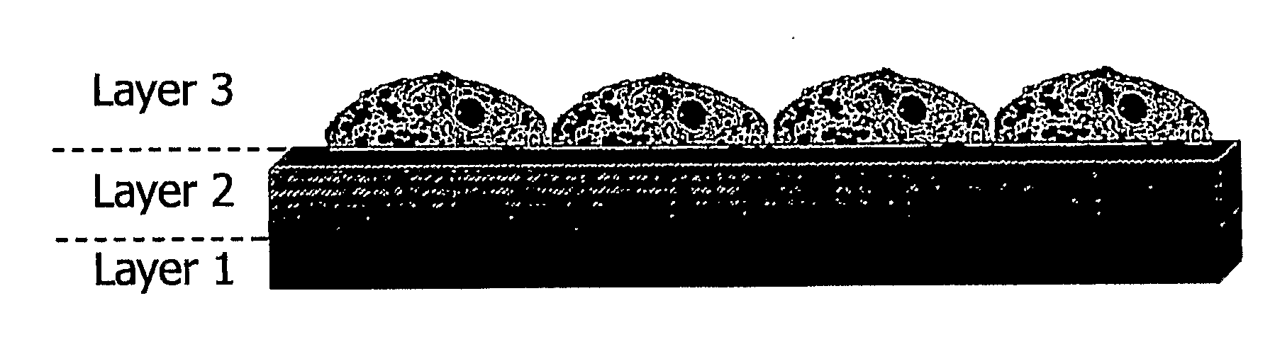
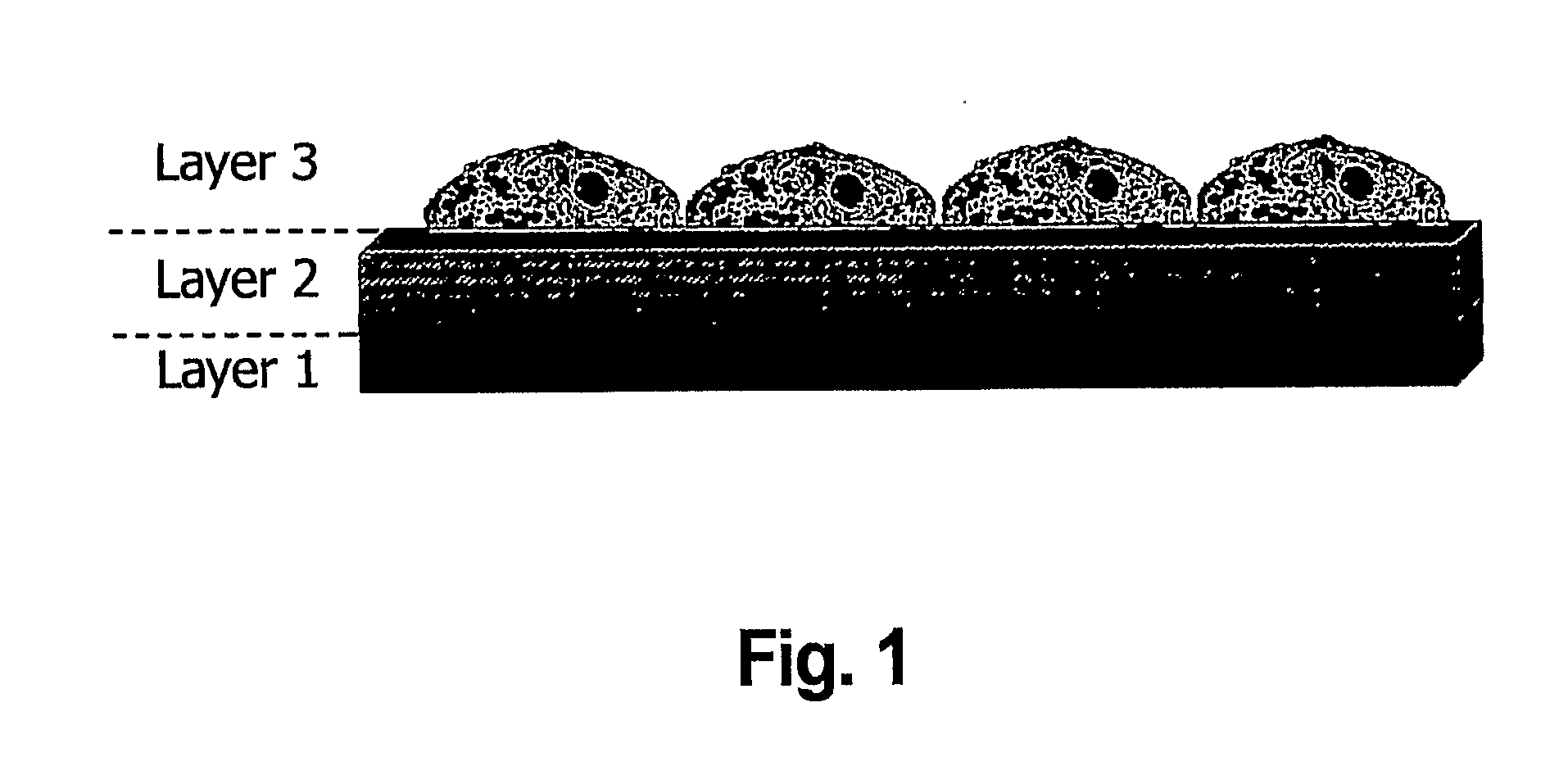
Layered bio-adhesive compositions and uses thereof
a bio-adhesive composition and composition technology, applied in the direction of drug compositions, film/foil adhesives, impression caps, etc., to achieve the effect of facilitating wound healing and facilitating wound healing
- Summary
- Abstract
- Description
- Claims
- Application Information
AI Technical Summary
Benefits of technology
Problems solved by technology
Method used
Image

Examples
example 1
Preparation of Bruch's Membrane
[0098]Bruch's membrane explants were harvested from cadaver eyes. RPE-Bruch's membrane-Choroid complexes were dissected out under a microscope; washed with PBS three times and kept in 0.02N ammonium hydroxide for 30 minutes to lyse all cellular components. Three cycles of PBS washing followed by removal of the basal lamina mechanically as described before. Briefly, a full thickness circumferential incision was made posterior to the ora serrata and the anterior segment and vitreous were removed carefully. The posterior pole of each eye cup was inspected visually with direct and retroillumination under a dissecting microscope and globes were discarded if there was any evidence of subretinal blood, previous surgery or any extensive structural or vascular alteration of the posterior segment due to a disease process, such as proliferative diabetic retinopathy or proliferative vitreoretinopathy. The eyecups were put in carbon dioxide-free Media (Gibco) and a...
example 2
Retinal Pigment Epithelial (RPE) Cells
[0101]Primary RPE cultures were prepared from the posterior poles of human cadaver eyes. The eyes were cleaned of extraocular tissue. The space between the sclera and choroid (i.e. the suprachroidal space) of the posterior pole was sealed by sticking the choroid and sclera together with cyanoacrylate glue and a small scleral incision was made 3 mm posterior to the limbus until the choroidal vessels are exposed. Tenotomy scissors were introduced through the incision into the suprachoroidal space and the incision was extended circumferentially. Four radial relaxing incisions were made in the sclera and the sclera is peeled away from the periphery to the optic nerve with care to avoid tearing the choroid. The eye cup is then incubated with 25 u / ml of Dispase (Gibco, Grand Island, N.Y.) for 30 minutes, rinsed with carbon dioxide-free medium (CFM, Gibco) and a circumferential incision was made into the subretinal space along the ora serrata. The loos...
example 3
Tissue Bonding of Explants from Inner Collagenous Layer
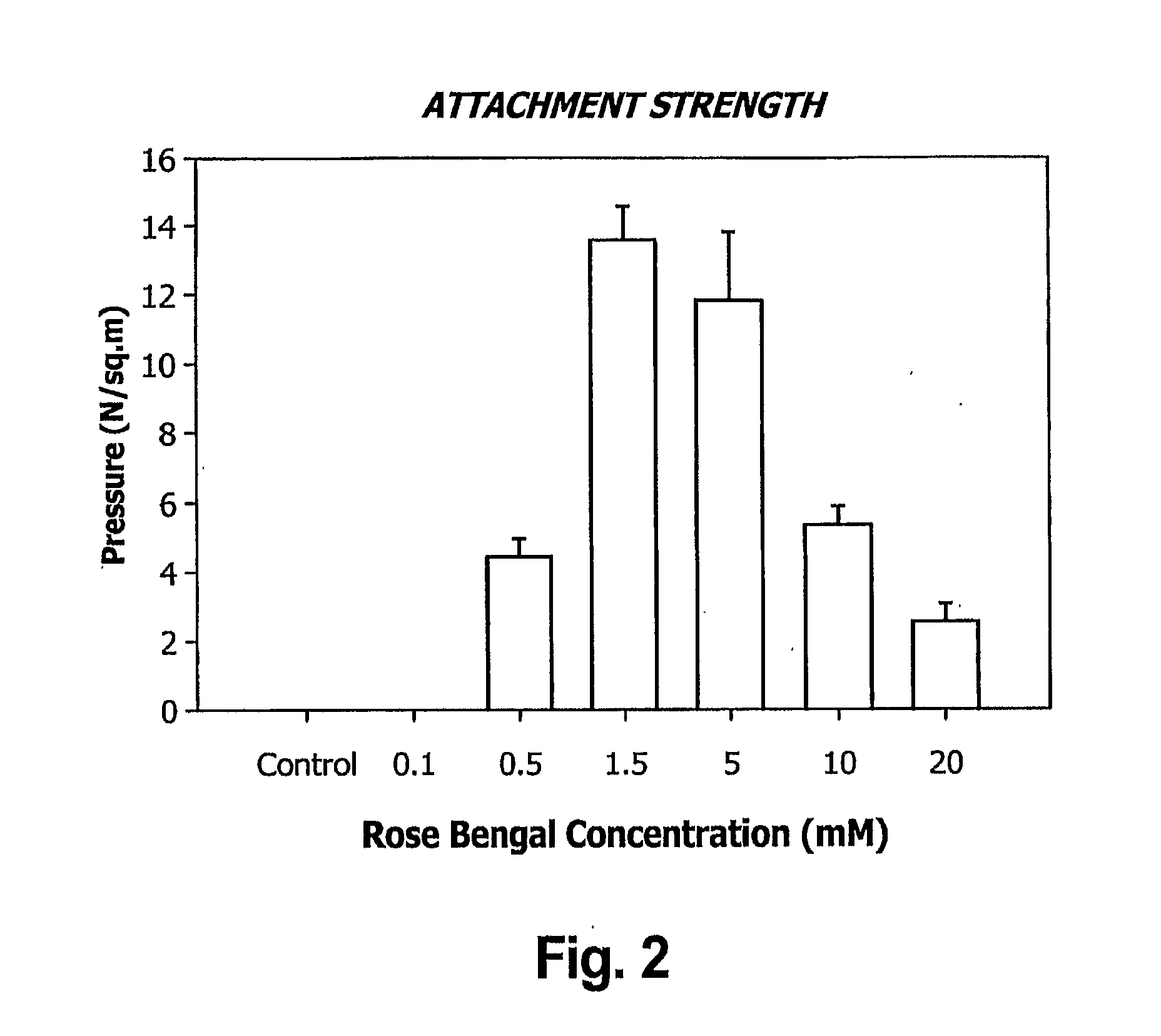
[0102]Six-millimeter explants of human peripheral inner collagenous layer were prepared from 10 aged (>60 years-old) cadaver eyes. Exposed inner collagenous layers were painted with rose bengal and photo-melded to each other by photo-exciting rose bengal with a white light source. The light intensity matched the vitrectomy endo illuminator and exposure was limited to 3 minutes to avoid retinal photo-toxicity. Attachment strength was measured in a buoyancy chamber using different concentrations of rose bengal (0.1-20 mM). Attached grafts were kept under 0.21 mN (milliNewtons) of constant traction for 3 days to test the stability of the melding process.
PUM
| Property | Measurement | Unit |
|---|---|---|
| Fraction | aaaaa | aaaaa |
| Thickness | aaaaa | aaaaa |
| Thickness | aaaaa | aaaaa |
Abstract
Description
Claims
Application Information