Navigation system for cardiac therapies using gating

a navigation system and cardiac therapy technology, applied in the field of image guided surgery, can solve the problems of not helping the catheter in actually guiding a medical device to a targeted tissue area for medical treatment, exposing both the patient and the operating room staff to radiation, and reducing the exposure to fluoroscopy. , to achieve the effect of improving outcomes, reducing radiation exposure, and ensuring accuracy

Inactive Publication Date: 2010-02-04
MEDTRONIC INC
View PDF25 Cites 277 Cited by
  • Summary
  • Abstract
  • Description
  • Claims
  • Application Information

AI Technical Summary

Benefits of technology

[0012]A navigation system is provided including a catheter carrying single or multiple localization sensors, a sensor interface, a user interface, a controller, and a visual display. Aspects of the present invention allow for the location of a catheter advanced within an internal space within the human body, for example within the cardiovascular structures, to be identified in two, three or four dimensions in real time. Further aspects of the present invention allow for accurate mapping of a tissue or organ, such as the heart or other soft tissue, and / or precise identification of a desired location for delivering a medical lead, or other medical device or therapy, while reducing the exposure to fluoroscopy normally required during conventional catheterization procedures. These types of therapies include, but are not limited to, drug delivery therapy, cell delivery therapy, ablation, stenting, or sensing of various physiological parameters with the appropriate type of sensor. In cardiac applications, methods included in the present invention compensate for the effects of respiration and the beating heart that can normally complicate mapping or diagnostic data. Aspects of the present invention may be tailored to improve the outcomes of numerous cardiac therapies as well as non-cardiac therapies, such as neurological, oncological, or other medical therapies, including lung, liver, prostate and other soft tissue therapies, requiring the use of a catheter or other instrument at a precise location.

Problems solved by technology

However, use of such fluoroscopic imaging throughout a procedure exposes both the patient and the operating room staff to radiation, and exposes the patient to contrast agents.
Such mapping catheters are useful in identifying an area of tissue that is either conducting normally or abnormally, however, some mapping catheters may not aid in actually guiding a medical device to a targeted tissue area for medical treatment.
Also, pacing lead procedures currently performed today for use in heart failure treatment are not optimized.
In this regard, the lead placement is not optimized due to the lack of having real-time anatomic information, navigation and localization information, hemo-dynamic data, and electro-physiological data.
Currently, pacing leads are simply “stuffed” into the heart without any optimization being performed due to lack of information that can be used for this optimization.
Advancement of a guide catheter or an over-the-wire lead through a vessel pathway and through cardiac structures requires considerable skill and can be a time-consuming task.
This type of procedure also exposes the patient to an undesirable amount of radiation exposure and contrast agent.
With regard to navigating an instrument through a moving body structure, difficulties arise in attempting to track such an instrument using known tracking technology as the instrument passes adjacent or through a moving body structure, since the virtual representation of the instrument may be offset from the corresponding anatomy when superimposed onto image data.
Other difficulties with cardiac procedures include annual check-ups to measure early indications for organ rejection in heart transplant patients.

Method used


Image

  • Navigation system for cardiac therapies using gating
  • Navigation system for cardiac therapies using gating
  • Navigation system for cardiac therapies using gating

Examples


Embodiment Construction

[0025]The following description of the preferred embodiment(s) is merely exemplary in nature and is in no way intended to limit the invention, its application, or uses. As indicated above, the present invention is directed at providing improved, non-line-of-site, image-guided navigation of an instrument, such as a catheter, balloon catheter, implant, lead, stent, needle, guide wire, insert, and / or capsule, that may be used for physiological monitoring, delivering a medical therapy, or guiding the delivery of a medical device in an internal body space, such as the heart or any other region of the body.

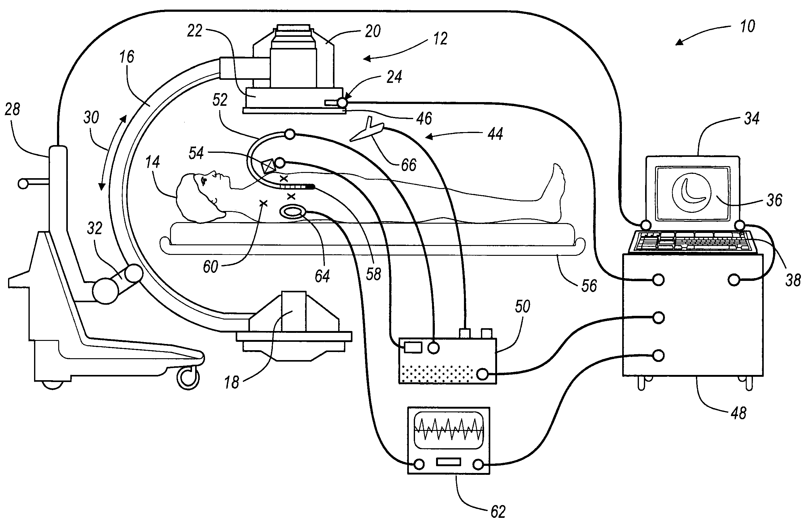
[0026]FIG. 1 is a diagram illustrating an overview of an image-guided catheter navigation system 10 for use in non-line-of-site navigating of a catheter during cardiac therapy or any other soft tissue therapy. It should further be noted that the navigation system 10 may be used to navigate any other type of instrument or delivery system, including guide wires, needles, drug delivery sys...


PUM


Abstract

An image guided navigation system for navigating a region of a patient which is gated using ECG signals to confirm diastole. The navigation system includes an imaging device, a tracking device, a controller, and a display. The imaging device generates images of the region of a patient. The tracking device tracks the location of the instrument in a region of the patient. The controller superimposes an icon representative of the instrument onto the images generated from the imaging device based upon the location of the instrument. The display displays the image with the superimposed instrument. The images and a registration process may be synchronized to a physiological event.

Description

FIELD OF THE INVENTION[0001]The present invention relates generally to image guided surgery, and more specifically, to systems and methods for using one or more medical images to assist in navigating an instrument through internal body structures, in particular for navigating a catheter in a moving body structure, such as the heart, during a surgical procedure.BACKGROUND OF THE INVENTION[0002]Image guided medical and surgical procedures utilize patient images obtained prior to or during a medical procedure to guide a physician performing the procedure. Recent advances in imaging technology, especially in imaging technologies that produce two, three, and four dimensional images, such as computed tomography (CT), magnetic resonance imaging (MRI), isocentric C-arm fluoroscopic imaging, positron emission tomography (PET), and ultrasound imaging (US), has increased the interest in image guided medical procedures.[0003]At present, cardiac catheterization procedures are typically performed...

Claims


Application Information

Patent Timeline
Patent Type & Authority Applications(United States)
IPC IPC(8): A61B5/055A61B5/352
CPCA61B5/0452A61B5/7285A61B6/541A61B6/5247A61B19/5244A61B2017/00703A61B8/543A61B34/20A61B6/5264A61B6/5288A61B5/349A61B5/352A61B2034/2063A61B2034/2065A61B2090/3735A61B2090/374A61B2090/3764A61B2090/3784A61B5/0066A61B5/036A61B5/4839A61B6/032A61B6/037A61B8/0841A61B8/12A61B18/1492A61B2018/00577A61B2018/00839A61M25/0108A61M25/09A61M2230/04A61N1/372
Inventor CANFIELD, MONTE R.NEIDERT, MICHAEL R.GARDESKI, KENNETH
Owner MEDTRONIC INC
Why Eureka
Try Eureka