

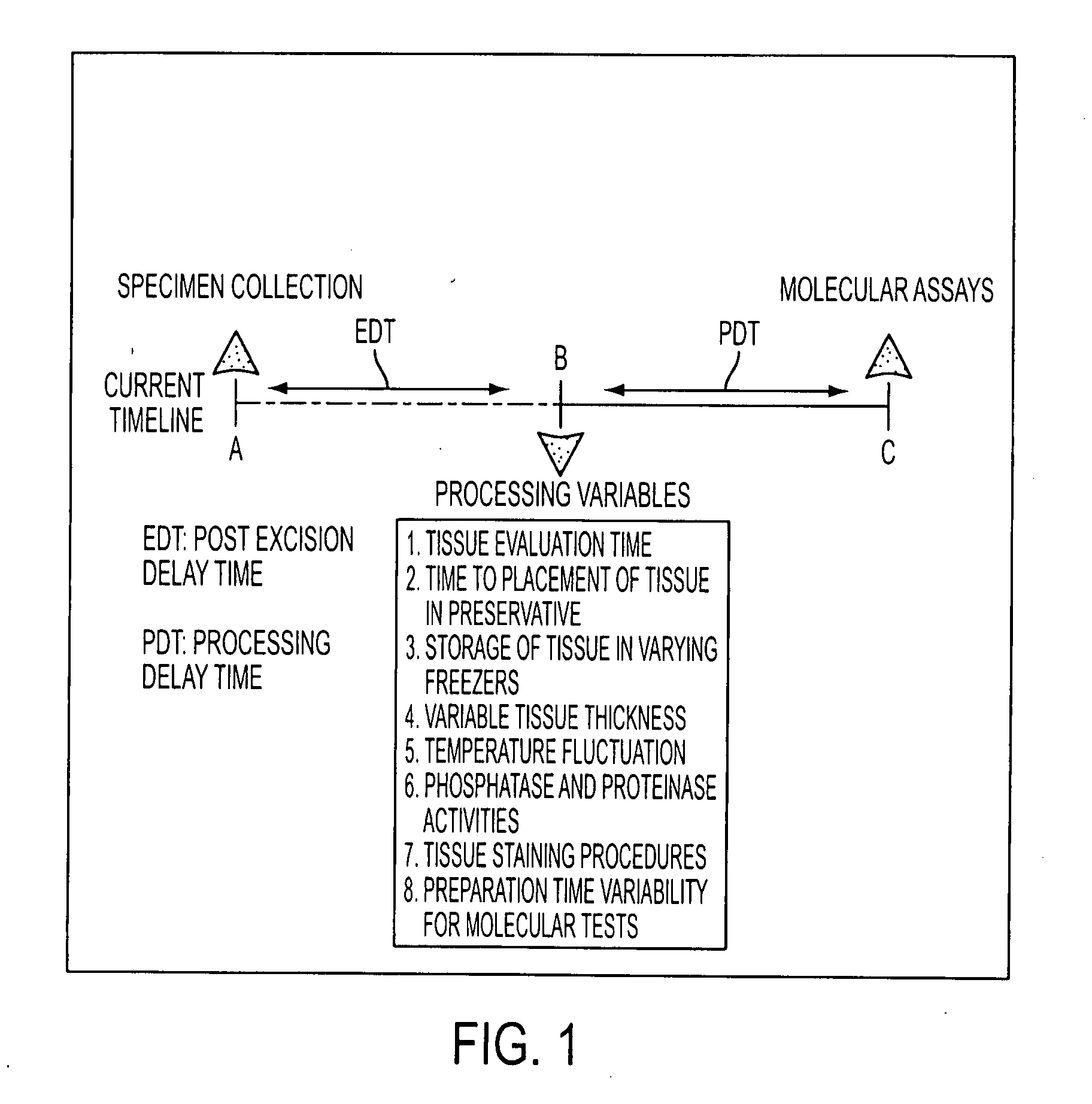
Tissue preservation and fixation method
a tissue and fixation technology, applied in the field of personalized cancer therapy, can solve the problems of time delay, inability to provide effective recapitulation of drug targets, and inability to immediately preserve procured tissue in liquid nitrogen
- Summary
- Abstract
- Description
- Claims
- Application Information
AI Technical Summary
Problems solved by technology
Method used
Image

Examples
example i
Materials and Methods
[0109]A. Reverse phase protein arrays (RPA)—quantitation of phosphoproteins in tissues and cells. The RPA format, originated by the present inventors and collaborators (see, e.g., Paweletz et al. (2001) Oncogene 20, 1981-1989), immobilizes an individual test sample in a miniature dilution curve such that an array contains hundreds of different patient samples, treatments, or time points. In the RPA format, each array is incubated with one detection protein (e.g. anti-peptide antibody), and a single analyte endpoint is measured and directly compared across multiple samples. Briefly, the lysates are printed on glass backed nitrocellulose array slides (FAST Slides Whatman, Florham Park, N.J. or ONCYTE slides Grace BioLabs, Bend, Oreg.) using a robotic array printing device (GMS 417 arrayer (Affymetrix, Santa Clara, Calif. or Aushon 2470, Aushon Biosystems, Burlington, Mass. equipped with 125-500 μm pins). Each lysate is printed in a dilution curve representing neat...
example ii
Human Tissue Phosphoprotein Stability Time Course Analysis (in a “Real-World” Hospital Setting)
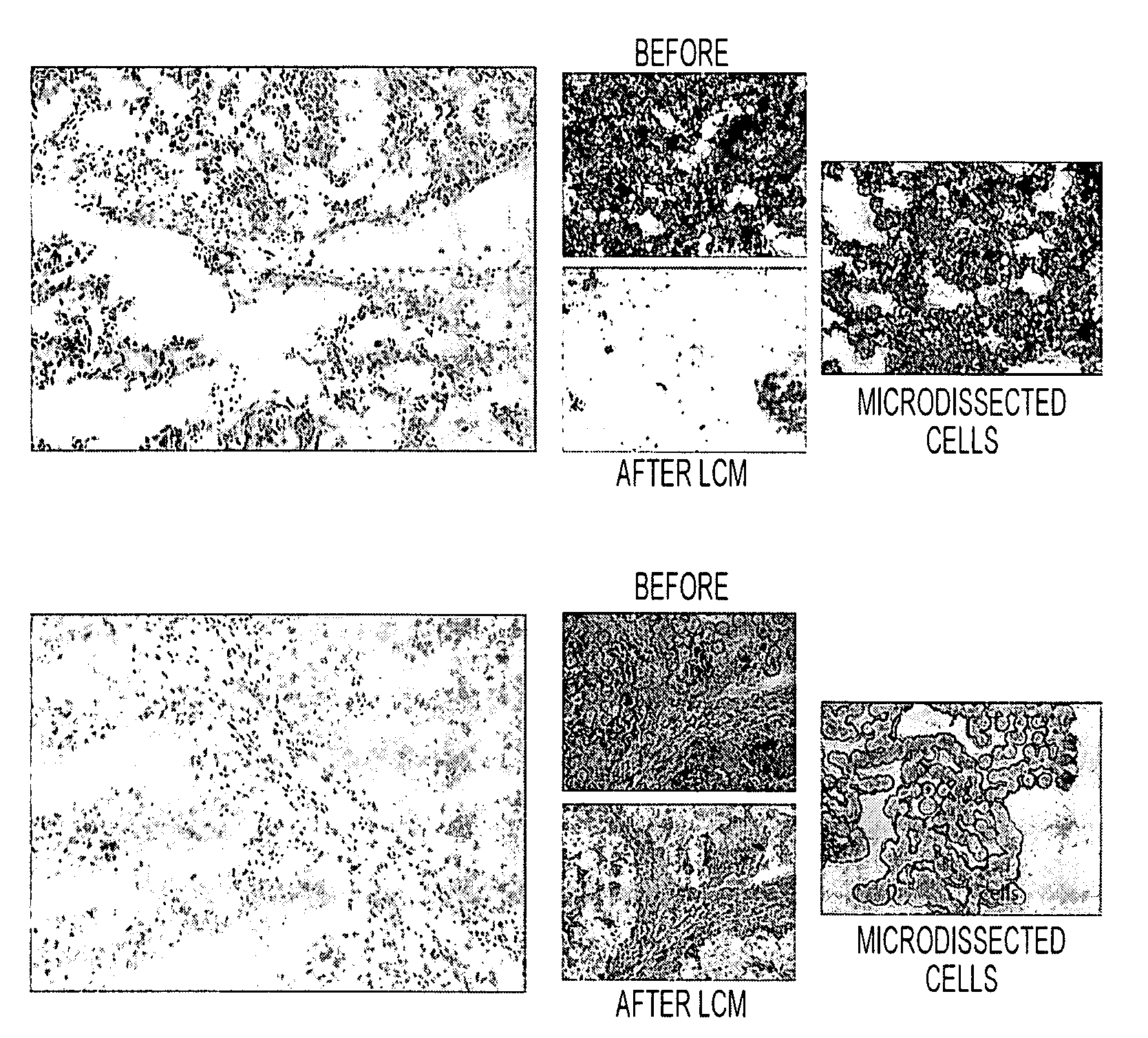
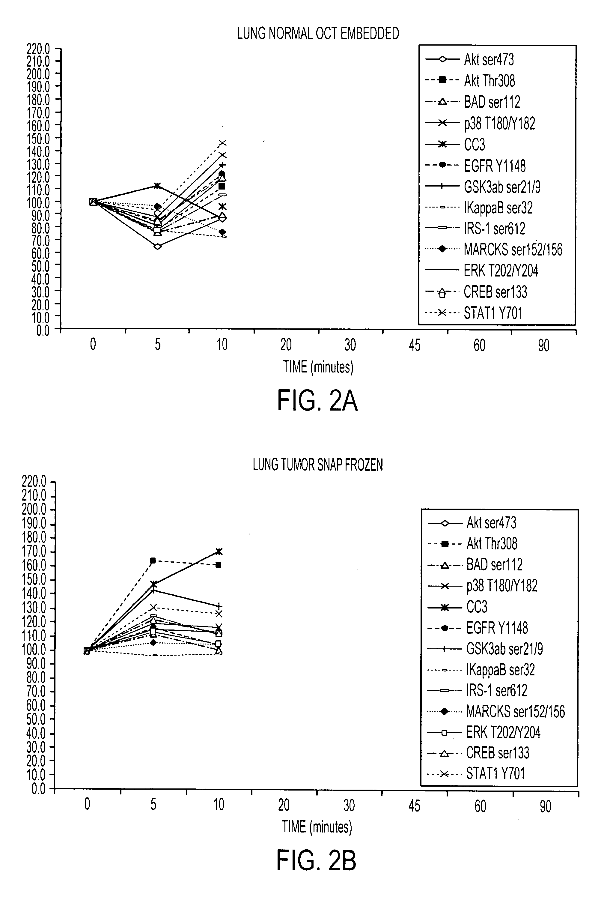
[0111]RPA microarray technology was employed to conduct a time course analysis of phosphoprotein residue end points on tissue procured from the following large tissue specimens: a) non-diseased uterus endometrium and myometrium, b) uterine leiomyomas, c) unaffected colonic mucosa and submucosa, d) unaffected lung and e) lung adenocarcinoma. Special emphasis was placed on the time period taking place immediately following procurement from the patient, e.g. 5 minutes, 10 minutes and 20 minutes, as a means to bracket the “time zero” window. This was done so that the earliest changes due to cooling of the tissue from 37° to room temperature could be observed. For uterus, we also sampled extended time points out to 90 minutes. End points were chosen to span growth factor receptor, pro-survival, and stress pathway related proteins (FIG. 2). The cellular content of each time point sample represen...
example iii
Identification of Tissue Phosphoprotein Stabilizers
[0113]Candidate preservative solutions were evaluated that contain combinations of one or more of the following fixatives and / or stabilizers: a) commercial phosphatase inhibitors, b) commercial kinase and proteinase inhibitors, c) 30% sucrose, 15% trehalose stabilizers, d) commercial PreservCyt, e) commercial CytoLyt, f) mild detergent, and g) precipitating fixatives (see Tables 3 and 4 and FIGS. 4 and 5). A suitable combination can be selected, based on this analysis, to generate a preservative candidate is effective for room temperature or 4° C. storage for 24 hours.
TABLE 3SuccessfulChemicalcutting frozenChemicalFormulaDiluentAdditivestissue sectionPolyethyleneH(OCH2CH2)8OHNoneNoneNoglycol (PEG 400)PolyethyleneH(OCH2CH2)8OHNonePhosphataseNoglycol (PEG 400)and proteaseinhibitors30% sucroseC12H22O11deionizedNoneMarginalH2O30% sucroseC12H22O11deionizedPhosphataseMarginalH2Oand proteaseinhibitorsAcetoneC3H6ONoneNoneNoAcetoneC3H6ONoneP...
PUM
| Property | Measurement | Unit |
|---|---|---|
| Fraction | aaaaa | aaaaa |
| Fraction | aaaaa | aaaaa |
| Fraction | aaaaa | aaaaa |
Abstract
Description
Claims
Application Information