Eureka AIR delivers breakthrough ideas for toughest innovation challenges, trusted by R&D personnel around the world.

Tissue preservation and fixation method

a tissue and fixation technology, applied in the field of personalized cancer therapy, can solve the problems of time delay, inability to provide effective recapitulation of drug targets, and inability to immediately preserve procured tissue in liquid nitrogen

Active Publication Date: 2013-06-11
GEORGE MASON UNIVERSITY
View PDF7 Cites 2 Cited by
  • Summary
  • Abstract
  • Description
  • Claims
  • Application Information

AI Technical Summary

Problems solved by technology

Nevertheless, while gene microarrays can provide important information about somatic genetic taxonomy, they are unable to provide an effective recapitulation of the drug targets themselves, which are embodied in the post-translational and fluctuating signaling molecular network events that occur at the proteomic level.
In the real world of a busy clinical setting, it may be impossible to immediately preserve procured tissue in liquid nitrogen.
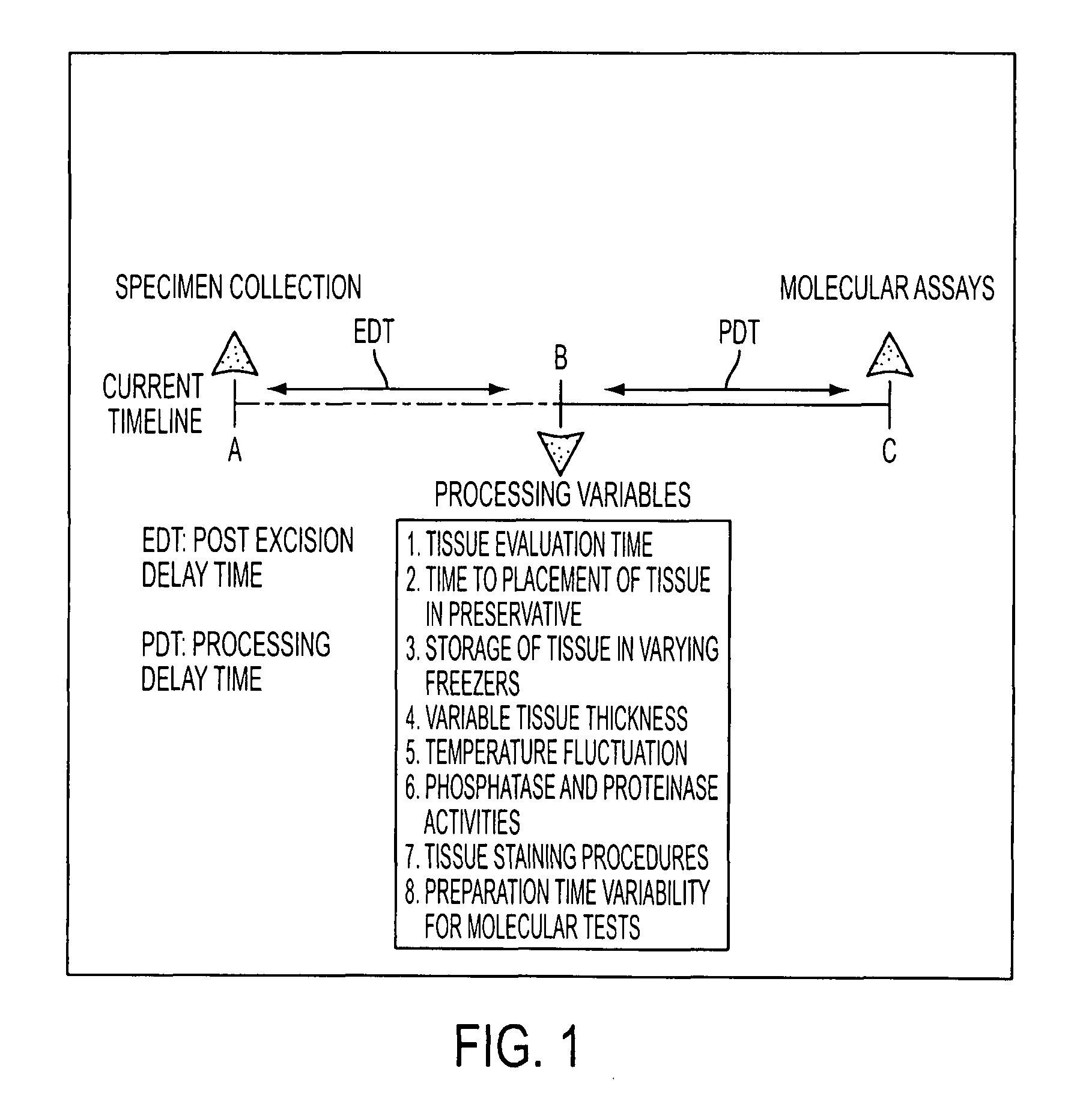
Moreover, the time delay from patient excision to pathologic examination and molecular analysis is often not recorded and may vary from 30 minutes to many hours depending on the time of day, the length of the procedure, and the number of concurrent cases.

Method used


Image

  • Tissue preservation and fixation method
  • Tissue preservation and fixation method
  • Tissue preservation and fixation method

Examples


example i

Materials and Methods

A. Reverse Phase Protein Arrays (RPA)—Quantitation of Phosphoproteins in Tissues and Cells.

[0112]The RPA format, originated by the present inventors and collaborators (see, e.g., Paweletz et al. (2001) Oncogene 20, 1981-1989), immobilizes an individual test sample in a miniature dilution curve such that an array contains hundreds of different patient samples, treatments, or time points. In the RPA format, each array is incubated with one detection protein (e.g. anti-peptide antibody), and a single analyte endpoint is measured and directly compared across multiple samples. Briefly, the lysates are printed on glass backed nitrocellulose array slides (FAST Slides Whatman, Florham Park, N.J. or ONCYTE slides Grace BioLabs, Bend, Oreg.) using a robotic array printing device (GMS 417 arrayer (Affymetrix, Santa Clara, Calif. or Aushon 2470, Aushon Biosystems, Burlington, Mass. equipped with 125-500 μm pins). Each lysate is printed in a dilution curve representing neat,...

example ii

Human Tissue Phosphoprotein Stability Time Course Analysis (in a “Real-World” Hospital Setting)

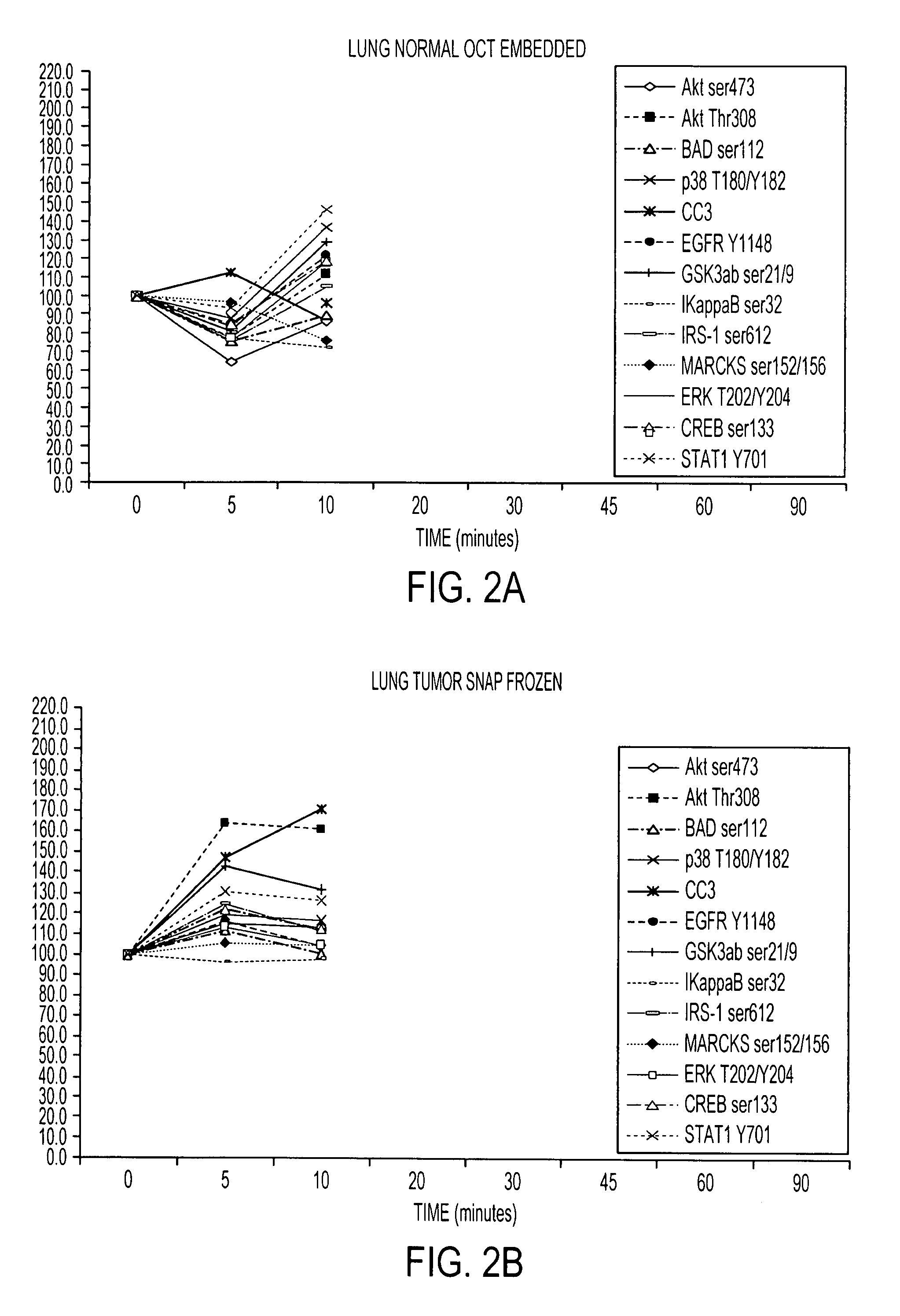
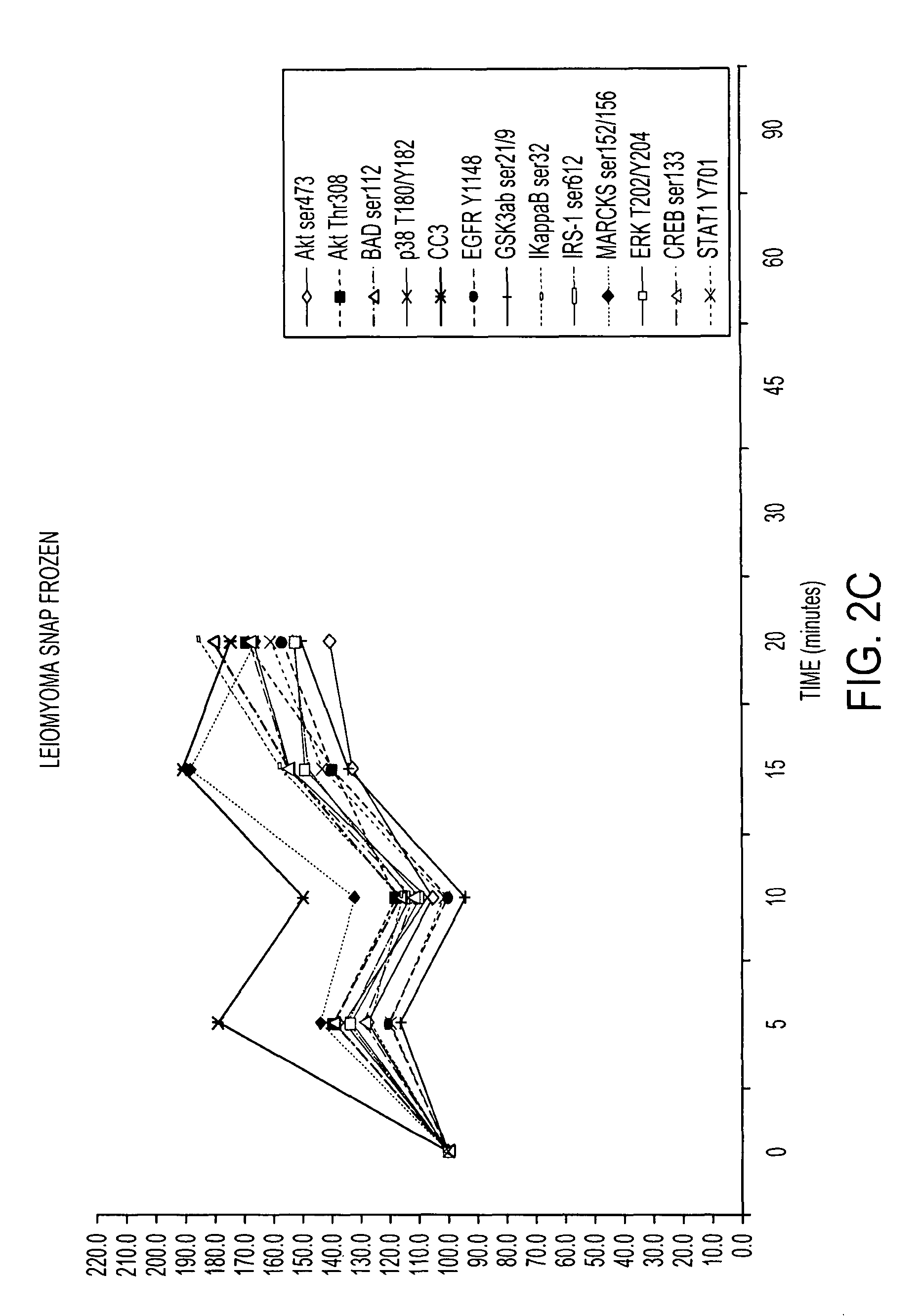
[0116]RPA microarray technology was employed to conduct a time course analysis of phosphoprotein residue end points on tissue procured from the following large tissue specimens: a) non-diseased uterus endometrium and myometrium, b) uterine leiomyomas, c) unaffected colonic mucosa and submucosa, d) unaffected lung and e) lung adenocarcinoma. Special emphasis was placed on the time period taking place immediately following procurement from the patient, e.g. 5 minutes, 10 minutes and 20 minutes, as a means to bracket the “time zero” window. This was done so that the earliest changes due to cooling of the tissue from 37° to room temperature could be observed. For uterus, we also sampled extended time points out to 90 minutes. End points were chosen to span growth factor receptor, pro-survival, and stress pathway related proteins (FIG. 2). The cellular content of each time point sample represen...

example iii

Identification of Tissue Phosphoprotein Stabilizers

[0119]Candidate preservative solutions were evaluated that contain combinations of one or more of the following fixatives and / or stabilizers: a) commercial phosphatase inhibitors, b) commercial kinase and proteinase inhibitors, c) 30% sucrose, 15% trehalose stabilizers, d) commercial PreservCyt, e) commercial CytoLyt, f) mild detergent, and g) precipitating fixatives (see Tables 3 and 4 and FIGS. 4 and 5). A suitable combination can be selected, based on this analysis, to generate a preservative candidate is effective for room temperature or 4° C. storage for 24 hours.

[0120]

TABLE 3SuccessfulChemicalcutting frozenChemicalFormulaDiluentAdditivestissue sectionPolyethyleneH(OCH2CH2)8OHNoneNoneNoglycol (PEG 400)PolyethyleneH(OCH2CH2)8OHNonePhosphataseNoglycol (PEG 400)and proteaseinhibitors30% sucroseC12H22O11deionizedNoneMarginalH2O30% sucroseC12H22O11deionizedPhosphataseMarginalH2Oand proteaseinhibitorsAcetoneC3H6ONoneNoneNoAcetoneC3H6...


PUM

PropertyMeasurementUnit
temperatureaaaaaaaaaa
sizeaaaaaaaaaa
particle sizeaaaaaaaaaa

Abstract

This invention relates, e.g., to a composition that, at room temperature, when contacted with a sample comprising phosphoproteins, can fix and stabilize cellular phosphoproteins, preserve cellular morphology, and allow the sample to be frozen to generate a cryostat frozen section suitable for molecular analysis. The composition comprises (1) a fixative that is effective to fix the phosphoproteins, and that has a sufficient water content to be soluble for a stabilizer and / or a permeability enhancing agent); (2) a stabilizer, comprising (a) a kinase inhibitor and (b) a phosphatase inhibitor and, optionally, (c) a protease (e.g., proteinase) inhibitor; and (3) a permeability enhancing agent (e.g. PEG). Methods are described for preserving phosphoproteins, using such a composition. Also described are endogenous surrogate markers for monitoring protein degradation, including the loss of posttranslational modifications (such as phosphorylation), e.g. the following removal of a cell or tissue from a subject; and exogenous molecular sentinels (e.g. phosphoproteins attached to magnetic nanoparticles) that allow one to evaluate the processing history of a cellular or tissue population sample.

Description

[0001]This application claims the benefit of the filing date of U.S. Provisional Application No. 60 / 855,120, filed Oct. 30, 2006, and U.S. Provisional Application No. 60 / 861,086, filed Nov. 27, 2006, both of which are incorporated by reference herein in their entirety.[0002]This invention was made with government support under the following grants awarded by the National Institutes of Health: 1R21CA155592-01, 1R33CA157403-01, 3R21CA125698-02S1, 1R21CA125698-01, 3R21CA125698-02S2, 5R21CA125698-02, and 5R33CA157403-03. The government has certain rights in the invention.BACKGROUND INFORMATION[0003]Phosphoprotein molecular profiling is an important component of the emerging field of individualized cancer therapy. Molecularly targeted anti-cancer therapeutics often involves the mitigation or modulation of defective kinase signaling networks. Elucidation of deranged signaling networks within tumors offers tremendous promise as a means to individualize molecular targeted cancer therapy and...

Claims


Application Information

Patent Timeline
Patent Type & Authority Patents(United States)
IPC IPC(8): A01N1/00
CPCG01N1/30A01N1/00B82Y30/00
Inventor ESPINA, VIRGINIA A.LIOTTA, LANCE A.GEHO, DAVID
Owner GEORGE MASON UNIVERSITY
Why Eureka
Eureka Blog
Learn More