Somatotransgenic bioimaging

a bioimaging and somatosensor technology, applied in the field of model pathologies, can solve the problems of poor drug candidates, toxicity, toxicity, and toxicity of cyps, and achieve the effect of monitoring the progression of a pathology and avoiding the effect of position effects

Inactive Publication Date: 2010-07-08
UCL BUSINESS PLC
View PDF3 Cites 0 Cited by
  • Summary
  • Abstract
  • Description
  • Claims
  • Application Information

AI Technical Summary

Benefits of technology

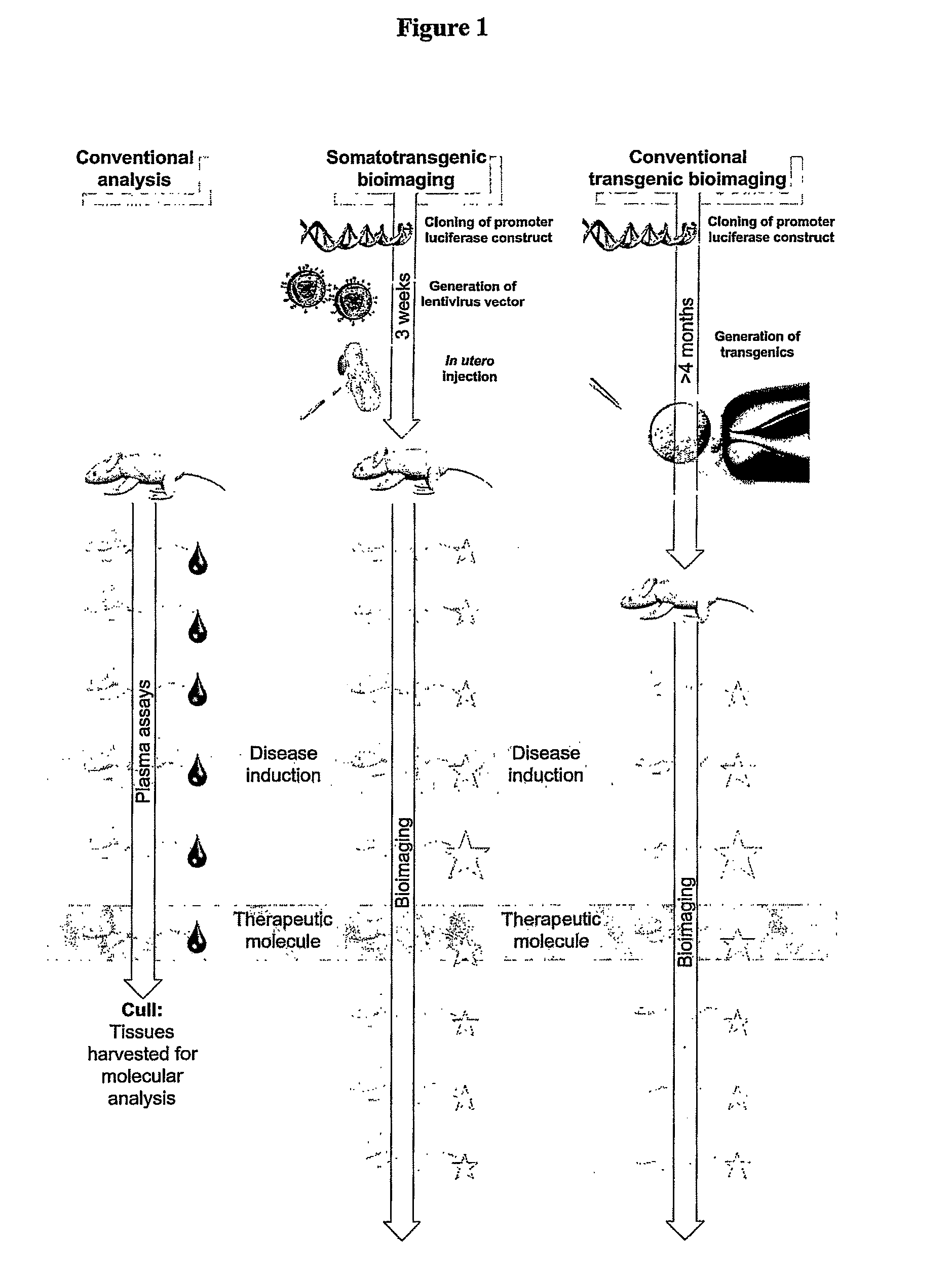
[0022]Also, in conventional transgenesis, all the cells of the transgenic animal ultimately arise from the same transformation event in the same cell, i.e. the transgene is in the same place and orientation in the genome of every cell. In the present invention, the transduction is carried out at the tissue level (somatotransgenesis) so there will be many different individual transformation events in many different individual cells. This means that position effects are avoided. In a conventional germline transgenic, if the vector integrates in an unfavourable location, that unfavourable result will exist in all the animal's cells and may give a misleading impression in any analysis. In a somatotransgenic animal according to the invention, any unfavourably positioned insertions will be compensated for by other, favourably positioned ones.
[0023]A further advantage is that, according to the invention, non-integrating vectors may be used where appropriate, whereas in a conventional transgenic an integration event would always be required, otherwise the transgene would not be replicated into every cell of the resulting animal
[0024]Another advantage of somatotransgenesis is that the luciferase can be introduced into any transgenic or knockout mouse model or background strain. In contrast, conventional germline transgenics have to be crossed on

Problems solved by technology

Metabolism of drugs by CYPs influences drug clearance, toxicity, activation and, potentially, adverse interactions with other drugs.
Compounds that are turned over and cleared from the body rapidly or that are

Method used


Image

  • Somatotransgenic bioimaging
  • Somatotransgenic bioimaging
  • Somatotransgenic bioimaging

Examples


example 1

[0098]Experiments were conducted to determine the possibility of achieving long-term tissue-specific transgene expression in mice.

Vector Production and Validation

[0099]The gp64- and vsvg-pseudotyped luciferase vector used for long-term analysis was produced as previously described by Seppen et al (Seppen J, Rijnberg M, Cooreman M P, Oude Elferink R P. Lentiviral vectors for efficient transduction of isolated primary quiescent hepatocytes. J Hepatol 2002; 36: 459-465).

[0100]Lentivectors were prepared as follows: Producer 293T cells were seeded at 2×10⁷ cells per T-150 flask. The next day, plasmid DNA was mixed in the following amounts per T-150 flask; vector construct (pHR.SINcpptSEW) 40 μg, pMDG.2 / pHCMVwhvGP64 10 μg, pCMVΔ8.74 30 μg to a final volume of 5 ml in OptiMEM (Invitrogen, Paisley, UK). Polyethylenimine (PEI, 25 kDa) (Sigma, Poole, UK) was added to 5 ml of OptiMEM to a final concentration of 2 μM and filtered through a 0.22 μm filter. The DNA was added dropwise to the PEI s...

example 2

[0106]In order to assess long-teen transgene expression, a single intra-amniotic dose of gp64 / HIV-luciferase (˜3×10⁷ iu) was administered to neonatal mice at day 1 (n=5). Mice were subjected to bioimaging over the course of one year and beyond and luciferase bioluminescence compared to controls (n=2). In vivo luciferase bioimaging was carried out as in Example 1. Luciferase expression was substantially above background throughout the analysis and persisted throughout this study (FIG. 6). The results demonstrate that significant expression is detectable up to one year after application.

example 3

[0107]Genetic bioeffectors useful in animal models were tested in vitro (Examples 3 to 5).

[0108]NIH-3T3 cells were transfected with plasmids containing TGF-β responsive elements driving luciferase expression. These cells were then transduced with a retroviral vector expressing TGF-β3. The SBE4 responsive element is specific to TGF-β activation via smad2 / 3 mediated transcriptional activation. This Smad activation can be further delineated to Smad2 specific transcriptional activation using the ARE responsive element in conjunction with the xenopus Fast-1 transactivator (ARE alone is only Smad2 / 3 specific). The BMP-specific responsive element activates through Smad1 / 5 / 8 activation and should not be responsive to TGF-β3 activity. Finally, Smad7 is an inhibitor Smad and is known to be upregulated in a negative feedback loop by TGF-β3 activation. The experiment was conducted as follows:

[0109]NIH-3T3 cells pre-plated at 1×10⁶ cells / well and transfected with 10 μg of reporter plasmid by sta...


PUM

PropertyMeasurementUnit
Responsivityaaaaaaaaaa
Toxicityaaaaaaaaaa
Bioluminescenceaaaaaaaaaa

Abstract

The invention relates to modelling diseases, to screening for compounds that modulate such diseases and to as-saying drug metabolism and toxicity in non-human transgenic animals, by a novel technique developed by the inventors known as “somatotransgenic bioimaging”. The invention thus provides: a method for determining whether the expression of a reporter gene is modulated by a compound or a method of evaluating the metabolism and/or toxicity of a compound, said method comprising: (a) administering said compound to a non-human transgenic animal, generated by gene transduction of one or more specific tissues when in utero or neonatal, with a vector comprising a bioluminescent reporter gene operably linked to a genetic element responsive to a pathology or therapy or to a genetic element responsive to drug metabolism and/toxicity; and (b) determining whether or not of said compound has an effect on the expression of said reporter gene in said specific tissue or tissues and/or determining the extent of any such effect, said determination comprising detecting from the animal bioluminescence caused by the activity of the gene product of the reporter gene. In some embodiments cells pre-transduced with vectors of the invention may also be introduced into the animals instead of delivering the vectors directly.

Description

FIELD OF THE INVENTION[0001]The invention relates to modelling pathologies, screening for compounds that modulate such pathologies and to evaluating drug metabolism and toxicity in non-human transgenic animals by a novel technique termed “somatotransgenic bioimaging”.BACKGROUND OF THE INVENTIONDrug Validation[0002]Potential therapeutics are generally identified using high-throughput in vitro technologies to begin with. It is then desirable to validate successful candidate compounds in vivo, for example in rodent models, before progressing to full-scale pre-clinical primate studies or clinical trials.[0003]Traditional pharmacological assays rely on taking measurements from peripheral, secreted or excreted body fluids or tissue biopsies and often rely on endpoint analyses. Measurements from fluids or tissues rely on an appropriate experimental variable, i.e. they can only work if there is something in the fluid or tissue that changes in response to the administration of the candidate ...

Claims


Application Information

Patent Timeline
IPC IPC(8): G01N33/00A01K67/00
CPCA01K2267/0337A01K2267/035C12Q1/6897C12N15/8509A01K2267/0393
Inventor WADDINGTON, SIMON NICHOLASMCKAY, TRISTAN ROWNTREE
Owner UCL BUSINESS PLC
Why Eureka
Try Eureka