Real-time multimode neurobiophysiology probe

a neurobiophysiology and probe technology, applied in the field of neurobiology, biotechnology and medical instruments, can solve the problems of aliasing spectral data, device has no means of sample particulate or molecular size selectivity, and no means for concentration or amplification of desired analyte(s)

Inactive Publication Date: 2010-09-23
BLUMENFELD WALTER +2
View PDF6 Cites 40 Cited by
  • Summary
  • Abstract
  • Description
  • Claims
  • Application Information

AI Technical Summary

Benefits of technology

[0012]Combinations of two or more of these techniques, applied simultaneously or sequentially at the sam

Problems solved by technology

Another confounding effect on the ATR analysis is the possibility of fouling the semi-permeable membrane with a local concentration of small molecules or an adherent fluid-borne substance,

Method used


Image

  • Real-time multimode neurobiophysiology probe
  • Real-time multimode neurobiophysiology probe
  • Real-time multimode neurobiophysiology probe

Examples


first embodiment

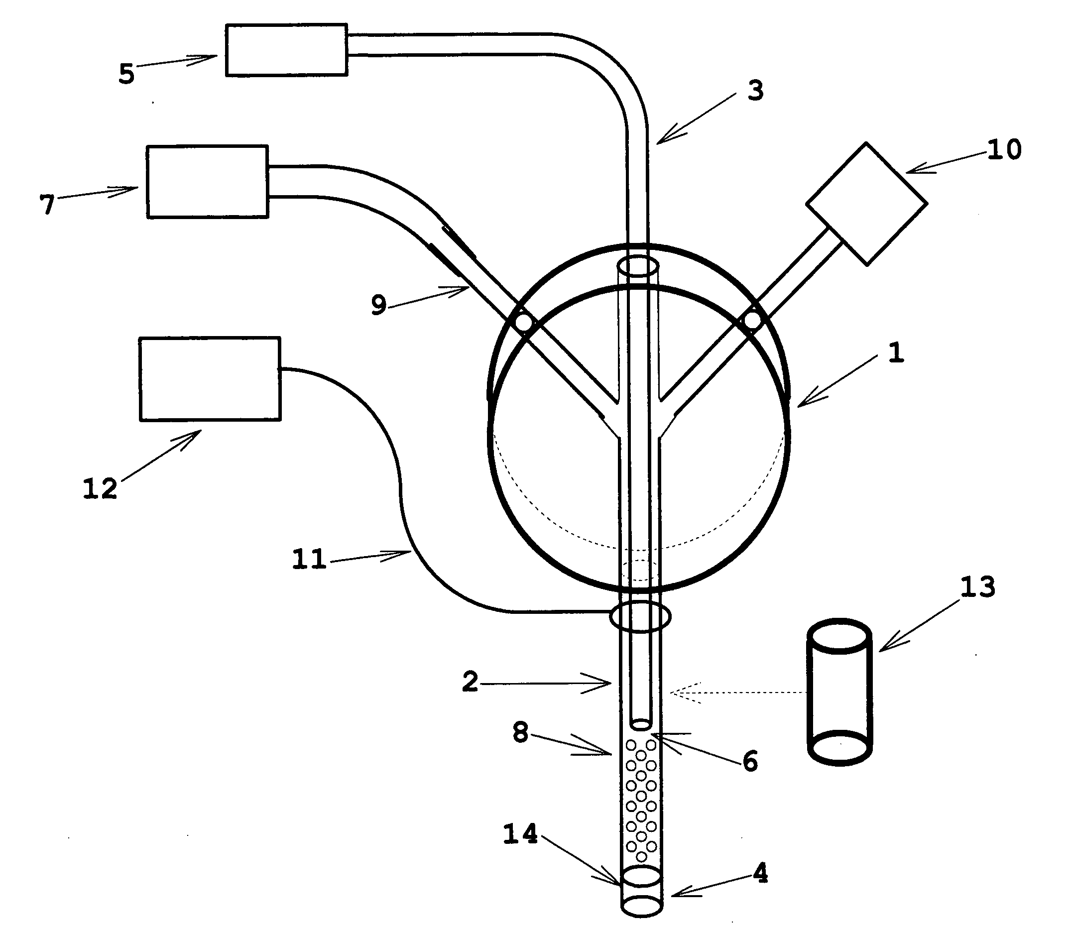
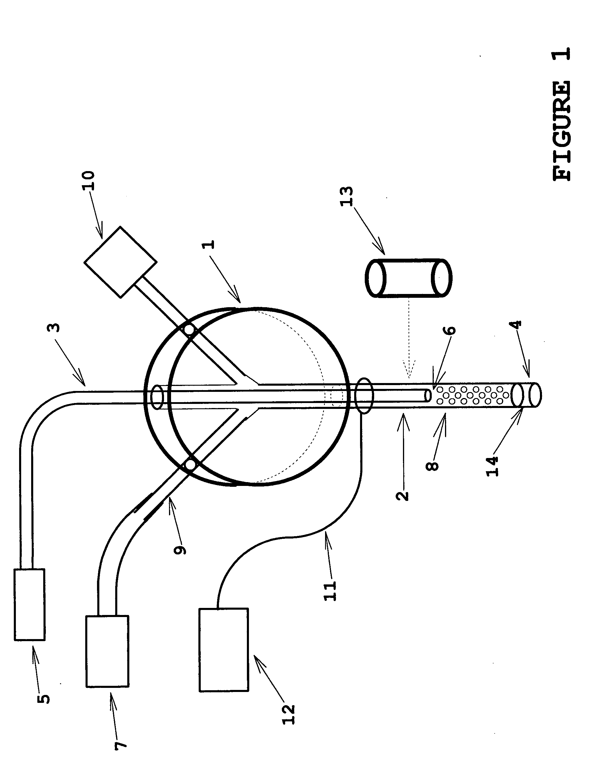
[0022]the invention is shown in FIG. 1. The functional part of the device is the microtube 2, typically a section of stainless steel or titanium hypodermic tubing (typically 100 to 300-micron internal diameter and having a typical working length from 25 mm to 100 mm) which is inserted into the tissue site of interest. Optical interrogation (via an optical fiber 3) of tissue or interstitial fluid proximal to the microtube tip 4 may be used to characterize the present tissue or fluid in real time, to detect transitions of tissue or fluid type as the microprobe insertion proceeds, or as real-time feedback information to control depth of insertion to a desired site. The optical interrogation may be done directly from the tissue or fluid by any of the well-known spectroscopy technologies in an optical spectroscopy system 5, or it may be done via a chemical sensor coating 6 at the tip of the optical fiber (such as a Ruthenium Dioxide coating whose fluorescence properties are responsive to...

second embodiment

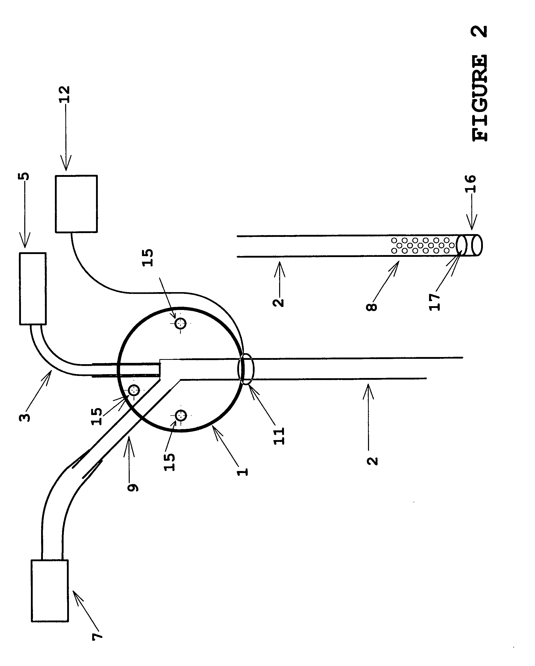
[0023]the invention is illustrated in FIG. 2. A standard beveled hypodermic needle tip is shown; in addition to the features referenced as in FIG. 1, it also shows three mounting through-holes 15 which facilitate mounting the device to the experimental subject or to a separate stereotactic apparatus by means of pins, screws or adhesive. In this embodiment, the optical fiber 3 is mounted to extend only to the center of the multiport manifold body 1, so that the internal passage of fluid in the interior volume of the microtube 2 is unobstructed. This allows fluid delivery or sample extraction with minimum resistance, and thereby reduces fluidic phase lag for sampling or delivery. A variation of this embodiment is also shown in partial illustration, in which the open beveled end of the microtube 2 is replaced by an impermeable tip seal 16, and an aperture array of perforations 8 is located in the sidewall of the microtube 2. This configuration allows the interior bore of the microtube ...

fifth embodiment

[0026]FIG. 5 depicts the invention, which requires an optical power source 21 (for example, and ultraviolet or infra-red laser) separate from that used for optical interrogation, and a multiple-lumen optical fiber 3. For applications involving optically activated diagnostic or therapeutic agents, controlled illumination of desired wavelength, timing and intensity may be delivered to the tissue site by the optical fiber. This configuration may also be applied, for example, to stimulus-response experiments in which a nerve is optically stimulated by mid-range IR laser energy delivered via the optical fiber 3 to nerve tissue in the field of view of the optical fiber end surface 6, and the response is characterized by the simultaneous probe tip electrical signals carried over conductive lead 11 to a detector-amplifier in the electrical sensing and excitation system 12 and estimated oxygen uptake based on time-domain fluorescence measurements of ruthenium dioxide coatings on inert micros...


PUM


Abstract

Apparatus and methods in which very small volumes of material may be extracted, delivered, interrogated or stimulated via optical, electromagnetic or mechanical means, in vivo or in vitro, for site-specific detection, characterization, stimulation, diagnostics or therapy, comprising optical, fluidic, chemical, electromagnetic and biological techniques applied via a microprobe in a single intra-parenchymal tissue perforation procedure in the brain. The primary use of the device is in neuroscience research, clinical diagnostics and therapeutics applications in the brain, however, the device may also be beneficially applied to other organs and biological systems. Human clinical applications may include neurosurgical intra-operative monitoring, extra-operative chronic monitoring of devices introduced in an operation, and diagnostic monitoring combined with simultaneous neuroimaging.

Description

BACKGROUND [0001]1. Field of the Invention[0002]This invention relates generally to the field of neuroscience, biotechnology and medical instrumentation, and particularly to molecular sampling, delivery and characterization methods applied in conjunction with optical, electromagnetic or electrochemical interrogation or excitation by means of a minimally-invasive probe at a designated site in the brain.[0003]2. Related Art[0004]U.S. Pat. No. 6,584,335 to Hans-Peter Haar describes an end-sealed hollow needle having a permeable area allowing size-limited fluid-borne molecules to be coupled via evanescent field effects through a semi-permeable coating to an optical fiber or waveguide positioned in the needle cavity. This allows optical interrogation by quantum-cascadelaser-excited multiple-wavelength attenuated total reflectance spectroscopy (ATR) in the 7 to 13-micron wavelength region. This enables detection and quantification of blood glucose concentration, which, in principle, might...

Claims


Application Information

Patent Timeline
IPC IPC(8): A61M37/00A61B5/00
CPCA61B5/0075A61B5/0084A61B5/031A61B5/14553A61N1/0534A61M25/007A61M2025/0042A61M2210/0693A61B5/6849A61B5/4064A61B5/4094
Inventor BLUMENFELD, WALTERSEPKUTY, JEHUDA PETERSOLYAR, GREGORY
Owner BLUMENFELD WALTER
Why Eureka
Try Eureka