

Biocompatible polymers and Methods of use
a biocompatible polymer and polymer technology, applied in the field of biocompatible polymers and methods of use, can solve the problems of reducing interest in the use of protein biopolymers, such as fibrin elastomers, and affecting the development of bioresorbable implants. the inability of these polymers to degrade in response to cellular invasion and promote the in-growth of host tissues, and the profound limitation of bioresorbable implants
- Summary
- Abstract
- Description
- Claims
- Application Information
AI Technical Summary
Benefits of technology
Problems solved by technology
Method used
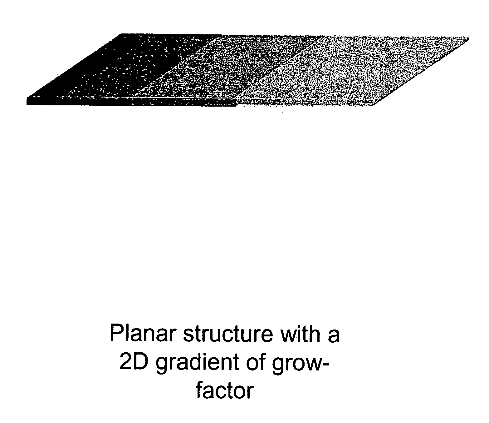
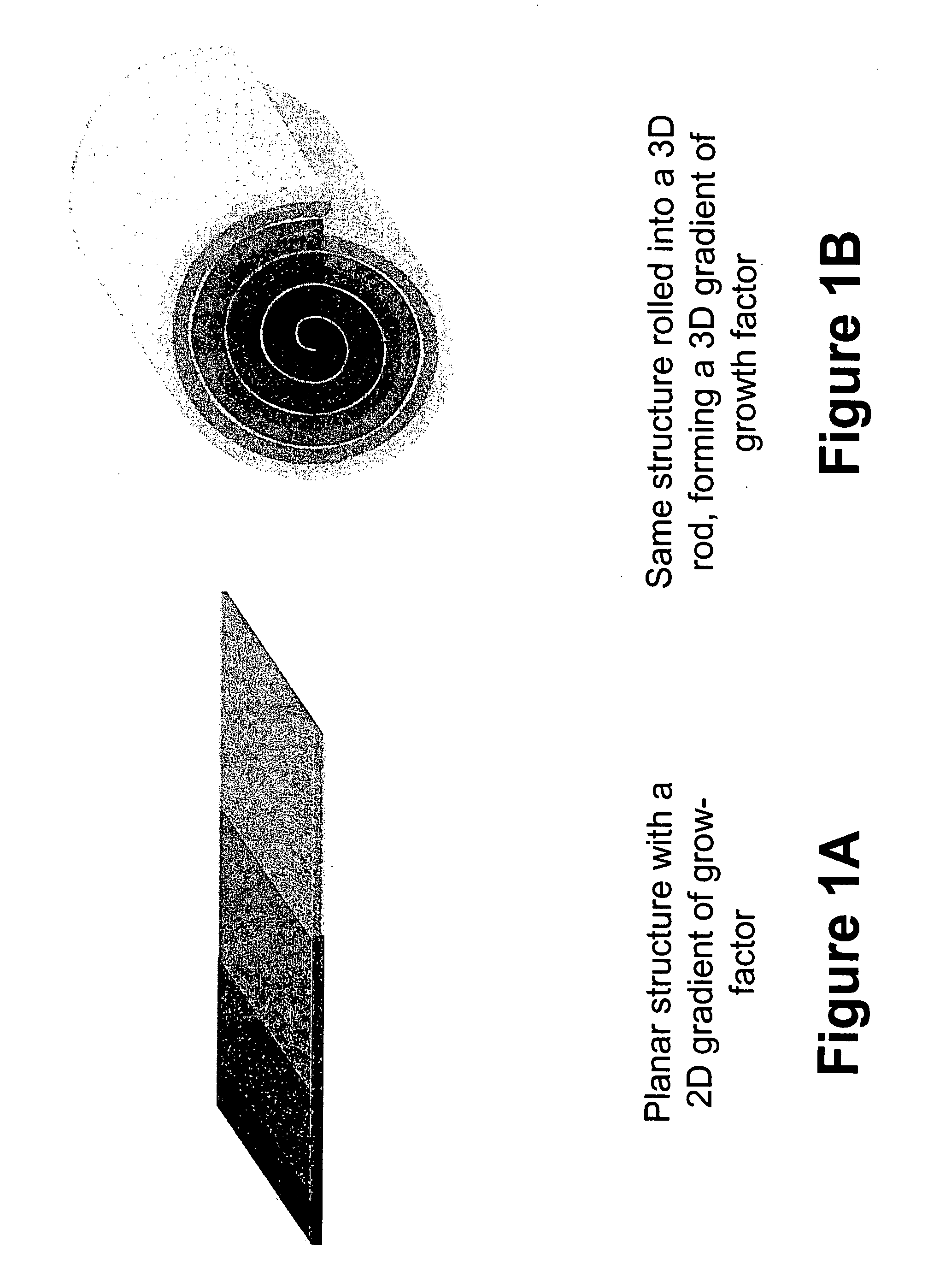
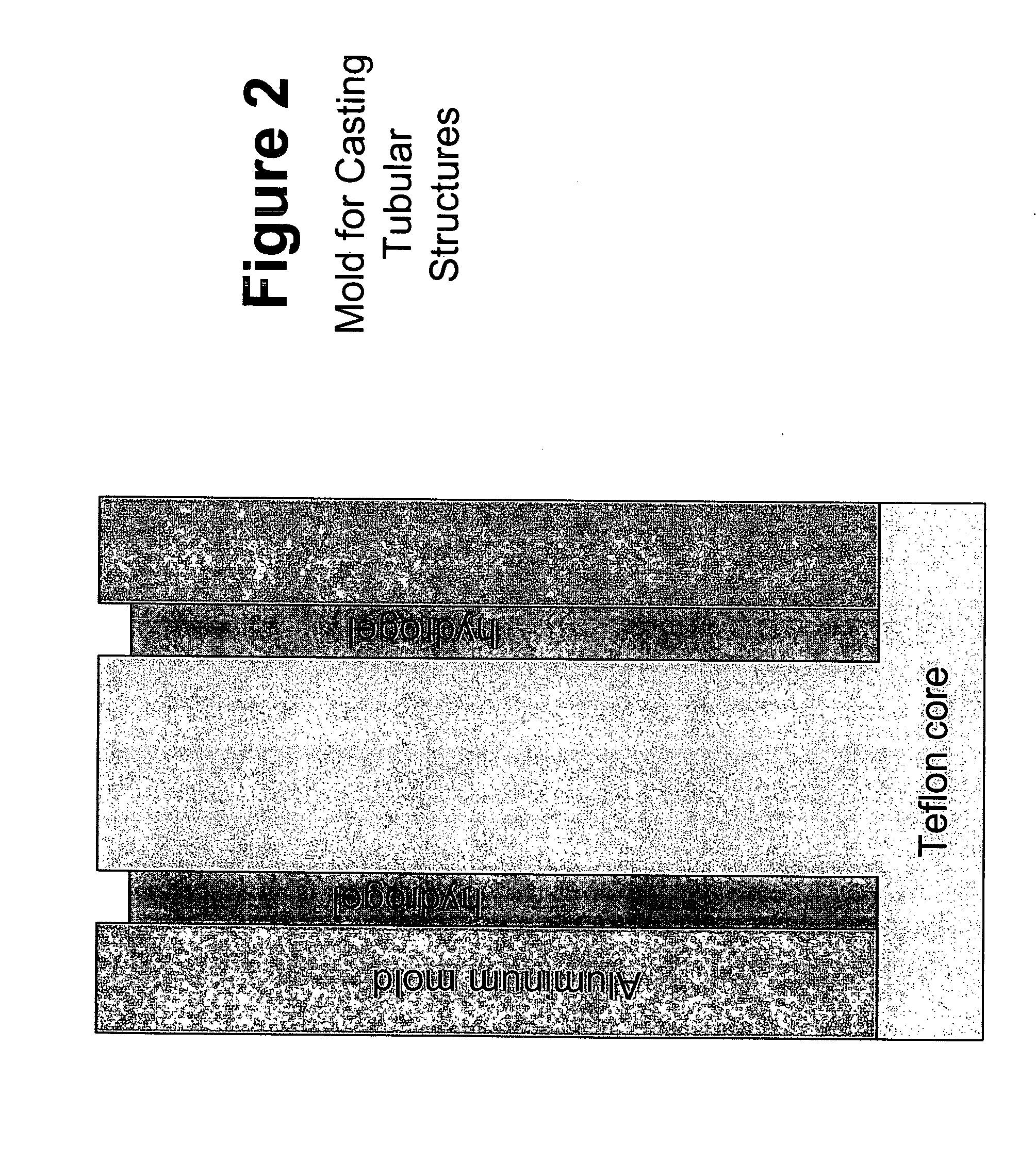
Image

Examples
example 2
Vacuum Dehydration of Fibrin Gel
[0214]Upon removal from the gel molds, the fully-hydrated fibrin gels were lyophilized in order to remove water and reduce the thickness of the fibrin gel films from 2.25 mm to approximately 100 μm. Lyophilization was performed in a gel dryer (commercially available from BioRad, Hercules, Calif.). The lid of the gel dryer was opened and the silicon rubber gasket was peeled back. Spectrapor 1 dialysis tubing (6-8k MWCO, Spectrum Laboratories, Rancho Dominguez, Calif.) was soaked in PBS and cut along one side to allow the tubing to be opened up into a sheet. The opened tubing had 3 cm×3 cm pieces cut into it. The resulting dialysis “sheets” were placed onto the gel dryer. The fibrin gel was placed in the center of the dialysis sheet. A 2.5 cm×2.5 cm×3 cm TEFLON™ segment with a 20 mm×20 mm square cut out of the center was placed around the gel. A 2.5 cm×2.5 cm×0.075 cm TEFLON™ segment was placed on top of the 2.5 cm×2.5 cm×3 cm TEFLON™ segment. The silic...
example 3
Osmotic Dehydration of Fibrin Gel
[0217]An alternative method to that disclosed in Example 2 was also used to process the fibrin gels upon gelation and removal from the gel molds. In this method, an osmotic dehydration process was used (based upon the method of Müller and Ferry, U.S. Pat. No. 4,548,736). Fibrin gels were prepared the same as in the method of Example 1. The fibrin gels were placed on a coverslip inside a 60 mm Petri dish. Approximately 500 μL of a 35% high molecular weight polyvinyl alcohol solution was added to the inside of an approximately 3 inch segment of SPECTRAPOR-1™ (6-8,000 MWCO) dialysis membrane tubing soaked in PBS. The ends of the tubing were clamped off. The tubing was placed on top of the gel making sure the polyvinyl alcohol solution rested directly on top of, and in complete contact with, the entire gel to ensure that water evenly diffused from the gel and entered into the tubing along the osmotic gradient. A lid was placed on the Petri dish and it wa...
example 4
Fibrin Film Biocompatibility Assay
[0220]Fibrin films fabricated according to the method described in Examples 1 and 2, and Examples 1 and 3, were placed on 12 day old chick embryos and biocompatibility was tested using the chick chorioallantoic membrane (CAM) assay. The tested films did not exhibit any objective signs of incompatibility. CAM blood vessels underneath the fibrin film were visualized by intravital injection of fluorescent quantum dots (QDs). Two days post-placement of the film, the embryo was injected with 655 nm emitting QDs and blood vessel fluorescence was imaged on a M2BIO stereoscope using a 1.6× objective (1× zoom), Retiga Exi CCD camera, and a (Ex:Em) 450spuv:655 / 20 filter set. The films induced no observable ill effects on the underlying blood vessels.
PUM
| Property | Measurement | Unit |
|---|---|---|
| diameter | aaaaa | aaaaa |
| diameter | aaaaa | aaaaa |
| temperature | aaaaa | aaaaa |
Abstract
Description
Claims
Application Information