Device, system and method for in-vivo analysis

a technology of in-vivo body fluids and devices, applied in the field of point of care analysis, can solve the problems of inability to localize or identify the origin of abnormal substances, cumbersome and complicated automatic machines, and inability to detect abnormal substances in the lab using simple and automatic machines,

Inactive Publication Date: 2010-12-23
GIVEN IMAGING LTD
View PDF21 Cites 14 Cited by
  • Summary
  • Abstract
  • Description
  • Claims
  • Application Information

AI Technical Summary

Benefits of technology

[0013]Embodiments of the invention may provide additional and / or other benefits and / or advantages.

Problems solved by technology

Typically, these in vitro methods of detection are carried out in the lab using cumbersome and complicated automatic machines due to the complicated requirements of such procedures which may involve a plurality of reactions and perhaps different conditions for each reaction.
Also, these methods of detection, when carried out in vitro, may not easily enable the localization or identification of the origin of an abnormally occurring substance.
Devices and systems for in-vivo imaging may be used, for example, to acquire in vivo images of the GI tract however, imaging alone may typically not enable early detection of pathology, such as cancer.

Method used


Image

  • Device, system and method for in-vivo analysis
  • Device, system and method for in-vivo analysis
  • Device, system and method for in-vivo analysis

Examples


example 1

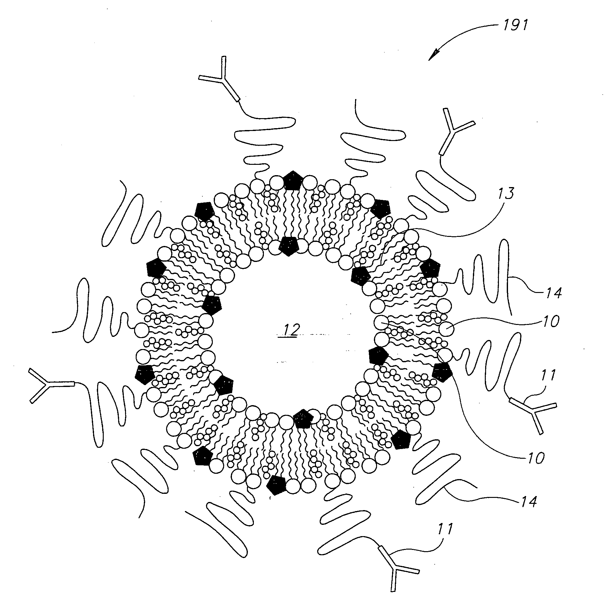
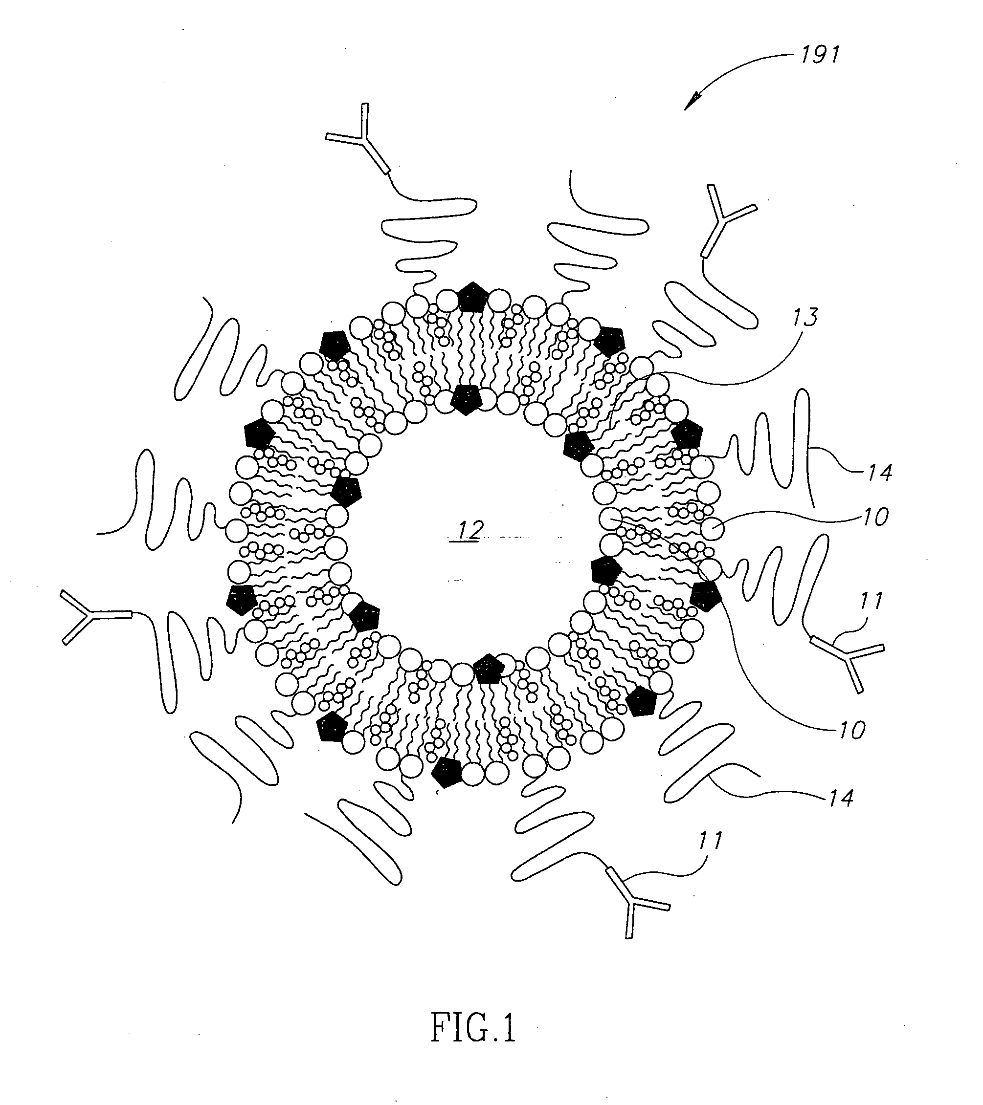
[0040]Trypsin inhibitor is a known biomarker for acute renal failure. Typsin is an enzyme known to cleave proteins containing the basic amino acid lysine. According to one embodiment of the present invention, a diagnostic kit can be prepared. The diagnostic kit, according to one embodiment, may include

[0041]A: liposomes based on phospholipids containing lysine or a mixture of polymersomes with proteins containing lysine in a solution with a pH of about 3.5 or lower;

[0042]B: pH indicator, for example bromocresol green having a yellow color in a pH lower than 3.8 and a blue color in a pH higher than 5.5. Other suitable indicators can be used. The indicator is entrapped in the liposomes;

[0043]C: Trypsin, which is typically inactive at a low pH, may be mixed with the liposome or attached to polysaccharides coating the liposome using technologies well known in the art. The above may be mixed and may be lyophilized and introduced into a test tube containing a urine sample (the sample can ...

example 2

[0044]Secretory Phospholipase A2 (sPLA2) is an enzyme that is active in the intercellular space. It is over expressed in inflammatory as well as cancerous tissues. The diagnostic kit, according to one embodiment, may include

[0045]A: liposomes based on phospholipids containing for example a fatty acid with an acryl group at 2-lysophospholipid or similar phospholipase that can be hydrolyzed by ePLA2, The liposomes are prepared in a solution with a pH of about 3.5 or lower;

[0046]B: pH indicator for example Chlorophenol red having a yellow color in a pH lower than 5.2 and a purple color at a pH higher than 6.8. Chlorophenol is nontoxic. The indicator is entrapped in the liposomes;

[0047]C: the liposome is conjugated with a specific antibody to the specific inflammation for example IBD in the colon or cancerous polyp.

[0048]In one embodiment the liposomes are encapsulated in a pill and are released only when the colon is reached e.g. liposomes are contained in a pill that is coated with a ...

example 3

[0051]Liposomes are prepared as described in Example 2 except that the signaling agent is an IR sensitive material. Said liposomes are suspended in physiological solution and introduced into the systemic blood stream by injection by a syringe or by intravenous (IV) injection or any other suitable delivery mechanisms as known in the art.

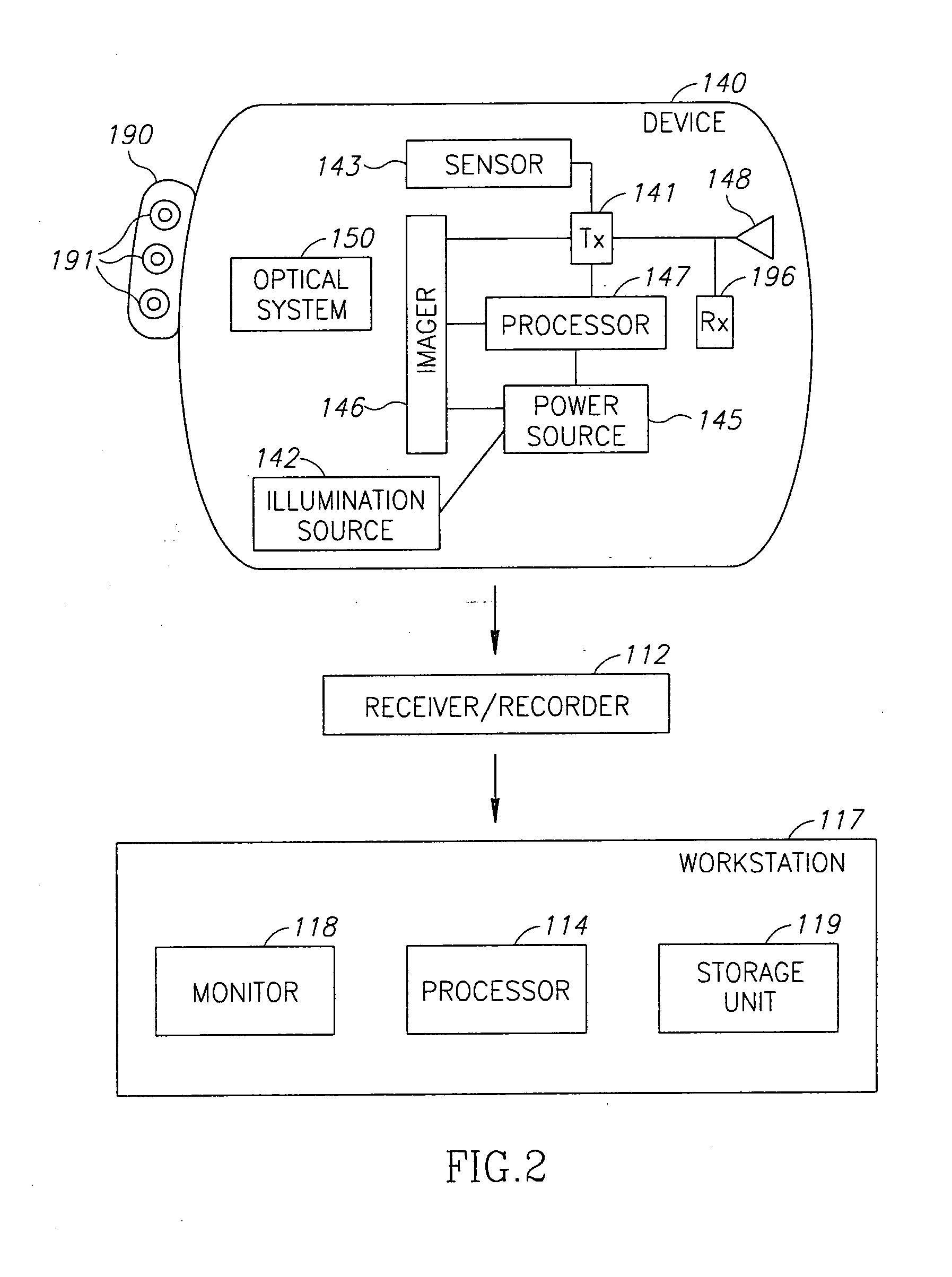
[0052]Once the liposomes are released they spread, in the body but specifically accumulate in tumors enriched with the targeted antigen. An in-vivo imaging device such as an imaging capsule or endoscope equipped with an IR source may be inserted into the patient's body, optionally after waiting a pre-defined period to allow the liposomes or nano-containers to spread and reach the tumor. The IR light may penetrate the tissue and excite the IR signaling material even if the tumor is sub facial. The light emitted by the excited IR signaling material can thus be detected by the imager of the capsule or the endoscope enabling the detection of deep tissues ...


PUM

PropertyMeasurementUnit
inner diameteraaaaaaaaaa
pHaaaaaaaaaa
timeaaaaaaaaaa

Abstract

A method for detecting the presence of a substance such as an antigen, the method including the steps of: maintaining a signaling material at a first pH within a liposome; contacting the liposome with a sample having a second pH such that if the substance is present in the sample the liposome will rupture thereby exposing the signaling material to the second pH; and detecting a change in an optical property, such as color, of the signaling material, the change caused by the exposure to the second pH.

Description

FIELD OF THE INVENTION[0001]The present invention relates to point of care analysis in general and specifically to in vivo body fluids analysis utilizing liposomes and / or nano-containers.BACKGROUND OF THE INVENTION[0002]In some cases diseases, such as cancer, can be detected by analyzing the blood for tumor specific markers, typically, specific antibodies. In vitro testing of body fluid samples for the presence of a suspected substance is common. For example, immunoassays are widely used for the determination, either qualitative or, quantitative, of a variety of substances, such as peptides, proteins, enzymes, hormones, vitamins, drugs, carbohydrates, etc. Typically, these in vitro methods of detection are carried out in the lab using cumbersome and complicated automatic machines due to the complicated requirements of such procedures which may involve a plurality of reactions and perhaps different conditions for each reaction.[0003]In some cases the appearance of antibodies in the b...

Claims


Application Information

Patent Timeline
Patent Type & Authority Applications(United States)
IPC IPC(8): A61K49/00G01N21/00
CPCA61B1/041A61B1/042A61B1/043A61B5/14539A61B5/0084A61B5/073A61B5/0071
Inventor RABINOVITZ, ELISHA
Owner GIVEN IMAGING LTD
Why Eureka
Try Eureka