
Lentiviral vectors pseudotyped with a sindbis virus envelope glycoprotein

a lentiviral vector and envelope glycoprotein technology, applied in the field of targeted gene delivery, can solve the problems of svgmu, unsuitable for therapeutic use, severe attenuation of mouse pathogenicity, and poor growth of permissive cell lines, and achieve the effect of facilitating infection of dendritic cells

Inactive Publication Date: 2011-03-17
IMMUNE DESIGN CORP
View PDF11 Cites 65 Cited by
  • Summary
  • Abstract
  • Description
  • Claims
  • Application Information

AI Technical Summary

Benefits of technology

This patent application is directed to pseudotyped lentiviral vectors that comprise a genome having a sequence of interest and an envelope comprising a glycoprotein of an arbovirus. The arbovirus glycoprotein can be from Sindbis virus, Dengue virus, and Venezuelan equine encephalitis virus. In particular, when the glycoprotein is an E2 protein from Sindbis virus, the E2 protein has at least one amino acid alteration at residue 160 compared to SEQ ID NO: 1. The amino acid alteration can be a deletion or an amino acid other than glutamic acid. The glycoprotein facilitates infection of dendritic cells. In all cases the E2 glycoprotein is not part of a fusion protein with Sindbis virus E3. In some embodiments, the E2 glycoprotein or variant binds DC-SIGN. The lentiviral vector also comprises a lentiviral genome that comprises a sequence of interest.

Problems solved by technology

All of these attempts however, involve the labor-intensive preparation of a patient-specific therapy that includes the loading of autologous DCs ex vivo with specific antigens, which are then administered to the patient.
There are a number of challenges however to achieving a safe and effective system.
The latter is a particular challenge in developing laboratory scale systems into products that can be produced by the pharmaceutical industry.
Though the SVGmu pseudotyped viral particles were able to selectively transduce cells expressing the DC-SIGN antigen, several aspects of the system make it unsuitable for therapeutic use.
For example, the E3 / E2 fusion protein displays an antigenic epitope of influenza hemagglutinin, and significantly, Sindbis virus strains with a mutation preventing correct processing of E3 from the E2 glycoprotein (so-called “pE2 mutants”), such as SVGmu, grow poorly in permissive cell lines and are severely attenuated in mouse pathogenicity.

Method used


Image

  • Lentiviral vectors pseudotyped with a sindbis virus envelope glycoprotein
  • Lentiviral vectors pseudotyped with a sindbis virus envelope glycoprotein
  • Lentiviral vectors pseudotyped with a sindbis virus envelope glycoprotein

Examples


example 1

Engineering of a Sindbis Virus Envelope Variant

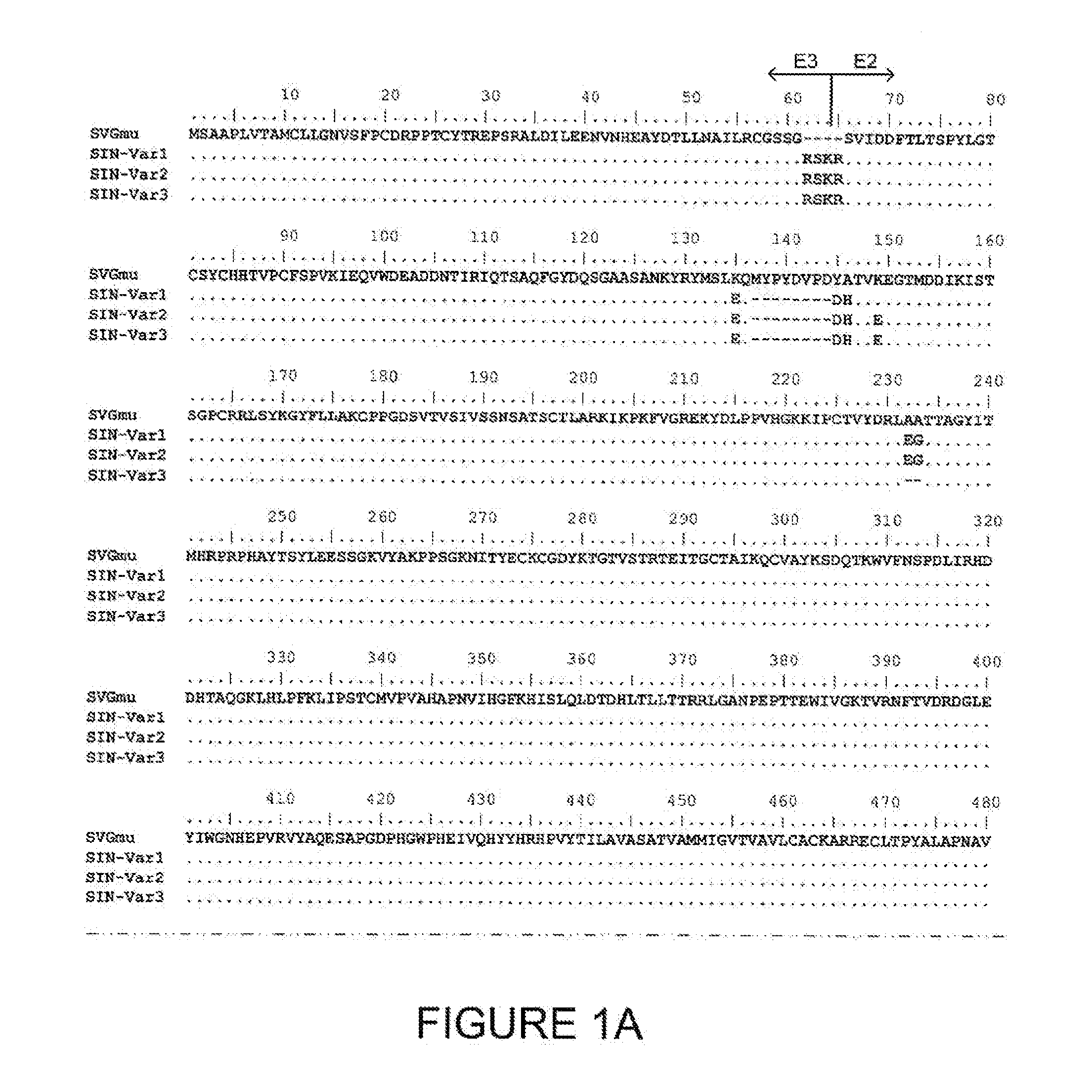
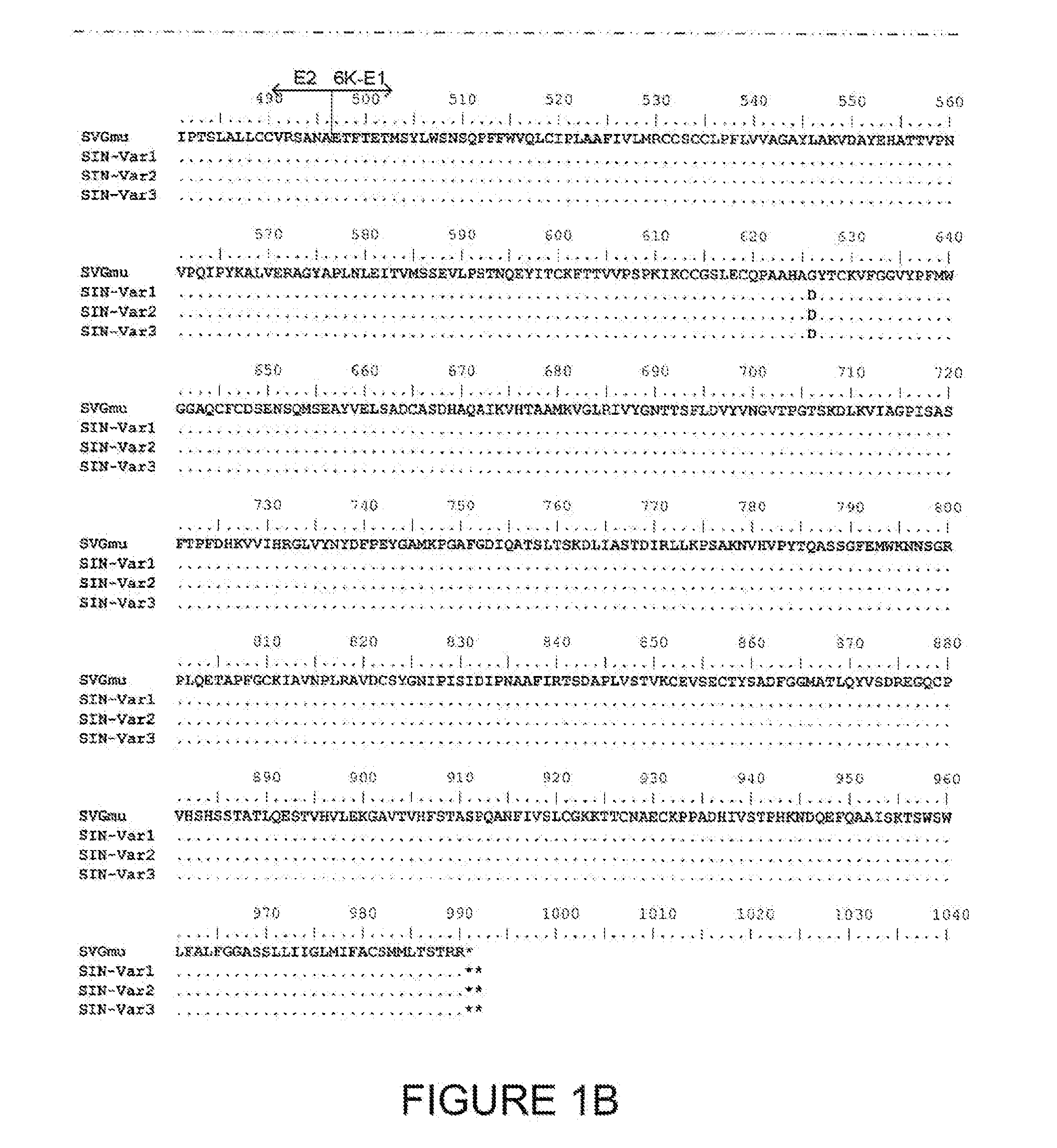
Sindbis virus (SV)—a member of the Alphavirus genus and the Togaviridae family—is able to infect DCs, probably through DC-SIGN (Klimstra, W. B., et al. 2003. J. Virol. 77: 12022-12032, which is incorporated herein by reference in its entirety). The canonical viral receptor for the laboratory strain of SV however, is cell-surface heparan sulfate (HS), which is found on many cell types (Strauss, J. H., et al. 1994. Arch. Virol. 9: 473-484; Byrnes, A. P., and D. E. Griffin. 1998. J. Virol. 72: 7349-7356, each of which is incorporated herein by reference in its entirety). To try to reduce heparan sulfate binding, a mutant E2 envelope (called SVGmu) was constructed by Wang et al. (US 2008 / 0019998, incorporated in its entirety). Part of their strategy involved deleting four amino acids at the E3 / E2 pro-protein junction and a deletion of two amino acids with a subsequent addition of a 10 amino acid sequence from hemagglutinin (see FIGS. 1A and...

example 2

Preparation of a Viral Vector Particle Comprising

A Sindbis Virus E2 Envelope Glycoprotein Variant

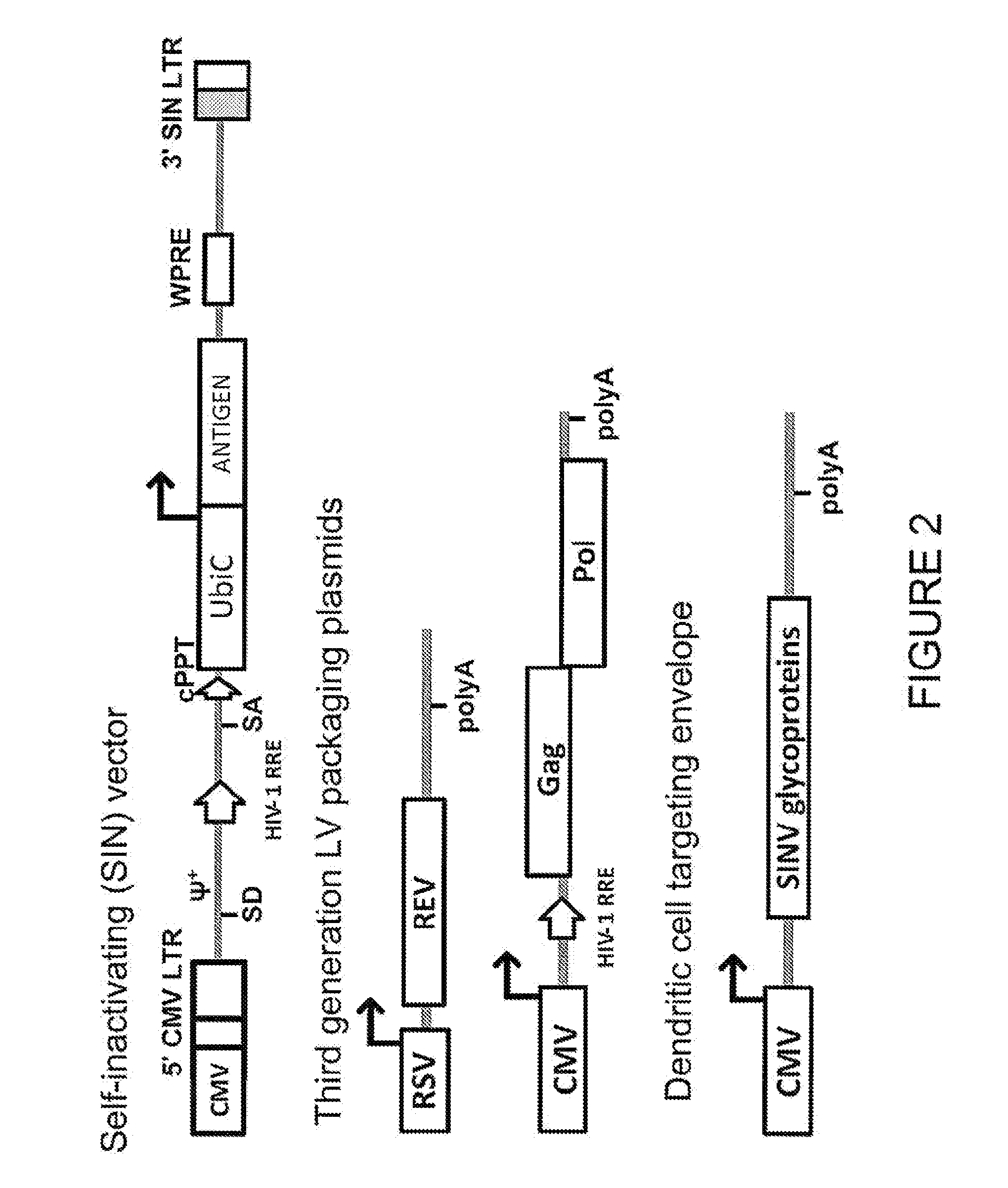
A Sindbis envelope-pseudotyped virus is prepared by standard calcium phosphate-mediated transient transfection of 293T cells with a lentiviral vector, such as FUGW or its derivatives, packaging plasmids encoding gag, pol and rev, and a variant Sindbis virus envelope sequence. FUGW is a self-inactivating lentiviral vector carrying the human ubiquitin-C promoter to drive the expression of a GFP reporter gene (Lois, C., et al. 2002. Science 295: 868-872, which is incorporated herein by reference in its entirety). The lentiviral transfer vectors (FUGW and its derivatives) are third generation HIV-based lentiviral vectors (see generally, Cockrell and Kafri Mol. Biotechnol. 36: 184, 2007), in which most of the U3 region of the 3′ LTR is deleted, resulting in a self-inactivating 3′-LTR.

Production of recombinant lentivirus vectors was accomplished by calcium phosphate (CaPO4)-mediated transient ...

example 3

Production of Lentiviral Vector Particles Comprising Sindbis Virus Envelope Glycoproteins

In this example, titers are determined for lentivirus vectors pseudotyped with different Sindbis virus envelopes. The E2 glycoproteins used were those contained in the sequences SIN-Var1 (SEQ ID No. 3), SIN-Var2 (SEQ ID No. 4), SIN-Var3 (SEQ ID No. 5), SVGmu (SEQ ID No. 2), HR (SEQ ID No. 18).

Sindbis virus glycoprotein pseudotyped lentiviral vector particles were generated by transfection of 293T cells as described in Example 2. Crude supernatants were harvested 48 hours post-transfection and used to transduce 293T cells expressing human DC-SIGN (293-DCSIGN) that had been plated in 6-well dishes the previous day at 2E5 cells / well. Titer was determined following a 72 hr incubation at 37° C. by analyzing transduced cells on a Guava Easy-Cyte cytometer (Millipore). 25,000 total events were counted to determine the percentage of GFP+ transduced cells, which was in turn used to calculate the GFP tite...


PUM

PropertyMeasurementUnit
Electric chargeaaaaaaaaaa
Acidityaaaaaaaaaa

Abstract

Lentiviral vector particles comprising a Sindbis virus E2 glycoprotein variant and a lentiviral vector genome comprising a sequence of interest are provided. A lentiviral vector particle comprising: (a) an envelope comprising a Sindbis virus E2 glycoprotein variant; and (b) a lentiviral vector genome comprising a sequence of interest; wherein the E2 glycoprotein variant facilitates infection of dendritic cells by the lentiviral vector particle, and wherein the E2 glycoprotein variant has reduced binding to heparan sulfate compared to a reference sequence (HR strain).

Description

REFERENCE TO SEQUENCE LISTINGThe sequence listing of this patent application is provided separately in a file named “IDC203_SEQ_LISTING_ST25.txt”. The content of this file, which was created on 22 Jul. 2010 and consists of 150,110 bytes, is incorporated in its entirety.TECHNICAL FIELDThis patent application relates generally to targeted gene delivery, and more particularly to the use of a pseudotyped lentivirus comprising an envelope that targets dendritic cells and can thus be used for dendritic cell vaccination.BACKGROUNDDendritic cells (DCs) are essential antigen presenting cells for the initiation and control of immune responses. DCs can capture and process antigens, migrate from the periphery to a lymphoid organ, and present the antigens to resting T cells in a major histocompatibility complex (MHC)-restricted fashion. These cells are derived from bone marrow (BM) and display dendritic morphology and high mobility. The discovery of DCs as specialized antigen-presenting cells (A...

Claims


Application Information

Patent Timeline
IPC IPC(8): A61K39/12C12N15/63C07H21/04C12N5/00A61P31/12A61K39/00
CPCA61K2039/5256C12N15/86C12N2740/15043C12N2740/15022A61K39/0011C12N7/00C12N2770/36145Y02A50/30A61K39/001195A61K39/001186A61K39/001156A61K39/001188A61K39/001191A61K39/001194A61K39/001192A61K39/001154A61K39/001184A61K39/4622A61K2239/50A61K39/4615A61K39/464499A61P31/12A61P31/14A61P31/16A61P31/18A61P31/20A61P31/22A61P35/00A61P35/02A61P37/04
Inventor ALLEN, JAMES M.DUBENSKY, JR., THOMAS W.LI, JIN ZHONGSLOAN, DEREK D.VAN HOEVEN, NEAL S.
Owner IMMUNE DESIGN CORP
Learn More