
Acoustic assisted phase conjugate optical tomography

a phase conjugate and optical tomography technology, applied in the direction of optical radiation measurement, spectral modifiers, radiation measurement, etc., can solve the problems of limited real-time information obtainable in vertebrate animal models by current methods, limited real-time amount, and general limited ability to perform biochemical imaging. to achieve the effect of optimizing the efficiency of modulation or frequency shifting

Active Publication Date: 2011-05-12
LONDON SCHOOL OF HYGIENE & TROPICAL MEDICINE +1
View PDF6 Cites 41 Cited by
  • Summary
  • Abstract
  • Description
  • Claims
  • Application Information

AI Technical Summary

Benefits of technology

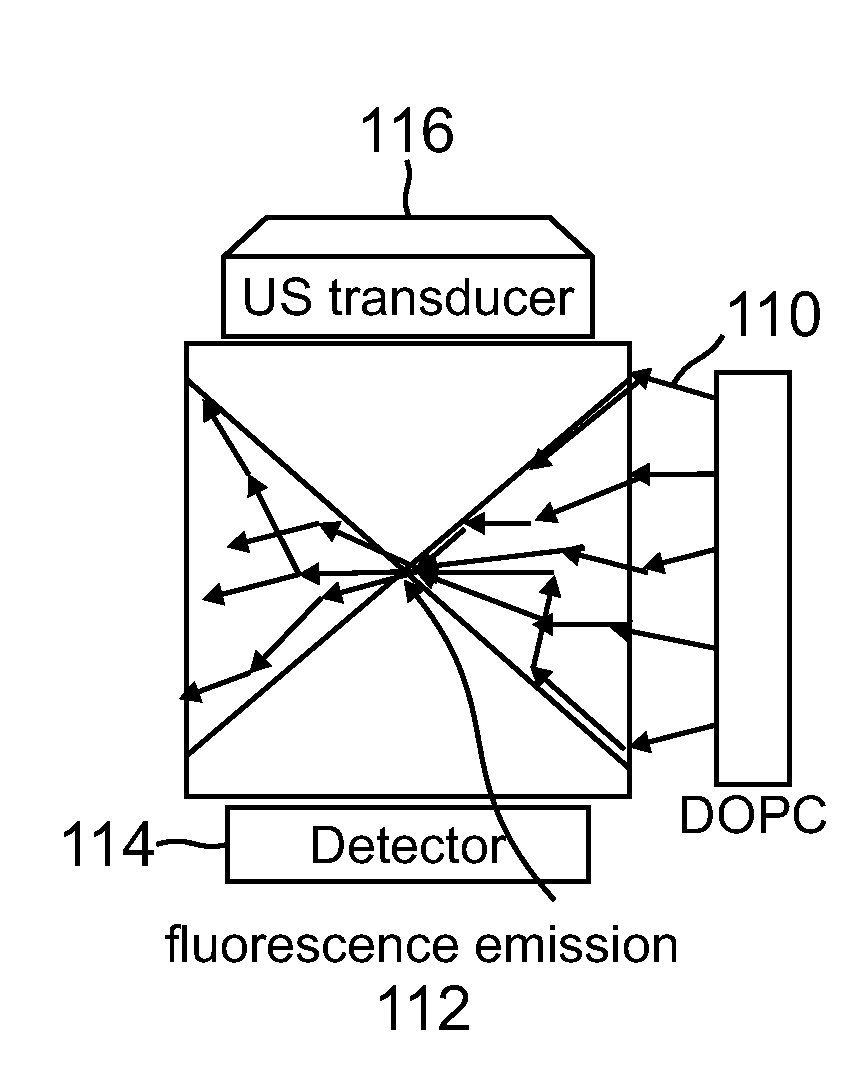
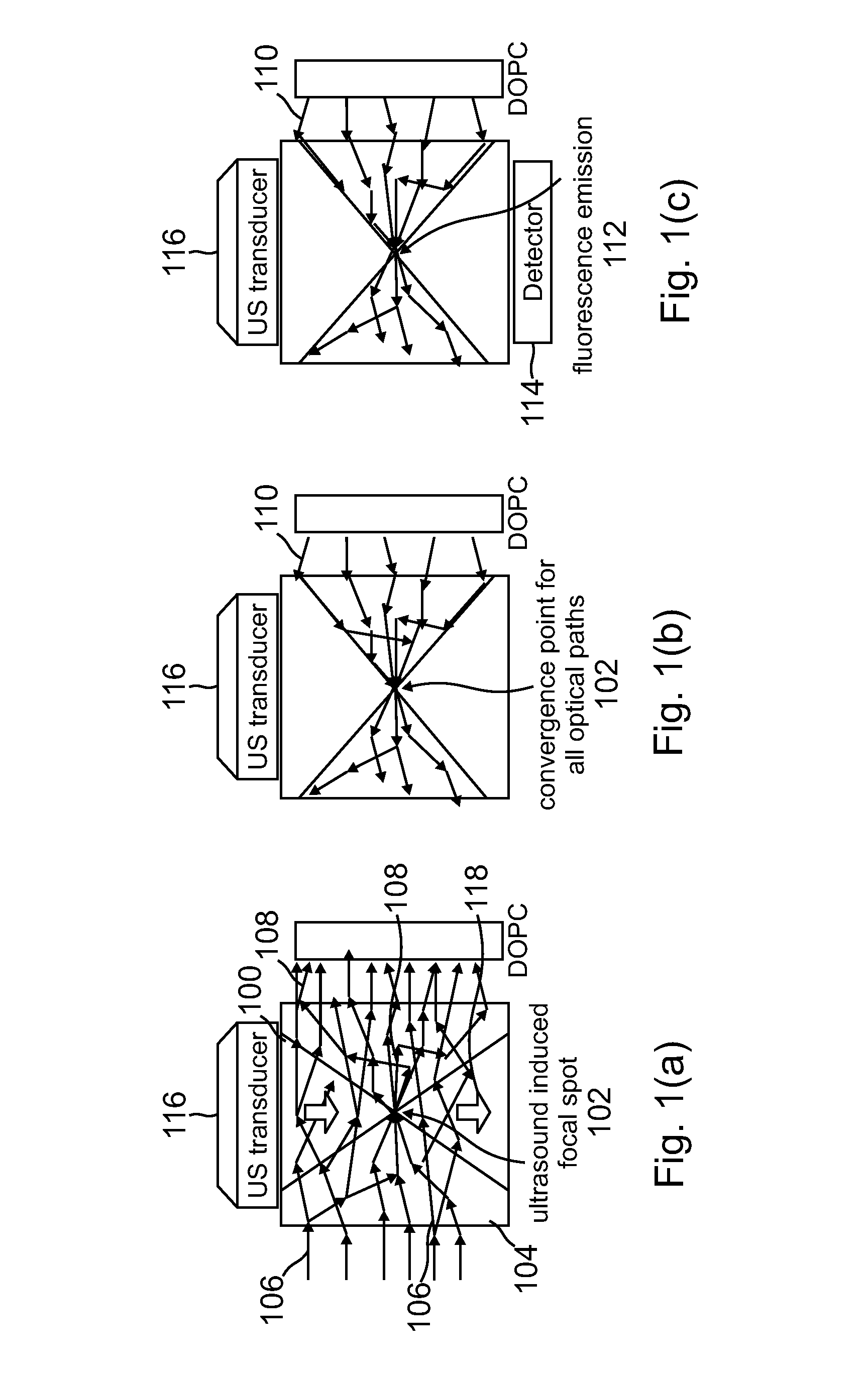
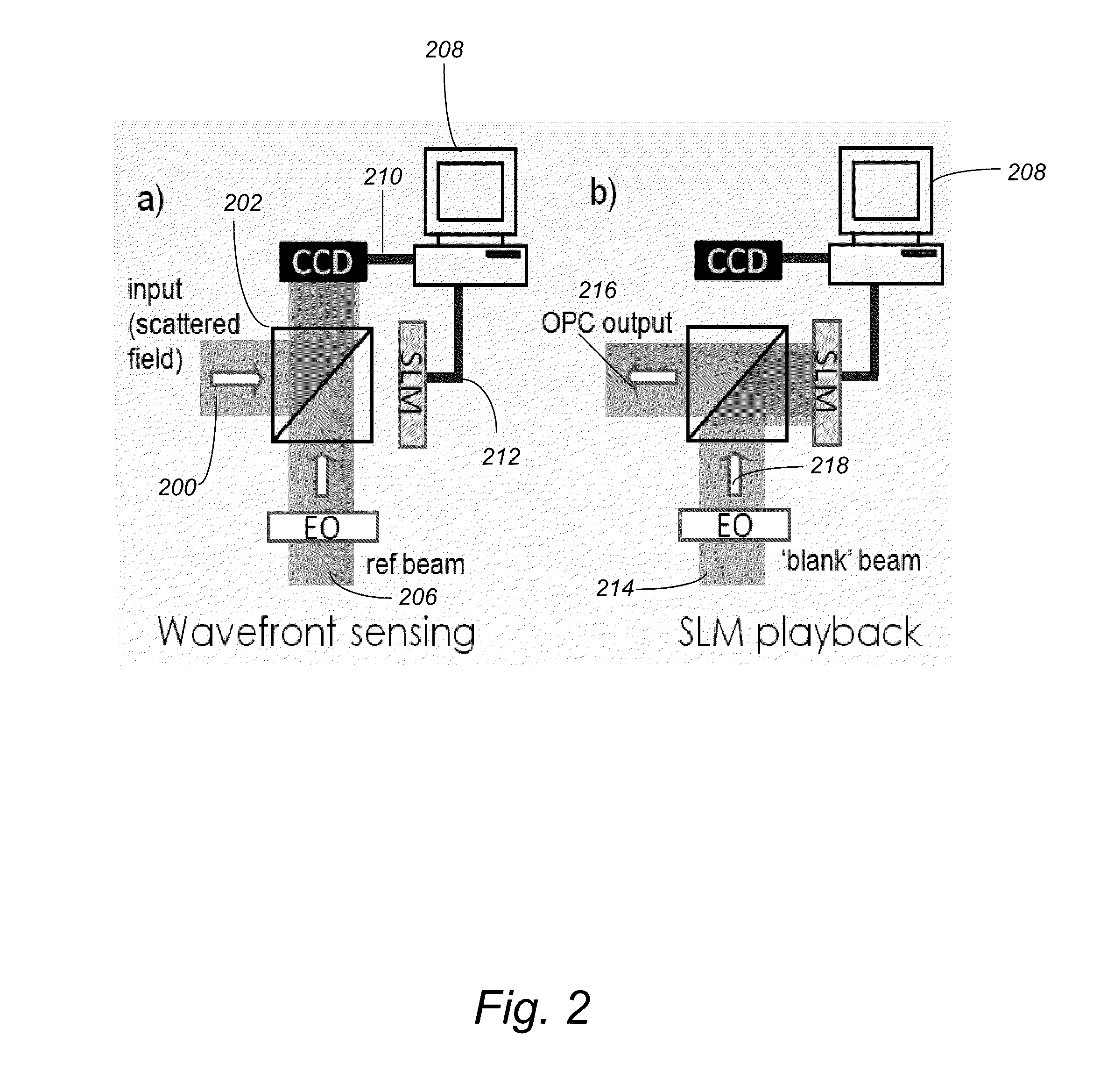
"The present invention provides an imaging system for imaging a sample using acoustic waves and optical signals. The system includes a microscope or optical microscope with a digital optical phase conjugation (DOPC) device and one or more detectors. The DOPC device modulates input light into modulated light, which is then detected by the detectors. The detected light is then used to create an image of the sample. The system can be used in optical coherence tomography to measure aberrations or distortions in the sample. The system can also use acoustic waves to perform photodynamic therapy by inducing biochemical reactions only at a single focal point in the sample. The system can be controlled to optimize imaging efficiency and can detect one or more resulting outputs that are based on an interaction between the output light and the sample."

Problems solved by technology

While these methods are excellent at rendering contrast based on the structural characteristics of the tissues, their general limited ability to perform biochemical imaging poses a significant limitation to their realizable diagnostic potentials.
It is often difficult to judge if a mass is simply a benign calcified accumulation or a developing tumor [1].
Similarly, the amount of real-time, in vivo information obtainable in vertebrate animal models by current methods is also limited.
Despite the biochemical sensing advantage, the conventional optical methods are unable to accomplish optical imaging with a resolution better than 100 microns in tissues that are thicker than a couple of millimeters.
Optical-based deep tissue imaging is largely impeded by the fact that biological tissues scatter light very strongly.
1. Optical Coherence Tomography (OCT) [6]. OCT has excellent resolution (˜ microns) but relatively limited imaging depth (typically 1 mm). Additionally, OCT renders mostly structural information-based or flow-based images and is not well suited to collect fluorescence or Raman information.
2. Diffuse optical tomography (DOT) [7]. This approach comprises a wide range of techniques and innovations. Broadly speaking, DOT sends light through the target tissue and carefully measures the resulting transmission from a number of exit points. DOT then renders a relatively low resolution ‘best-guess’ image of the tissue. DOT can work with thick tissues, but its resolution is fairly low (>1 mm). The biochemical-associated information collected is largely absorption spectrum based.
3. Ultrasound-modulated optical tomography (UOT) [8]. In this method, an ultrasound beam is brought to a focus within the target tissue which is illuminated by light. The transmitted light field will carry a modulated component, which correlates to the light field component that has passed through that ultrasound focal point. By scanning the ultrasound focus through the tissue and measuring the modulation strength in the transmitted light field, an image of the sample can then be rendered. The imaging depth for such a strategy is high (˜ cm) and the resolution corresponds to the ultrasound focal spot size (˜10's to 100 microns). Unfortunately, the much sought modulation is associated with a high background signal that significantly degrades sensitivity.
4. Photoacoustic tomography (PAT) [3]. In PAT, the target tissue is illuminated with a pulsed laser source. The laser pulse is absorbed by absorbers in the tissue and induces rapid thermal-expansion at the absorber site. The generated acoustic waves are then detected and measured by an array of ultrasound transducer at the tissue's boundaries. This method has a large imaging depth (˜1 cm) and the resolution achieved can also be high (˜100 microns). The biochemical information gathered is largely absorption-based.

Method used


Image

  • Acoustic assisted phase conjugate optical tomography
  • Acoustic assisted phase conjugate optical tomography
  • Acoustic assisted phase conjugate optical tomography

Examples


Embodiment Construction

[0067]In the following description of the preferred embodiment, reference is made to the accompanying drawings which form a part hereof, and in which is shown by way of illustration a specific embodiment in which the invention may be practiced. It is to be understood that other embodiments may be utilized and structural changes may be made without departing from the scope of the present invention.

[0068]Overview

[0069]Despite the rapid progress in biomedical optics in the past few decades, high tissue turbidity in the optical domain remains a difficult challenge that impedes high-resolution deep optical tissue imaging. Due to elastic scattering, conventional fluorescence imaging methods are severely limited in imaging depth (typically hundreds of microns at most). Fundamentally, the problem lies in the fact that it is not possible to focus light tightly in deep tissues using conventional optics.

[0070]Focusing light in a scattering medium is not an impossible proposition. Simplisticall...


PUM

PropertyMeasurementUnit
mean absorption lengthaaaaaaaaaa

Abstract

A light microscope for imaging a sample containing one or more fluorescent agents, comprising a source for generating acoustic waves that are focused at a focus in the sample, wherein the acoustic waves frequency shift a frequency of light passing through the focus, thereby creating a frequency shifted light beam; at least one spatial light modulator (SLM) positioned to illuminate the sample with an output beam that is an optical phase conjugate of the frequency shifted light beam, wherein the output beam is a reflection of a first reference beam off one or more pixels of the SLM, and the pixels are for modulating the first reference beam to create the output beam; and a detector positioned to detect fluorescence generated by the output beam exciting the fluorescent agents at the focus in the sample, thereby imaging the sample.

Description

CROSS REFERENCE TO RELATED APPLICATIONS[0001]This application claims the benefit under 35 U.S.C. 119(e) of the following co-pending and commonly-assigned U.S. provisional patent applications, which are incorporated by reference herein:[0002]Provisional Application Ser. No. 61 / 355,328, filed on Jun. 16, 2010 by Meng Cui, Ying Min Wang and Changhuei Yang, entitled “ACOUSTIC ASSISTED PHASE CONJUGATE OPTICAL TOMOGRAPHY,” attorneys' docket number 176.59-US-P1 (CIT-5626-P);[0003]Provisional Application Ser. No. 61 / 259,975, filed on Nov. 10, 2009, by Changhuei Yang and Meng Cui, entitled “APPROACHES FOR BUILDING COMPACT FLUORESCENCE MICROSCOPES,” attorneys' docket number 176.58-US-P1 (CIT-5473-P1); and[0004]Provisional Application Ser. No. 61 / 260,316, filed on Nov. 11, 2009, by Changhuei Yang and Meng Cui, entitled “APPLICATIONS OF TURBIDITY SUPPRESSION BY OPTICAL PHASE CONJUGATION,” attorneys' docket number 176.58-US-P2 (CIT-5473-P2); and[0005]Provisional Patent Application Ser. No. 61 / 37...

Claims


Application Information

Patent Timeline
Patent Type & Authority Applications(United States)
IPC IPC(8): H01L27/00G01N21/64
CPCG02B21/0056Y10T29/49826G02B21/0092A61B5/0066G01N2021/1708G02B21/0004A61B5/0059G02B21/00G01N21/6456G02B21/0068G01N21/4795G01N21/1717G01N2021/1727
Inventor CUI, MENGWANG, YING MINDIMARZIO, CHARLESYANG, CHANGHUEI
Owner LONDON SCHOOL OF HYGIENE & TROPICAL MEDICINE
Learn More