Luminescence assay method

a technology of luminescence and assay method, which is applied in the field of bioassay method, can solve the problems of inability to easily construct or miniaturize instruments, inability to detect sensitively, and inability to universally employ, and achieve the effect of reducing fluorescen

Active Publication Date: 2012-01-12
OY ARCTIC PARTNERS
View PDF2 Cites 13 Cited by
  • Summary
  • Abstract
  • Description
  • Claims
  • Application Information

AI Technical Summary

Benefits of technology

[0040]ii) chelate discomplementation, i.e. said lanthanide ion carrier chelate carrying said lanthanide is separated from said antenna ligand, and accordingly in decreased fluorescence.

Problems solved by technology

Currently available label technologies suitable for homogeneous, non-separative monitoring of nucleic acid hybridization still suffer from interference of sample matrices, the technologies cannot be universally employed, e.g. are not suitable for 5′ nuclease assays [U.S. Pat. No. 5,210,015], or they simply do not enable detection sensitive enough to be performed using a rapid read-out required, or the instrumentation required for detection is too complex or expensive to be feasibly constructed or miniaturized.
Although FRET is a widely employed and an essential technique in many applications, it has severe performance limitations [Hemmilä, I (1985) Clin Chem 31: 359-370.] and, in practice, the RET probes fail to comply with the strict requirements of true proximity probes.
Thus, conventional fluorophores and RET probes do not provide the specificity in signal generation required for the proximity probe binding-principle.
Further, it is difficult to measure more than two parameters simultaneously in a multiparametric FRET-based assay due to wide spectral coverage of an individual donor-acceptor pair.
Detection sensitivity of conventional fluorescence based techniques is limited by autofluorescence, scattered excitation light and absorbance of biological sample matrices, and in acceptor-based resonance energy transfer based assays also by crosstalk of the emission of the donor at acceptor-specific emission wavelength and direct excitation of the acceptor at the donor-specific excitation wavelength.
Many compounds and proteins present in biological fluids or serum are naturally fluorescent, and the use of conventional fluorophores leads to serious limitations of sensitivity [Soini E and Hemmilä I (1979) Clin Chem 25: 353-361; Wu P and Brand L (1994) Anal Biochem 218:1-13].
Another major problem when using homogeneous fluorescence techniques based on intensity measurements is the inner filter effect and the variability of the optical properties of a sample.
Sample dilution has been used to correct this drawback, but always at the expense of analytical sensitivity.
The development of a stable lanthanide chelate structure containing an efficient light-harvesting antenna originally turned out difficult.
The stability of the lanthanide(III) ion carrier chelates used for dissociation enhancement, however, should be only moderate and the dissociation kinetics quite rapid especially at low pH, as otherwise the ion is not released fast enough for fluorescence enhancement.
Both of these methods provide significant advantages compared to the conventional methods, but the specificity in signal generation is still limited by the radiative energy transfer (absorption of donor emission), especially when the labeled probes are present in high concentration (e.g. to achieve a large dynamic range, or to facilitate binding in case of weak interactions) [H. Bazin, M. et al.
The light collection efficiency of individual ions and chelated ligands without light harvesting structure is poor and requires relatively high excitation light intensity.
Although, fluorescence resonance energy-transfer (FRET) is an extremely versatile technology, especially the energy-transfer probe-based assay is limited by energy-transfer efficiency (relatively low signal) and background through reabsorption of the donor emission (limited dynamic range).

Method used


Image

  • Luminescence assay method
  • Luminescence assay method
  • Luminescence assay method

Examples


example 1

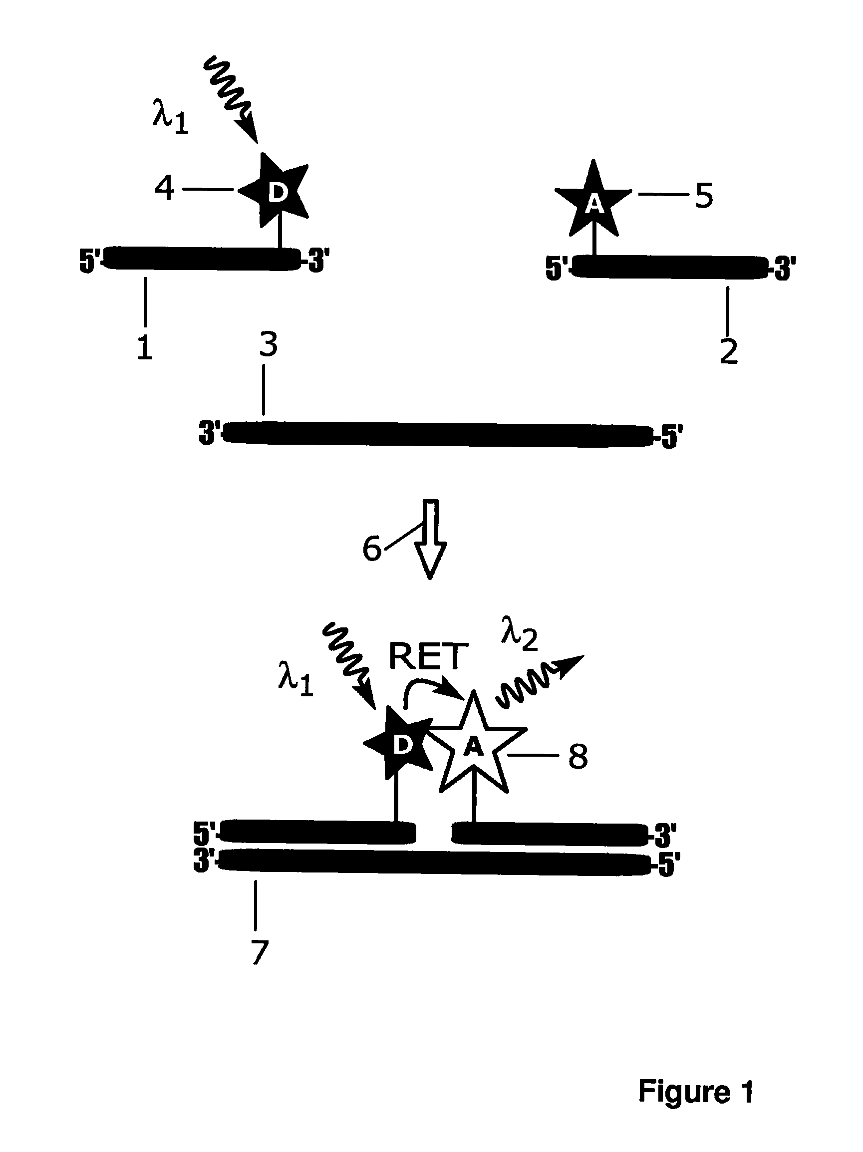
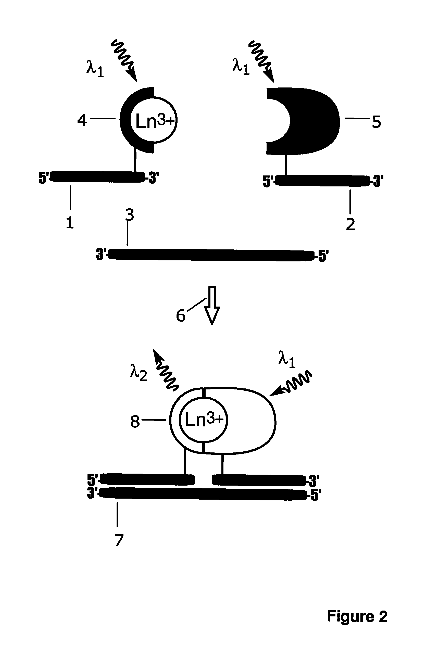
Homogeneous Proximity Probe-based Hybridization Assay

[0160]Synthetic target DNA oligonucleotide (5′-GATGCAGTAGCAGGAAGAGGATCG-TAGCAATG-3′; SEQ ID NO: 1), amino-modified probe A oligonucleotide (5′-CAT-TGCTACGATCC(C6dT)C-3′; SEQ ID NO: 2) and amino-modified probe B oligonucleotide (5′-T(C2dT)CCTGCTACTGCATC-3′; SEQ ID NO: 3) were purchased from Sigma-Aldrich (St. Louis, Mo.). Probe A was labelled with Eu3+ ion carrier chelate, (N1-(4-isothiocyanatobenzyl)diethylenetriamine-N1,N2,N3,N3-tetrakis(acetato)europium(III) [Mukkala, V.-M. et al.(1989) Anal. Biochem., 176: 319], Eu3+-N1) at the primary amino group modification located near the 3′-end and probe B was labelled with light harvesting antenna ligand (4-((isothio-cyanatophenyl)ethynyl)pyridine-2,6-dicarboxylic acid, 3d-antenna) near the 5′-end. Probe A, 25 nmol, was incubated with 20-fold molar excess of Eu3+-N1 in 50 mM carbonate buffer, pH 9.8, at +37° C. over night. The total volume of the labelling reactions was 50 μL. For labell...

example 2

Specificity in Signal Generation

[0166]Non-complementary target oligonucleotide (5′- CTGCTCTATCCACGGCG-CCCGCGGCTCCTCTC-3′; SEQ ID NO: 4) was purchased from Biomers.net (Ulm, Germany). The experiment described in Example 1 was repeated by replacing the target oligonucleotide with the non-complementary target oligonucleotide. Replacement of the complementary target oligonucleotide with variable concentrations of a noncomplementary 32-mer oligonucleotide resulted in the same fluorescence signal as in the absence of complementary target oligonucleotide; no detectable signal differences were observed in presence of variable concentrations non-complementary target oligonucleotides compared to zero concentration of target oligonucleotides; i.e. blank control. This indicates that the signal generation mechanism in the present invention is highly specific and dependent on the two simultaneous biomolecular recognition events.

example 3

Emission Spectrum and Fluorescence Lifetime

[0167]Additional probe, amino-modified probe C oligonucleotide (5′- CATTGCTACGAT-CC(C2dT)C-3′; SEQ ID NO: 5) was purchased from Sigma-Aldrich (St. Louis, Mich.) and labelled with intrinsically fluorescent 2,2′,2″,2′″[[4-[(4-isothiocyanatophenyl)-ethynyl] pyridine-2,6-diyl]bis(methylenenitrilo)]tetrakis(acetato)europium(III) (Eu3+-7d; schematic structure in FIG. 8c). Probe C, 5 nmol, was incubated with 20-fold molar excess of Eu3+-7d in 50 mM carbonate buffer, pH 9.8, at +37° C. over night and purified as described for A-Eu3+-N1 in Example 1.

[0168]Fluorescence spectrum and emission lifetime of an intrinsically fluorescent Eu3+-chelate labelled probe C-Eu3+-7d, and separately the target oligonucleotide directed complex of the probe A-Eu3+-N1 and probe B-3d-antenna were measured with a Varian Cary Eclipse fluorescence spectrophotometer (Varian Scientific Instruments, Mulgrave, Australia). The target oligonucleotide (0 or 10 nM) was mixed with ...


PUM

PropertyMeasurementUnit
visible wavelengthsaaaaaaaaaa
temperaturesaaaaaaaaaa

Abstract

A bioassay employing a first group including a lanthanide ion carrier chelate and a first recognition element, a second group including an antenna ligand and a second recognition element; where the lanthanide ion carrier chelate binds strongly to lanthanide, or the lanthanide ion carrier chelate binds moderately to lanthanide, and an agent complexing the lanthanide ion is additionally employed at a concentration of at least 1 pmol/l. The antenna ligand binds weakly to the lanthanide ion. Analyte recognition by the first recognition element and by the second recognition element results in either chelate complementation and increased fluorescence, or chelate discomplementation and decreased fluorescence.

Description

FIELD OF THE INVENTION[0001]This invention relates to a bioassay method for detecting and / or quantitating an analyte.BACKGROUND OF THE INVENTION[0002]The publications and other materials used herein to illuminate the background of the invention, and in particular, cases to provide additional details respecting the practice, are incorporated by reference.[0003]A number of assays based on bioaffinity binding reactions or enzymatically catalyzed reactions have been developed to analyze biologically important compounds from or their activity or their biological effect or its modulation induced by various biological samples, samples in environmental studies, industrial processes and compound libraries. Some of these assays rely on specific bioaffinity recognition reactions, where e.g. natural biological binding components, artificially produced binding compounds or moulded plastic imprints (molecular imprinting) are used as recognition elements to form the specific binding assay. Other a...

Claims


Application Information

Patent Timeline
Patent Type & Authority Applications(United States)
IPC IPC(8): G01N21/64C12Q1/02C12Q1/70C12Q1/68
CPCC12Q1/6818G01N33/542G01N33/582C12Q2563/137
Inventor SOUKKA, TEROLAMMINMAKI, URPO
Owner OY ARCTIC PARTNERS
Why Eureka
Try Eureka