Liposome Compositions and Methods of Use Thereof

a technology of liposomes and compositions, applied in the field of compositions and methods for delivery of molecules to cells, can solve the problems of difficult to rely on any single tumor biomarker, difficult to identify and treat cancer cells while sparing non-cancerous cells, and difficult to reliably identify and treat cancer cells. , to achieve the effect of reducing phlip binding/insertion and phlip labeling or targeting of cells

Active Publication Date: 2012-02-16
UNIV OF RHODE ISLAND BOARD OF TRUSTEES +2
View PDF1 Cites 24 Cited by
  • Summary
  • Abstract
  • Description
  • Claims
  • Application Information

AI Technical Summary

Benefits of technology

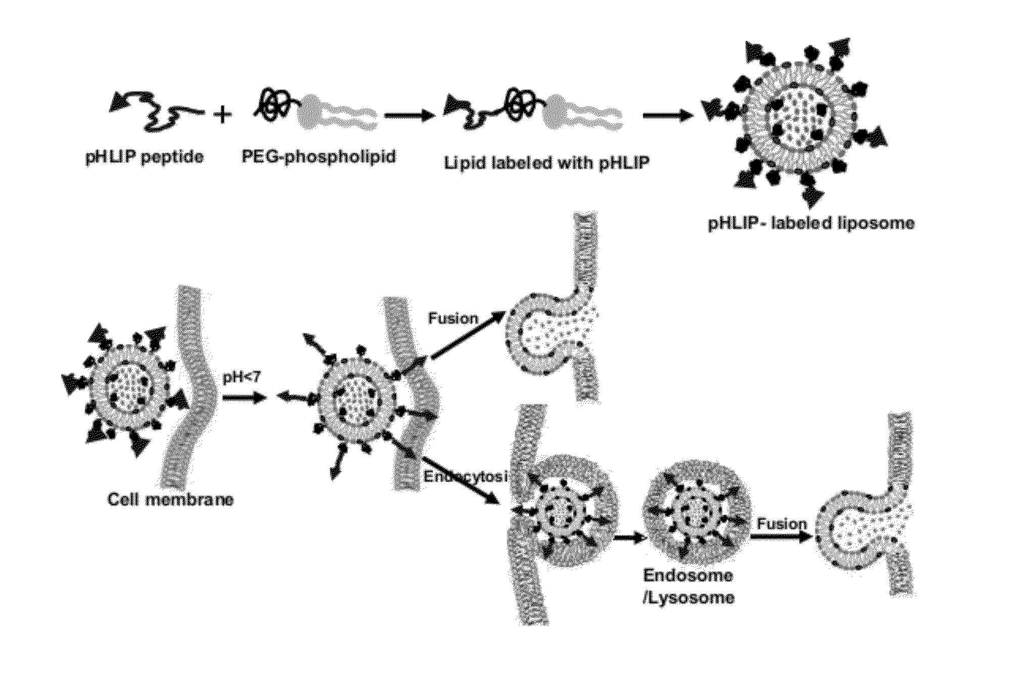
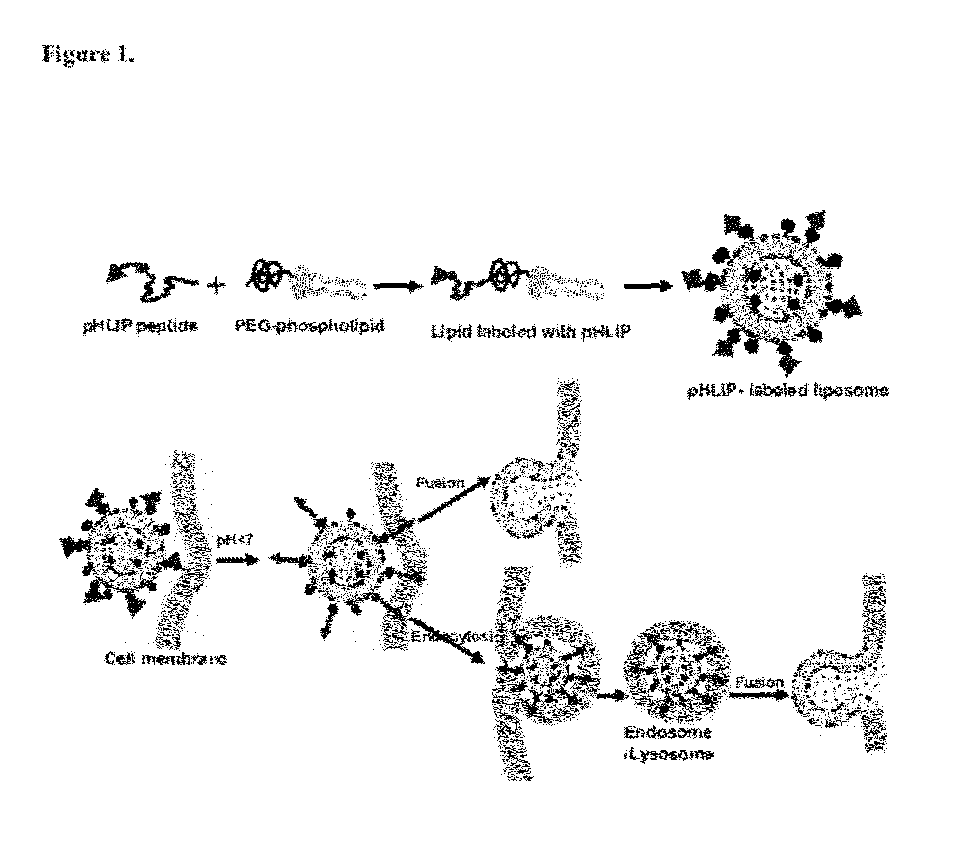
[0012]A method of delivering a cargo into a target cell is carried out by contacting the target cell with a cargo-loaded pHLIP-decorated (pHLIP+) liposome. Preferably, hydrophobic region of the lipid bilayer of the pHLIP+ liposome is substantially free of pHLIP polypeptide. The use of pHLIP-containing liposomes leads to at least 1%, 5%, 10%, 25%, 50%, or 2-fold, 5-fold, 10-fold or more of cargo delivered to the cytoplasm of the target cell compared to the amount delivered using pHLIP” liposome (i.e., a liposome that does not contain a pHLIP). pHLIP+ liposomes deliver their cargo to cells by fusion with the cell membrane, by endocytosis, or both. The target cell may be characterized by a microenvironment comprising a low pH.
[0013]In some embodiments, the pHLIP+ liposome fuses with a cell membrane of the target cell. In some cases, the pHLIP+ liposome both fuses with a cell membrane of the target cell and is taken up by the cell by endocytosis. For example, the pHLIP+ liposome preferentially fuses with a membrane of an endosomal and/or a lysosomal compartment of the target cell after uptake by endocytosis.
[0014]For example, the target cell is a tumor cell or other cell characterized by a local microenvironment of low pH, e.g., cells of a diseased tissue with a naturally acidic extracellular environment or cells of a tissue with an artificially induced acidic extracellular environment relative to normal physiological pH. Many diseased tissues are characterized by an acidic microenvironment. However, acidity in tumors or non-tumor target tissues is optionally induced by co-injection of glucose or a diluted solution of acid at the tissue site at which therapy using the compositions is desired. For example, an acidifying composition (e.g., glucose or dilute acid) is administered, e.g., injected subcutaneously, before delivery of the pH sensitive compositions (30 s, 1 min., 5 min., 10 min., 30 min., 1 hr., 2 hrs, 6 hrs, 12 hrs, 24 hrs, 48 hrs, or more prior to administration of the environmentally sensitive composition to the target tissue site). Alternatively, the tissue acidifying agent and the pHL

Problems solved by technology

Despite many advances in the field of cancer diagnosis and treatment, a reliable method of identifying and treating cancer cells while sparing non-cancerous cells has been elusive.
One of the limitations is the heterogeneit

Method used


Image

  • Liposome Compositions and Methods of Use Thereof
  • Liposome Compositions and Methods of Use Thereof
  • Liposome Compositions and Methods of Use Thereof

Examples


example 1

pHLIP-Mediated Delivery of Liposomes

[0085]The following regents and methods were used to generate the data described herein.

Lipids

DOPE

(1,2-dioleoyl-sn-glycero-3-phosphoethanolamine)

[0086]

DOPC

(1,2-dioleoyl-sn-glycero-3-phosphocholine)

[0087]

DSPE-PEG(2000) Maleimide

1,2-distearoyl-sn-glycero-3-phosphoethanolamine-N-[maleimide(polyethylene glycol)-2000] (ammonium salt)

[0088]

DSPE-PEG(2000)

1,2-distearoyl-sn-glycero-3-phosphoethanolamine-N-[methoxy(polyethylene glycol)-2000]

[0089]

Fluorescein DHPE

N-(fluorescein-5-thiocarbamoyl)-1,2-dihexadecanoyl-sn-glycero-3-phosphoethanolamine, triethylammonium salt

[0090]

Rhod PE

1,2-dioleoyl-sn-glycero-3-phosphoethanolamine-N-(lissamine rhodamine B sulfonyl

[0091]

R18 (Octadecyl Rhodamine B Chloride)

[0092]

Conjugation of pHLIP with Lipids

[0093]pHLIP containing Cys on the N-terminus and DSPE-PEG(2000) Maleimide lipids. Standard maleimide chemistry reaction was applied in methanol. (Reaction of thiol with maleimide.)

where

R1—DSPE-PEG(2000) Maleimide (MW 2941)

[009...

example 2

pHLIP Enhances Uptake of Liposomes by Cells

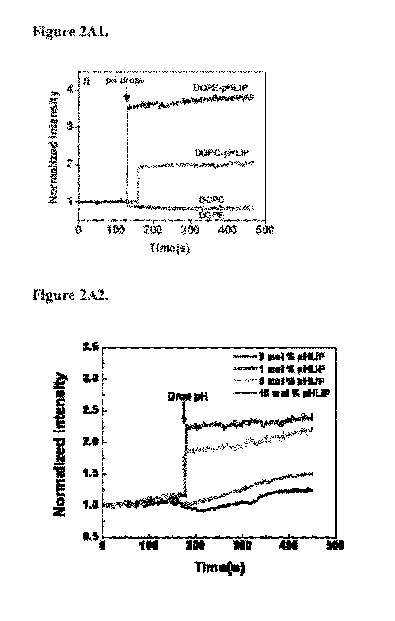
[0106]pHLIP (pH-Low-Insertion-Peptide) insertion into membrane occurs as a result of protonation of Asp / Glu residues due to a decrease of pH. Protonation enhances peptide hydrophobicity and increases its affinity for a lipid bilayer, which triggers peptide insertion and formation of transmembrane helix. Since many pathological states are associated with the development of elevated level of extracellular acidity (or low extracellular pH), pHLIP-liposomes are ideally suited for selective delivery of diagnostic and therapeutic agents to the cancer cells. Attachment of cargo molecules to the N-terminus (FIG. 14).

[0107]One approach to deliver gold material (or cytotoxic compounds) to tumor is to use liposomes. In contrast to fusogenic liposomes developed before for delivery, which can fuse with cellular membrane only in the absence of PEG coating, pHLIP mediates fusion between lipid bilayer of plasma membrane or membrane of endosome / lysozome and...

example 3

pHLIP Promotes Distortion of Plasma and Endosome Membranes, the Release of R18-Labeled FA into the Cytoplasm, and Targeting of Mitochondria

[0110]After cellometer counting, cells were reseeded in collagen-coated cell dishes for microscopy imaging. Cellular localization of fluorescent fatty acids (R18) incorporated into liposomes containing PEG polymers and pHLIP or no pHLIP on the surface (non-fusogenic DOPC lipids were used in study). In case of liposomes, fluorescent signal was mostly localized in endosomes, while pHLIP promotes distortion of plasma and endosome membranes and release of R18-labeled FA into cytoplasm and targeting of mitochondria. FIG. 9 shows the localization of Rho-labeled liposome in cells.

[0111]FIG. 11 shows images of Fluorescein-labeled liposomes fused with a cellular membrane. FIG. 11(a) phase contrast; (b) FITC. Co-localization of FITC-liposome (c) and plasma membrane staining of red-fluorescent Alexa Fluor594 wheat germ agglutinin (d). The data demonstrate t...


PUM

PropertyMeasurementUnit
Fractionaaaaaaaaaa
Compositionaaaaaaaaaa
Hydrophobicityaaaaaaaaaa

Abstract

The present application relates to compositions comprising and methods of using a liposome comprising a pHLIP polypeptide, wherein a lipid bilayer of the liposome is substantially free of the pHLIP polypeptide.

Description

RELATED APPLICATIONS[0001]This application claims the benefit of priority under 35 U.S.C. §119(e) to U.S. Provisional Application NO: 61 / 373,660, filed Aug. 13, 2010, which is incorporated herein by reference in its entirety.STATEMENT AS TO FEDERALLY SPONSORED RESEARCH[0002]This invention made with U.S. Government support under Grant Number BCRP CDMRP BC061356 and W81XWH-07-1-0498 from the Department of Defense, and RCA 125280A, RO1 133890, 5R21CA125280-02, and GM073857 from the National Institutes of Health. The Government has certain rights in the invention.FIELD OF THE INVENTION[0003]The invention relates to compositions and methods for delivery of molecules to cells.BACKGROUND[0004]Despite many advances in the field of cancer diagnosis and treatment, a reliable method of identifying and treating cancer cells while sparing non-cancerous cells has been elusive. One of the limitations is the heterogeneity of human cancers. It has therefore been problematic to rely on any single tum...

Claims


Application Information

Patent Timeline
IPC IPC(8): A61K9/127A61K31/7105A61P35/00A61K31/337A61K31/20A61K31/711A61K31/704
CPCA61K9/127A61K31/20A61K31/337A61K31/704A61K31/7105A61K31/711A61P35/00A61K47/6911A61K31/713A61K9/1271
Inventor RESHETNYAK, YANA K.ANDREEV, OLEG A.ENGELMAN, DONALD M.
Owner UNIV OF RHODE ISLAND BOARD OF TRUSTEES
Why Eureka
Try Eureka