Novel polypeptides and use thereof

Inactive Publication Date: 2012-03-22
ALLIGATOR BIOSCI
2 Cites 0 Cited by

AI-Extracted Technical Summary

Problems solved by technology

S. aureus can potentially infect any tissue of the body and occasionally spreads from the primary site of ...

Benefits of technology

[0159]FIG. 3—Human anti-CHIPS IgG inhibits detection of CHIPS by capture ELISA. Recovery of 2.5 ng·mL−1 CHIPS spiked into various concentrations pooled human serum and measured by capture ELISA (a). Depletion of IgG from human serum by passage over Protein-G-Seph...

Abstract

The present invention provides a polypeptide having a biological activity of the Chemotaxis Inhibitory Protein of Staphylococcus aureus (‘CHIPS’), the polypeptide comprising or consisting of the amino acid sequence of SEQ ID NO: 2, or a fragment or variant thereof having a biological activity of CHIPS, wherein the fragment or variant retains amino acid substitutions K40E, D42V, N77H, K100R, K105R, N111 K and/or G112A relative to the wildtype CHIPS protein of SEQ ID NO:1. In one embodiment, polypeptide consists of the amino acid sequence of SEQ ID NO: 2. Related aspects of the invention provide pharmaceutical compositions comprising a polypeptide of the invention, together with methods or making and using the same.

Application Domain

Antibacterial agentsFungi +14

Technology Topic

Molecular biologyAmino acid +6

Image

  • Novel polypeptides and use thereof
  • Novel polypeptides and use thereof
  • Novel polypeptides and use thereof

Examples

  • Experimental program(3)

Example

EXAMPLE B
Directed Evolution of CHIPS to Generate Functional Variants with Reduced Interaction with Human Antibodies
Abstract
[0227]Chemotaxis Inhibitory Protein of Staphylococcus aureus (CHIPS) is a protein that binds and blocks the C5a receptor (C5aR) and formylated peptide receptor, thereby inhibiting the immune cell recruitment associated with inflammation. If CHIPS was less reactive with existing human antibodies, it would be useful as an anti-inflammatory drug. Therefore, we applied directed evolution and computational/rational design to the CHIPS gene in order to generate new CHIPS variants displaying lower interaction with human IgG, yet retaining biological function. The optimization was performed in four rounds; one round of random mutagenesis to add diversity into the CHIPS gene and three rounds of DNA recombination by Fragment INduced Diversity (FIND®). Every round was screened by phage selection and/or ELISA for decreased interaction with human IgG and retained C5aR binding. The mean binding of human anti-CHIPS IgG decreased with every round of evolution. For further optimization, new amino acid substitutions were introduced by rational design, based on the mutations identified during directed evolution. Finally, seven CHIPS variants with low interaction with human IgG and retained C5aR blocking capacity could be identified.
Introduction
[0228]Inflammation is the tissue response to injury or infection by pathogens. The attraction of immune cells and soluble molecules to the site of damage or infection initiates the healing process. Even though the ability to raise an inflammatory response is crucial for survival, the ability to control inflammation is also necessary for health. Anti-inflammatory drugs aim at blocking key events in inflammation for treatment of disorders with excessive or uncontrolled inflammation. Examples of such drugs are Remicade® and Kineret®, approved for treatment of rheumatoid arthritis.
[0229]Many bacteria have evolved strategies to evade the human immune system, for example by avoiding recognition, or by secreting proteins that neutralize the antibacterial effects mediated by the immune system. Chemotaxis Inhibitory Protein of Staphylococcus aureus (CHIPS) is a 14.1 kDa protein which is a potent inhibitor of immune cell recruitment and activation associated with inflammation, through binding and blocking the C5a receptor (C5aR) and the formylated peptide receptor (De Haas et al., 2004; Postma at al., 2004). This way, CHIPS is a promising anti-inflammatory protein for treatment of several inflammatory diseases, e.g. sepsis (Rittirsch et al., 2008) or ischemia-reperfusion injury and immune complex disease (Heller et al., 1999) However, most individuals have pre-formed titers of antibodies specific for CHIPS (Wright et al., 2007). These antibodies might neutralize the function of CHIPS or induce an immune reaction, hence the CHIPS molecule would benefit from optimization to function well as a drug in the human circulation.
[0230]Directed evolution is an established approach for improving proteins. It has been utilized to improve many protein functions such as stability, activity or affinity (Johannes et al., 2006). Importantly for the development of protein therapeutics, directed evolution has proven to be a useful tool for generating protein variants with enhanced therapeutic potential (Yuan et al., 2005). The directed evolution approach is particularly efficient as it does not require prior knowledge of the structure of the protein. Instead of using inefficient and time consuming methods based on site-directed mutagenesis, rounds of gene recombination and high-throughput screening can be performed to identify improved variants. The process can be repeated and beneficial mutations will be accumulated while mutations not required for the property of interest will be excluded, as reviewed by (Yuan et al., 2005) and (Zhao, 2007).
[0231]Several distinct methods for directed evolution have been described in the literature; among them DNA shuffling (Stemmer, 1994a; Stemmer, 1994b) and the Staggered Extension process (StEP) (Yuan et al., 2005; Zhao et al., 1998). Another DNA recombination technology called Fragment INduced Diversity (FIND®), has previously proven to be useful in the optimization of thermostability of carboxypeptidase U (Knecht et al., 2006) and the activity of IL-1 receptor antagonists (Dahlen et al., 2008).
[0232]Even though directed evolution has been successfully utilized to identify new and improved protein variants, a limitation with this type of technology is the incapability of screening the entire sequence space of a protein. However, sequence space can be explored more efficiently if directed evolution is combined with computational tools and rational design (Wong et al., 2007; Zhao, 2007).
[0233]In this example, FIND® was used in combination with rational/computational design of the CHIPS gene with the aim to create new protein variants with lower interaction with pre-existing specific human IgG. An improved CHIPS molecule would be characterized by decreased reactivity with pre-existing antibodies, but also preserved activity towards the C5aR. Therefore, receptor binding was monitored in parallel with the screening process for decreased IgG interaction. This way, we were able to isolate new CHIPS variants with significantly reduced interaction with human anti-CHIPS IgG yet preserved C5aR blocking activity.
Materials & Methods
Cloning, Expression and Purification of Recombinant Proteins
[0234]Wild-type (Wt) CHIPS1-121 was cloned, expressed and purified as described earlier (De Haas et al., 2004). CHIPS with truncated C-terminus (CHIPS AC) was 112 amino acids long with two additional non-relevant amino acids included in the C-terminal end of the expressed protein as a result of cloning (CHIPS1-112). Genes encoding CHIPS AC and its corresponding single mutants K61A, K69A and K100A as well as CHIPS ΔN/C(CHIPS31-113) were created from the gene encoding wt full-length CHIPS1-121 by truncation and site-directed mutagenesis. These CHIPS variants were then cloned and expressed as described above. Single mutants were used for structural analysis by Haas et al. (Haas et al., 2005), but were also screened for anti-CHIPS IgG binding and mutants K61A, K69A and K100A showed decreased binding (data not shown).
[0235]CHIPS variants selected from libraries in this study were expressed in the same way, but purified from inclusion bodies (Gustafsson et al., 2009) or expressed by the Expressway Cell-Free E. coli Expression System (Invitrogen, Carlsbad, Calif.) as recommended by the manufacturer.
Library Construction
Random Mutagenesis
[0236]To create diverse libraries of CHIPS variants in Round 1 (see FIG. 14), two different methods of random mutagenesis were used to create in total four libraries. Error-prone PCR was performed as described previously (Leung et al., 1989). One library with high mutation frequency (Library 1.1) and one with low mutation frequency (Library 1.2) were created. A 20 cycle PCR was performed using primers (Fw: 5′-TCGCGGCCCAGCCGGCCATGGCCTTTACTTTTGAACCG-3′ [SEQ ID NO: 3] and Rev: 5′-GCCTGCGGCCGCAGATCTACCATTAATTACATAAG-3′ [SEQ ID NO: 4]) in the presence of 7.5 mM MgCl2 and 0.64 mM MnCl2. 2.5 U AmpliTaq Thermostable DNA polymerase (Applied Biosystems, Foster City, Calif.) was added and the reaction was performed using the program 94° C., 5 min/(94° C., 30 s/55° C., 30 s/72° C., 40 s) 20 times and finally elongation at 72° C. for 10 minutes. GeneMorph II (Stratagene, La Jolla, Calif.) was used as recommended by the manufacturer. 10 μg DNA (a mixture of CHIPS ΔC and the corresponding K61A, K69A and K100A single mutants) was used for the design of the low mutation frequency library (library 1.4) and 1 ng DNA for the library with higher mutation frequency (library 1.3). The PCR reaction contained the primers described above and the PCR program was 95° C., 2 min/(95° C., 1 min/60° C., 1 min and 72° C., 1 min) 40 times and finally elongation at 72° C. for 10 minutes. To increase the mutation frequency in the 1 ng library, it was subjected to one more round of Genemorph II mutagenesis. This time, the amount of DNA in the PCR reaction was 10 ng. After purification, the PCR products were sub-cloned into the pGEM-T vector (Promega, Madison, Wis.) according to the manufacturer's recommendations and the sequences were analyzed and base exchanges evaluated.
FIND®
[0237]FIND® recombinations were performed in Rounds 2 to 4 to create diverse libraries of recombined clones, as described in e.g. patents EP 1 341 909 and EP 1 504 098. Briefly, single-stranded DNA was prepared by generating PCR products using one biotinylated and one regular primer. The PCR product was immobilized on a column containing streptavidin-conjugated magnetic beads (Miltenyi Biotec GmbH, Bergisch Gladbach, Germany) and placed in the magnetic field of a pMACS separator. The PCR product was denatured with 0.1 M NaOH and the eluted non-biotinylated DNA strand was collected and purified by agarose gel electrophoresis using Recochips (Takara Bio Inc., Shiga, Japan) according to the manufacturer's recommendations.
[0238]The FIND® experiments were initiated by fragmenting 200 ng sense and antisense ssDNA, respectively, with Exonuclease I (Exo I). (New England Biolabs, Ipswich, Mass.) (100 U/μg DNA) for 10 minutes, Exonuclease V (Exo V) (USB, Cleveland, Ohio) (25 U/μg DNA) for 45 minutes and Exonuclease VII (Exo VII) (USB) (10 U/μg DNA) for 30 minutes in separate tubes in buffers as recommended by the manufacturers. The ssDNA fragments resulting from the exonuclease digestions were recombined in a PCR-like reaction, without added primers, followed by amplification using a standard PCR protocol. After purification, PCR products were subcloned into the pGEM-T vector (Promega) according to the manufacturer's recommendations and sequences were analyzed.
Protein Expression in Plate Format
[0239]CHIPS libraries created by random mutagenesis or FIND® were cloned into a modified pRSET B vector (Invitrogen) in BbsI and BglII sites for expression in E. coli. Libraries were transformed into E. coli BL21 star DE3 pLysS (Invitrogen), plated on 20 cm Qtray plates with LB agar supplemented with 50 μg/ml ampicillin and 34 μg/ml chloramphenicol and incubated at 37° C. overnight. The following day, E. coli colonies were picked and inoculated in 96 well round bottom plates containing 150 μl Luria Broth (LB) supplemented with 50 μg/ml ampicillin and 34 μg/ml chloramphenicol using a colony picker robot. The cultures were incubated at 37° C. with 78% humidity and shaking at 700 rpm in a Multitron plate shaker (Infors HT, Bottmingen, Switzerland) overnight. Day cultures were prepared from the overnight cultures by 1/100 dilution into fresh medium with 50 μg/ml ampicillin and incubation was continued at 37° C. as above. To induce protein expression, 0.5 mM IPTG (isopropyl β-D-thiogalactoside) was added to the cultures after three hours, and the cultures were then cultivated for another three hours. E. coli cultures were pelleted by centrifugation and pellets were frozen at −20° C. Lysates were prepared by freeze-thawing the E. coli pellet in PBS 0.05% Tween 20 with Complete EDTA-free protease inhibitor (Roche, Basel, Switzerland), 25 U/ml Benzonase (Sigma-Aldrich, St Louis, Mo.) and 1 KU/ml rLysozyme (EMD Chemicals, Darmstadt, Germany) and incubation for 10 min at room temperature with shaking
Site-Directed Mutagenesis
[0240]Site-directed mutagenesis was performed using the QuikChange II mutagenesis kit (Stratagene) according to the manufacturer's recommendations with primers carrying the specific mutation. The new CHIPS variants were sequence verified and transformed into E. coli BL21 Star(DE3)pLysS (Invitrogen) for protein expression.
Affinity Purification of Human Anti-CHIPS31-113 IgG
[0241]Purified CHIPS31-113 was coupled to CNBr activated Sepharose 4B (Amersham Biosciences, Uppsala, Sweden) and packed on a Tricon 5/20 column (Amersham Biosciences) according to the manufacturer's instructions. Affinity purification was performed on an ÄKTA Prime system (Amersham Biosciences) according to the manufacturer's protocol. Total human IgG (1 g) (IV-IgG) (Sanquin, Amsterdam, The Netherlands) was run over the column and then bound human IgG was eluted with 0.1 M glycine pH 3.0 and the pH neutralized with 1 M Tris, pH 8.0. Eluted fractions containing protein were pooled and buffer was changed to PBS on PD-10 columns (Amersham Biosciences).
Phage Selection
[0242]Random mutagenesis libraries and FIND® libraries were cloned into the SfiI and NotI sites of the phagemid pFAB75 (Johansen et al., 1995) and transformed into E. coli TOP10 F′ (Invitrogen) for expression on phage particles. Phage stocks were prepared according to standard protocols, using VSCM13 (Stratagene) as helper phage (Cicortas Gunnarsson et al., 2004). Positive selections were performed on a biotinylated C5aR peptide with sulfated tyrosines consisting of amino acids 7-28 (biotin-C5aR peptide) (AnaSpec, San José, Calif.) at a final concentration of 10−7 M and streptavidin-coated magnetic Dynabeads (Invitrogen). The mixture was incubated for 1 hour on rotation at room temperature, followed by extensive washing in PBS 0.05% Tween 20 with 1% bovine serum albumin (BSA) (selection buffer). Elution of peptide binders was performed with 1M Glycine 0.1% BSA, pH 2.2, followed by addition of 1M Tris pH 9.0 to neutralize the eluate. The selection protocol was then repeated once as described above. Directly after the second round of positive selection, CHIPS phage stocks were subjected to a round of negative selection for human anti-CHIPS31-113 IgG binding. Estapor 0.83 μm magnetic beads (Bangs-Laboratories Inc., Fishers, Ind.) coated with human anti-CHIPS31-113 IgG were washed three times in selection buffer and then blocked in selection buffer for 1 hour on rotation at room temperature. The eluate from the positive selection was added to the beads and they were incubated for another 15 minutes at room temperature. After separation on a magnet, the supernatant was saved and used for infection of exponentially growing E. coli TOP10 F′ and phagemids were purified from the E. coli.
ELISA
[0243]ELISA was used for screening and characterization of binding throughout the study. Maxisorb clear or white 96 or 384 well plates (Nunc, Roskilde, Denmark) were coated overnight at 4° C. with the specific protein or antibody in PBS. Incubations were carried out in a volume of 100 or 25 μl for 1 hour at room temperature if not described differently, always followed by washing three times with PBS 0.05% Tween 20. Super Signal ELISA Pico Chemiluminescent Substrate (Pierce, Rockford, Ill.) was used and luminescence was measured.
Analysis of Protein Expression
[0244]For quantification of expressed proteins, plates were coated with 3 μg/ml monoclonal anti-CHIPS antibody 2H7 recognizing a peptide of CHIPS amino acids 24-30 (Haas et al., 2004). Plates were blocked in PBS 0.05% Tween 20 with 3% milk powder, washed and incubated with dilutions of lysates from the ΔC CHIPS variants. Binding was detected with 3 μg/ml polyclonal rabbit anti-CHIPS N-terminal IgG (IgG produced by immunization of a rabbit with a KLH-coupled synthetic peptide corresponding to CHIPS N-terminal amino acids 1-14) and horseradish peroxidase (HRP) conjugated goat anti-rabbit IgG (Southern Biotech, Birmingham, Ala.).
Analysis of Anti-CHIPS IgG Binding
[0245]For detection of binding of human anti-CHIPS31-113 IgG to CHIPS AC variants, plates were coated, blocked and incubated with E. coli lysates as described for analysis of protein expression. Affinity purified human anti-CHIPS31-113 IgG was added and binding was detected with goat-anti-human IgG HRP (Jackson ImmunoResearch, West Grove, Pa.). During initial screenings, single point measurements were performed on the CHIPS lysates and were compared to a titration curve of Wt CHIPS1-121. Results were correlated to the results from the expression ELISA. Full titration curves were made for a limited number of variants in later screenings/characterizations.
Analysis of Peptide Binding
[0246]In order to measure the binding of CHIPS variants towards the C5aR peptide, 5 μg/ml Streptavidin (Sigma-Aldrich) was coated. Furthermore, the biotin-C5aR peptide (Anaspec) was added to a final concentration of 0.3 μg/ml after washing and blocking the plates (2% BSA in PBS 0.05% Tween 20). Plates were then incubated with CHIPS lysates and detection was performed with 1 μg/ml mAb 2H7 and HRP-conjugated rabbit anti-mouse IgG (Dako, Glostrup, Denmark).
Analysis of Anti-CHIPS IgG Binding in Competition with CHIPS1-121
[0247]Five-fold dilution series of the CHIPS variants were preincubated with 60 ng/ml affinity purified human anti-CHIPS31-113 polyclonal IgG in a polypropylene plate (Nunc) for 2 hours at room temperature. Purified wt CHIPS1-121 was coated in the ELISA plate. After blocking with 4% BSA in PBS-0.05% Tween-20, the IgG/CHIPS variant mixtures were added to the plate and further incubated for 2 hours at room temperature. Detection was performed with goat-anti-human IgG HRP and o-phenylenediamine dihydrochloride (OPD) substrate.
Analysis of Serum IgG Binding
[0248]IgG from human pooled serum was tested for reactivity with CHIPS variants in ELISA. The plate was coated with equimolar amounts of the proteins or PBS. After blocking in PBS-0.05% Tween-20 with 3% milk powder, serially diluted human serum was added. IgG binding to CHIPS variants was detected with rabbit anti-human IgG-HRP (Dako). The IgG titre reported was calculated by plotting the luminescence data against the dilution factor followed by analysis in a non linear curve fitting model. The titre was reported as the dilution factor of serum at which a cut-off value was reached. This cut-off value was set by coating wildtype CHIPS1-121 and analyzing binding of IgG in pooled human serum of different dilutions. The signal generated by serum diluted 1/40,000 was set as a cut-off value, since IgG binding to CHIPS1-121 at this dilution was shown to be in the dynamic interval of the binding curve.
Biological Assays
Binding to the Human C5aR
[0249]Human neutrophils were prepared from buffy coats obtained from Lund University Hospital (Lund, Sweden) The buffy coats were diluted 1/1 (v/v) with PBS with 2% new born calf serum (NBS) (Lonza), and added on top of Ficoll Paque plus (GE Healthcare, Uppsala, Sweden). neutrophils were collected and in PBS after centrifugation at 1000×g for 30 minutes. Then, erythrocytes were lysed by incubation with ice-cold H2O for 30 seconds and 4×PBS was added and the suspension was centrifuged at 600×g 4° C. for 7 minutes. Neutrophils were collected in PBS with 2% NBS and remaining erythrocytes were lysed by incubation with ice-cold H2O for 30 seconds and 4×PBS was added. Neutrophils were collected by centrifugation at 1000×g 4° C. for 5 minutes.
[0250]Binding to the human C5aR was studied on human neutrophils as well as on the stably transfected cell line U937/C5aR, a generous gift from Dr. E. Prossnitz (University of New Mexico, Albuquerque, N. Mex.). Cells were grown in 75 cm2 cell culture flasks in a 5% CO2 incubator at 37° C. and were maintained in RPMI 1640 medium with L-glutamine (Lonza) and 10% fetal bovine serum (FBS) (Lonza, Basel, Switzerland). Binding to the C5aR was analyzed in two ways by flow cytometry. In the first method, dilution series of ΔC CHIPS variants (expressed by the Expressway Cell-Free E. coli Expression System from Invitrogen) were incubated with cells and CHIPS binding was detected by the 2H7 monoclonal anti-CHIPS antibody, followed by a R-phycoerythrin (RPE) labeled goat anti-mouse immunoglobulin (Dako). In the second method, CHIPS ΔC variants were incubated with cells as above, then the degree of inhibition of binding was quantified by adding a monoclonal anti-C5aR antibody and the RPE-labeled goat anti-mouse immunoglobulin to the cells.
C5aR Blocking
[0251]C5a induced calcium mobilization in human neutrophils was studied by flow cytometry. 5×10⁶/ml neutrophils were incubated with 2 μM Fluo-3AM (Sigma-Aldrich) in RPMI 1640 medium with 0.05% BSA for 30 min at room temperature (RT), followed by washing and resuspension in RPMI 1640 with 0.05% BSA. Cells were then preincubated with a 3-fold dilution series of purified CHIPS variants (re-cloned into the ΔN/C format) at room temperature for 30 min and C5a (Sigma-Aldrich) (final concentration 0.3 nM) was added to induce calcium release. This was measured by means of fluorescence on a FACScalibur flow cytometer (BD Biosciences, San José, Calif.).
[0252]C5a induced migration of human neutrophils (chemotaxis) was measured in a transwell system (Neuro Probe, Gaithersburg, Md.). Therefore 5×10⁶/ml human neutrophils were labelled with 4 μM Calcein-AM (Sigma-Aldrich), washed in Hank's balanced salt solution (HBSS) with 1% human serum albumin (HSA) and resuspended in HBSS with 1% HSA. Cells were further incubated for 15 minutes at RT with a titration of purified CHIPS ΔN/C variants. C5a was added to the lower compartment of the wells to a final concentration of 1 nM. Labelled cells were added to the upper compartments. Plates were incubated for 30 minutes at 37° C. with 5% CO2. Then filters were rinsed with PBS to remove non-migrating cells and fluorescence was measured at an excitation of 485 nm and emission of 530 nm in a fluorescence plate reader. The data were fitted in a non-linear regression model (sigmoidal dose response curve 0-100 with variable slope).
Thermal Denaturation by Circular Dichroism (CD) Spectroscopy
[0253]The CD signal at 212 nm was monitored during thermal unfolding of the CHIPS variants from 4-85° C. at a scan rate of 1° C./min, response of 16 s and bandwidth of 1 nm. The protein concentration was 0.5 mg/ml in PBS pH 7.2 and a quartz cuvette with 1 mm pathlength was used. To investigate the reversibility, a thermal scan from 85-4° C. was monitored after the upward scan. Structural changes were determined from far-UV CD spectra, at 4 or 85° C., before and after each thermal scan. Spectra were recorded between 250-195 nm, the scan rate was 20 nm/min, the response 8 s and the bandwidth 1 nm. All CD spectroscopy was carried out on a Jasco (Jasco Inc., Easton, Md.) J-720 spectropolarimeter with a JASCO PTC-343 Peltier type thermostated cell holder. Since the thermal unfolding was irreversible for all variants no thermodynamic stability could be obtained. However, since unfolding was monitored at the same speed for all variants the Tm gives comparative thermal stabilities between the variants. The Tm was obtained by fitting eq. 1 to CD data.
ɛ obs = ( k N · T + b N ) + ( k U · T + b U ) · - ( A ( 1 - ( T / T m ) ) ÷ 3000 ( T - T m - T · In ( T / T m ) ) ) / RT ( 1 + - ( A ( 1 - ( T / T m ) ) ÷ 3000 ( T - T m - T · In ( T / T m ) ) ) / RT ) ( eq . 1 )
[0254]In eq. 1, εobs is the observed ellipticity at 212 nm, kN, bN, kU and bU define the baselines of the native and unfolded states respectively. A is a parameter in the fitting process but has no value for an irreversible unfolding, T is the temperature in Kelvin and R is the gas constant. In the equation, the protein is assumed to follow a two-state denaturation process and have a constant ΔC°p in the temperature region so that the denaturation follows Gibbs-Helmholtz equation. For such an unfolding the parameter A is ΔH° and 3000 is an estimated measure for ΔC°p but these parameters have no relevance for an irreversible unfolding.
Molecular Modelling
[0255]Modelling was performed by the use of the available CHIPS31-121 NMR structure (PDB code: 1XEE) (Haas et al., 2005) and the PyMol molecular graphics program (DeLano, 2008).
Results
[0256]Strategy to Create CHIPS Variants with Low IgG Binding
[0257]The evolution for decreased anti-CHIPS IgG interaction, yet preserved C5aR blocking activity was performed in one round of random mutagenesis and three rounds of FIND® recombination, followed by computational analysis and rational design (FIG. 14). A shorter CHIPS variant, truncated at both the N- and C-terminus (CHIPS ΔN/C) and comprising single mutants K61A, K69A and K100A previously shown to be less prone to bind human anti-CHIPS IgG (data not shown), was chosen as starting material for the optimization process. The first 30 N-terminal amino acids in CHIPS were kept as a recognition sequence (which was not subjected to mutagenesis or recombinations) for capture antibodies in ELISA. Selected clones were then re-cloned into the truncated (CHIPS ΔN/C) format before characterization of biological activity (C5aR inhibition).
[0258]In order to increase the probability to find new CHIPS variants with decreased IgG binding, several different ELISAs were applied for studying the interaction between CHIPS and affinity purified anti-CHIPS31-113 IgG. Several screening rounds were performed in ELISA for each of the libraries in Rounds 1, 3 and 4. The primary screening of each round was performed by one-point measurements, whereas the assays were performed more comprehensively in later rounds of screening by making full titration curves for each of the selected mutants.
[0259]Furthermore, to preserve the biological functionality of the new CHIPS variants during selections and screening, binding to a peptide of the C5aR N-terminal amino acids 7-28 with sulfated tyrosines was continuously monitored. Residues 10-18 of the C5aR have previously been shown to be the binding domain for CHIPS (Postma et al., 2005). Tyrosines 11 and 14 of the C5aR have been shown to be sulfated, which was shown to be critical for C5a dependent activation of the C5aR (Farzan et al., 2001). A recent study on CHIPS binding to peptides of the C5aR N-terminus stresses the role for sulfated tyrosines in positions 11 and 14 for CHIPS binding and shows that CHIPS binds with high affinity to sulfated peptides of the C5aR N-terminus (Ippel et al., 2009).
Random Mutagenesis Libraries and Screening
[0260]Diversity was introduced into the CHIPS ΔC sequence by random mutagenesis. Four libraries with different mutation frequencies were created (details of the libraries are described in Table 3). All four libraries were subjected to phage selection; first for C5aR peptide binding (positive selection), followed by selection for decreased anti-CHIPS IgG binding (negative selection). Supernatants from the negative selection were pooled, allowed to infect E. coli and phagemids were purified. The CHIPS encoding sequences from the pool of mutants were re-cloned into the expression vector pRSET B and 360 CHIPS variants were subsequently expressed in plate format and screened in ELISA for decreased anti-CHIPS31-113 IgG binding (FIG. 15A). The clones showed on average 70% anti-CHIPS31-113 IgG binding as compared to wt CHIPS1-121. The most improved clone showed 53% binding.
[0261]The 64 clones with lowest anti-CHIPS31-113 IgG binding were further analyzed for retained C5aR peptide binding in ELISA. The average value of C5aR peptide binding was 80% binding as compared to wt CHIPS1-121. The 30 clones with the highest C5aR peptide binding were selected for further analysis of decreased anti-CHIPS31-113 IgG binding by making full titration curves in ELISA. Finally, 9 clones with significantly reduced anti-CHIPS31-113 IgG binding, yet retained C5aR peptide binding were selected for DNA recombination by FIND®.
FIND® Libraries and Screening
FIND® Round 1
[0262]Two libraries with different recombination frequencies (i.e. different numbers of cross-overs) were created from the 9 clones (containing in total 18 amino acid substitutions) selected in the random mutagenesis step (Table 3). Library 2.1 was designed by FIND® with a short randomized oligonucleotide added to the reaction. This oligonucleotide was corresponding to amino acids 100-112 and was added to increase the number of mutations in the C-terminal end of CHIPS. Library 2.2 was designed by FIND® under error-prone conditions to increase the number of new mutations in the entire CHIPS sequence.
[0263]The libraries were subjected to phage selection as described above and supernatants from the negative selection were pooled, and phagemids were purified from E. coli. This pool of DNA was used as starting material for the second round of FIND®.
FIND® Round 2
[0264]In the second round of FIND®, one library (Library 3.1) was created (see Table 3). 6.3×10³ clones were expressed in plate format and screened in ELISA for both interaction with anti-CHIPS31-113 IgG and C5aR peptide. The average binding to the C5aR peptide was 92% of wt CHIPS1-121 binding. The 320 clones that showed maximum 70% of wt CHIPS1-121 binding to the anti-CHIPS31-113 IgG and at least 80% of wt CHIPS1-121 binding to the C5aR peptide were selected for a second round of ELISA-screening for lower anti-CHIPS31-113 IgG binding. The response was correlated to expression levels by analysis in a separate ELISA. The most improved clone showed 13% anti-CHIPS31-113 IgG binding as compared to wt CHIPS1-121, and the average value among the clones was 39% binding (FIG. 15 A).
[0265]Of these, 40 clones showed <40% binding compared to wt CHIPS1-121 and were then further analyzed in a dose-dependent set up in ELISA. The EC50 value (i.e. the concentration of each CHIPS variant mediating half-maximal binding) and plateau value of each variant were determined and compared to the values of wt CHIPS1-121. The 12 clones that were improved compared to the best clone from the random mutagenesis round were selected for a last round of FIND® recombination. These clones showed at least 2.4 higher EC50 and a maximum of 54% of wt CHIPS1-121 plateau value in anti-CHIPS31-113 IgG binding.
FIND® Round 3
[0266]Two libraries were created in the final round of FIND®. The first library was based on six of the selected clones with 14 amino acid changes represented (Library 4.1) and the second library was made from all 12 clones selected during the previous round of FIND® (in total 25 amino acid changes) (Library 4.2). Both libraries were designed by the use of two repeated rounds of FIND® without any selection or screening in between, which generated a higher frequency of recombined clones (92%) than in the previous libraries (Table 3).
[0267]9.6×10³ clones were expressed in plate format and screened in ELISA for decreased human anti-CHIPS31-113 IgG binding. 1000 clones showed maximum 10% of wt CHIPS1-121 binding to anti-CHIPS31-113 IgG and were further analyzed for C5aR peptide binding in ELISA. The mean value of C5aR peptide binding was 45% of wt CHIPS1-121 binding. The 96 clones that showed at least 95% binding to the C5aR peptide as compared to wt CHIPS1-121 (FIG. 15 A), were selected for further analysis in ELISA for decreased anti-CHIPS31-113 IgG binding and in flow cytometry for retained C5aR binding. The clones showed on average 7.1% binding of anti-CHIPS31-113 IgG as compared to wt CHIPS1-121. The most improved clone showed 2.5% binding as compared to wt CHIPS1-121 (FIG. 15B). The distribution of the 96 clones is shown in FIG. 15 C.
[0268]After sequencing, 42 unique clones were identified. All 42 clones showed <10% binding to the anti-CHIPS31-113 IgG as compared to wt CHIPS1-121 after the last round of screening. These clones were expressed by a cell-free protein expression system to improve the yield of protein and the products were characterized further by ELISA and flow cytometry. During this thorough characterization, a small number of clones showed higher binding towards the anti-CHIPS31-113 IgG than measured during the previous screening, hence the cut-off value was set to 13% instead of the previous 10%. The 10 clones showing the highest binding to the human C5aR as well as low binding to anti-CHIPS31-113 IgG (<13% of wt CHIPS1-121) (FIG. 16), were selected for further mutagenesis. These clones are shown in table 5.
Molecular Modelling and Rational Design
[0269]To decrease the interaction with anti-CHIPS31-113 IgG even further, 27 new CHIPS variants were created by site-directed mutagenesis of four of the 10 selected clones (Table 5 and 6). By introducing new mutations into these specific clones, all 12 mutated positions identified during directed evolution were represented. The site-directed mutagenesis was designed by analyzing the protein structure, with special attention paid to the structural role of the mutated amino acid residues generated during the directed evolution. New substitutions were suggested in some of these, mutated positions and these were also joined in new combinations compared to the clones generated during directed evolution.
Characterization of CHIPS Variants
[0270]The mutants were first analyzed by ELISA for binding to the C5aR peptide. The clones that showed at least 90% binding to the peptide were selected for further analyses. In subsequent binding experiments performed with human neutrophils, clone S3.09 with alanine in position 112 was found to display improved binding to the C5aR (105 of wt CHIPS1-121 binding) compared to the corresponding clone with valine in this position (F3.08; 84% of wt CHIPS1-121 binding). For this reason, V112 was substituted to an alanine in position 112 in most of the selected clones. In addition, the clones were recloned into the ΔN/C format and were subsequently purified from inclusion bodies through extensive washing of the inclusion bodies, solubilization, refolding (dropwise addition of the protein solution into PBS) and gel filtration as described previously (Gustafsson et al., 2009). The 16 clones that showed the highest C5aR blocking activity (blocking of Ca2+ release after C5a stimulation) in flow cytometry experiments on U937/C5aR cells were selected for further characterizations. After analyzing CHIPS binding to serum IgG by ELISA and biological functionality (C5aR blocking activity) by flow cytometry on human neutrophils, 7 clones could finally be selected as the most promising candidates (see Supplementary table III). This choice was based on the following criteria: Serum IgG titer at a maximum of 2.3% and C5aR blocking activity (in neutrophils) of at least 50%, of that observed for wt CHIPS1-121. These clones were further characterized by studying inhibition of neutrophil migration (chemotaxis) and by determining Tm values by CD spectroscopy (Table 4). The temperature denaturations show that all clones have a high melting temperature compared to CHIPS ΔN/C. Some variants show a minor transition at a low temperature and a major transition at a high temperature, indicating partial unfolding at the low temperature. CHIPS ΔN/C shows a reversible unfolding while all seven clones show an irreversible thermal unfolding. This suggests a higher aggregation propensity of the clones in the unfolded state compared to wt CHIPS1-121 and CHIPS ΔN/C. The theoretical pl values of the seven clones are given in Table 6. These values are slightly higher than the pl of wt CHIPS1-121 (pl of 9.36), hence wt CHIPS1-121 and the seven variants carry a net positive charge at a pH lower than 9.36.
[0271]FIG. 17 shows a sequence alignment of the top seven clones. The clones contain between five and eight mutations per sequence. Three mutated positions are located in the α-helix, one in the loop between the β1 and β2 strands, two in the loop between the β2 and β3 strands, one in β strand 3, one in the loop between the β3 and β4 strands and two in β strand 4. More specifically, positions K40, D42, N77, K100, N111 and G112 are mutated in four or more clones in different combinations with mutations in positions K50, K69, K92 and K105. Substitutions N77Y, N111K and G112A are the most common among the clones, represented in six out of seven clones, respectively.
[0272]Out of the seven clones, one variant (376) was identified as the most interesting and selected to be further characterized in future studies. FIG. 18 shows a surface representation of clone 376 with the mutations marked in green. Although this candidate does not have the lowest IgG titer among the top seven clones, it has a titer that is decreased almost 180 times compared to wt CHIPS1-121. The highly preserved ability of this mutant to block C5aR signalling and inhibit C5a induced chemotaxis was considered to be of greater importance.
Discussion
[0273]Although a certain characteristic of a protein can be of interest for drug development, other properties might need to be improved in order to design a promising drug candidate. Today, there are a number of drugs (approved or in clinical phase trials) that have been optimized by the use of protein engineering. Tissue plasminogen activator (t-PA) has been improved several times to finally have a longer half-life in serum as well as higher specificity for fibrin (Keyt et al., 1994). This engineered version of tPA (TNKase®) is now approved for the treatment of acute myocardial infarction. ANYARA is a superantigen coupled antibody with tumor specificity, currently in clinical trials. The antigenicity of the superantigen staphylococcal enterotoxin A, SEA, has been decreased to make ANYARA a more attractive anti-tumor drug candidate (Erlandsson et al., 2003).
[0274]In combination with a well designed screening method, directed evolution can be utilized to improve almost any characteristic of a protein, i.e. improved affinity, higher potency or decreased immunogenicity. However, when improving a specific property of interest, it is important to continuously monitor other significant characteristics of the protein that might also be altered during the optimization of the specific property.
[0275]In this study, we were able to decrease the interaction between CHIPS variants and human IgG to only 0.5% of wt CHIPS1-121 while still keeping the C5aR blocking activity. This was achieved by continuously monitoring the C5aR binding during the rounds of directed evolution and screening to ensure that this property was not lost during the optimization process. Moreover, to increase the probability to find new CHIPS variants with decreased IgG binding, several methods for verifying this property were applied during the rounds of screening.
[0276]Directed evolution (random mutagenesis and FIND®) was applied in combination with computational/rational design to improve the CHIPS molecule towards lower interaction with specific human IgG. Diversity was first introduced into the sequence by random mutagenesis, followed by three rounds of FIND® performed sequentially with selection and/or screening after each round. Without need for prior knowledge of the epitopes for pre-existing IgG in CHIPS, the mutations found to be beneficial in the previous round were recombined to form new CHIPS variants and IgG binding was shown to decrease with every round. After the last round of FIND®, the best clones displayed a binding of human anti-CHIPS31-113 IgG that was reduced to only 2.5% of the binding towards wt CHIPS1-121. This was a significant decrease in binding achieved by the application of directed evolution. However, to decrease the binding even further, site-directed mutagenesis was designed by molecular modelling and additional mutations were introduced. The most improved final clone showed 0.5% of the IgG binding observed for wt CHIPS1-121. This was accomplished by analyzing the structural distribution of the positions found to be of importance in the directed evolution process.
[0277]The combination of mutations in the top seven clones is responsible for the unique properties of these variants. In an attempt to investigate the contribution of the different mutated residues, a structural analysis of the most frequently mutated positions among the final seven clones; D42, N77, N111 and G112 was applied. D42 is an amino acid in the α-helix that seems to be important for intramolecular interactions. Substitution to a valine (V) potentially breaks the H—H bond formed between D42 and R46. This change may alter the structure of the CHIPS molecule and possibly also change an IgG epitope. The introduction of the hydrophobic valine at position 42 seems to increase the stability of the molecule. Most likely, this hydrophobic residue fits well into the interior of the structure and stabilizes the hydrophobic core and that may be the reason why it is represented in six out of the seven selected clones. However, the mutation might affect the reversibility and the aggregation propensity in the unfolded state due to increased hydrophobicity. N77, is mutated to a tyrosine (Y) in six of the clones and to a histidine (H) in one clone. It is exposed in the β2-β3 loop and could be directly involved in IgG binding. When comparing N77Y and N77H it appears that the tyrosine increases the stability compared to the histidine in this position. On the other hand clone 376, with a histidine in this position, has a better preserved biological function (inhibition of chemotaxis) as compared to clone 335 that is identical apart from a tyrosine in position 77. N111 is an exposed residue in β4. This position becomes more positively charged upon substitution to lysine (K), which is a significant change of the surface that was shown to be beneficial in six out of the seven clones. G112 in β4 is not particularly exposed. A small amino acid was found to be advantageous in this position. If a large amino acid, such as valine, is inserted in this position, it might collide with M93, and as a result the structure may be affected. Changing the G112V mutation, selected during directed evolution, to an alanine (A) was found to be beneficial for preserving C5aR blocking activity in all clones carrying the G112V mutation. Interestingly, three of the seven top clones (variants 335, 338 and 377) were the same as clones found during directed evolution, but with a substitution to A in position 112 instead of V.
[0278]Among the final seven clones, four out of 12 lysines are mutated to arginines. Arginine substitutions of the lysines in position 92 and 105 are found in three of the clones and in position 100 in four of the clones. There may be several explanations to why the four lysines have been substituted by arginines. The substitution from lysine to arginine can arise from only one base change and arginine has many similar properties to lysine, while several of the other amino acids, possible to achieve through one base change, are more different from lysine and therefore fit onto the surface of the protein with more difficulty. Arginines might stabilize the protein and are generally common in binding surfaces. In the CHIPS variants, arginines may contribute to the preserved C5aR binding.
[0279]The approach to combine random mutagenesis or directed evolution with computational/rational design has also been successfully applied by others (Buskirk et al., 2004). For example, mutagenesis can first been utilized to provide information on residues important to mutate. This way, mutagenesis can be directed from a randomized point of view instead of being based on rational choices (Lingen et al., 2002).
[0280]Our results demonstrate that epitopes for human IgG can be efficiently reduced in a protein of bacterial origin by the use of directed evolution and computational/rational design.
[0281]The removal of antibody epitopes is relevant in several disciplines within immunology. In allergy research, IgE epitopes are removed to create hypoallergenic allergen derivatives to be used as candidate vaccines (Linhart et al., 2008; Mothes-Luksch et al., 2008; Szalai et al., 2008; Vrtala et al., 2004). This work has been performed mainly by epitope mapping and subsequent genetic engineering or by the design of mosaic proteins or hybrid molecules, but there are also studies where hypoallergens have been created by the use of directed evolution. In a recent study (Gafvelin et al., 2007), directed evolution by multi-gene recombination to three group 2 mite allergen genes generated hypoallergen candidates with reduced IgE reactivity and preserved T-cell reactivity.
[0282]In conclusion, by the use of directed evolution, computational analysis and rational design we have generated new CHIPS molecules with decreased interaction with pre-existing specific human IgG without affecting the interaction between CHIPS and the C5aR to a high extent. This work has resulted in CHIPS variants that are better suited to therapeutic use than the wt CHIPS1-121 protein, because of a significantly reduced tendency to form complexes with pre-existing human IgG, and thereby better tolerated and functionally more efficient than the wt CHIPS1-121 protein as C5aR antagonists.
[0283]Out of these, one variant (376) was identified having unexpectedly advantageous properties. This clone was designated ADC-1004.
REFERENCES
[0284] Buskirk, A. R., Landrigan, A. and Liu, D. R. (2004)Chem Biol, 11, 1157-1163. [0285] Cicortas Gunnarsson, L., Nordberg Karlsson,E., Albrekt, A. S., Andersson, M., Holst, O. and Ohlin, M. (2004) Protein Eng Des Sel, 17, 213-221. [0286] Dahlen, E., et al. (2008) J Immunotoxicol, 5, 189-199. [0287] De Haas, C. J., Veldkamp, K. E., Peschel, A., Weerkamp, F., Van Wamel, W. J., Heezius, E. C., Poppelier, M. J., Van Kessel, K. P. and Van Strijp, J. A. (2004) J Exp Med, 199, 687-695. [0288] DeLano, W. L. (2008) The PyMOL Molecular Graphics System. Delano Scientific LLC, Palo Alto, Calif. [0289] Erlandsson, E., et al. (2003) J Mol Biol, 333, 893-905. [0290] Farzan, M., Schnitzler, C. E., Vasilieva, N., Leung, D., Kuhn, J., Gerard, C., Gerard, N. P. and Choe, H. (2001) J Exp Med, 193, 1059-1066. [0291] Gafvelin, G., Parmley, S., Neimert-Andersson, T., Blank, U., Eriksson, T. L., van Hage, M. and Punnonen, J. (2007) J Biol Chem, 282, 3778-3787. [0292] Gustafsson, E., Forsberg, C., Haraldsson, K., Lindman, S., Ljung, L. and Furebring, C. (2009) Protein Expr Puff, 63, 95-101. [0293] Haas, P. J., de Haas, C. J., Kleibeuker, W., Poppelier, M. J., van Kessel, K. P., Kruijtzer, J. A., Liskamp, R. M. and van Strijp, J. A. (2004) J Immunol, 173, 5704-5711. [0294] Haas, P. J., et al. (2005) J Mol Biol, 353, 859-872. [0295] Heller, T., et al. (1999) J Immunol, 163, 985-994. [0296] Ippel, J. H., de Haas, C. J., Bunschoten, A., van Strijp, J. A., Kruijtzer, J. A., Liskamp, R. M. and Kemmink, J. (2009) J Biol Chem. doi:10.1074/jbc.M808179200. [0297] Johannes, T. W. and Zhao, H. (2006) Curr Opin Microbiol, 9, 261-267. [0298] Johansen, L. K., Albrechtsen, B., Andersen, H. W. and Engberg, J. (1995) Protein Eng, 8, 1063-1067. [0299] Keyt, B. A., et al. (1994) Proc Natl Acad Sci USA, 91, 3670-3674. [0300] Knecht, W., et al. (2006) Febs J, 273, 778-792. [0301] Leung, D. W., Chen, E. and Goeddel, D. V. (1989) Technique, 11-15. [0302] Lingen, B., Grotzinger, J., Kolter, D., Kula, M. R. and Pohl, M. (2002) Protein Eng, 15, 585-593. [0303] Linhart, B., Mothes-Luksch, N., Vrtala, S., Kneidinger, M., Valent, P. and Valenta, R. (2008) Biol Chem, 389, 925-933. [0304] Mothes-Luksch, N., et al. (2008) J Immunol, 181, 4864-4873. [0305] Postma, B., Kleibeuker, W., Poppelier, M. J., Boonstra, M., van Kessel, K. P., van Strijp, J. A. and de Haas, C. J. (2005) J Biol. Chem. [0306] Postma, B., Poppelier, M. J., van Galen, J. C., Prossnitz, E. R., van Strijp, J. A., de Haas, C. J. and van Kessel, K. P. (2004) J Immunol, 172, 6994-7001. [0307] Reetz, M. T. (2004) Proc Natl Acad Sci USA, 101, 5716-5722. [0308] Rittirsch, D., et al. (2008) Nat Med, 14, 551-557. [0309] Sneeden, J. and Loeb, L. (2003) Random oligonucleotide mutagenesis. In Arnold, F. and Georgiou, G. (eds.), Directed evolution library creation. Methods and protocols. Humana Press, Totowa, N.J., Vol. 231, pp. 65-73. [0310] Stemmer, W. P. (1994a) Proc Natl Acad Sci USA, 91, 10747-10751. [0311] Stemmer, W. P. (1994b) Nature, 370, 389-391. [0312] Szalai, K., et al. (2008) Mol Immunol, 45, 1308-1317. [0313] Wong, T. S., Roccatano, D. and Schwaneberg, U. (2007) Environ Microbiol, 9, 2645-2659. [0314] Wright, A. J., Higginbottom, A., Philippe, D., Upadhyay, A., Bagby, S., Read, R. C., Monk, P. N. and Partridge, L. J. (2007) Mol Immunol, 44, 2507-2517. [0315] Vrtala, S., Focke-Tejkl, M., Swoboda, I., Kraft, D. and Valenta, R. (2004) Methods, 32, 313-320. [0316] Yuan, L., Kurek, I., English, J. and Keenan, R. (2005) Microbiol Mol Biol Rev, 69, 373-392. [0317] Zhao, H. (2007) Biotechnol Bioeng, 98, 313-317. [0318] Zhao, H., Giver, L., Shao, Z., Affholter, J. A. and Arnold, F. H. (1998) Nat Biotechnol, 16, 258-261.
TABLE 3 Library characteristics Mutations New Frequency Number of Frequency Library in starting mutations/ recombined recombinations/ unique size Round Library1 material2 clone2 sequences (%) recombined clone sequences (%) (clones) 1 (Random 1.1 (3) 6.3 (3.6) N/A4 N/A4 N/D3 2 × 10⁶ mutagenesis) 1.2 (3) 2.2 (1.5) N/A4 N/A4 N/D3 2 × 10⁶ 1.3 (3) 3.1 (2.5) N/A4 N/A4 90 2 × 10⁶ 1.4 (3) 1.4 (1.1) N/A4 N/A4 100 2 × 10⁶ 2 (FIND ® round 1) 2.1 26 (18) 0.2 28 1.4 81 2 × 10⁵ 2.2 26 (18) 2.4 48 1.6 97 7 × 10⁵ 3 (FIND ® round 2) 3.1 N/D3 N/A4 58 1.6 88 5 × 10³ 4 (FIND ® round 3) 4.1 19 (14) N/A4 92 1.7 96 5 × 10³ 4.2 36 (25) N/A4 92 1.8 96 1 × 10⁴ 1Sequence analysis was performed on 24 clones from each library. 2Mutations shown as base changes with amino acid changes in brackets. 3N/D Not determined 4N/A Not applicable
TABLE 4 Characteristics of the final seven clones in comparison to wt CHIPS1-121 and CHIPS ΔN/C. Inhibition of Ca2+ release 95% confidence Theoretical Comments on in human Chemotaxis interval chemotaxis IgG isoelectric temperature Clone neutrophils IC50 (nM) IC50 (nM) titer point (pl) Tm (° C.) denaturation Wt CHIPS1-121 ++++ 8.2 6.7-10 33667 9.36 60.51 one transition, reversibility not determined CHIPS ΔN/C +++ 51 38-68 1826 9.70 60.1 ± 0.4 one transition, reversible 332 ++(+) 796 568-1120 694 9.67 75.7 ± 1.8 one transition, irreversible 335 ++(+) 205 118-355 611 9.74 83.9 ± 0.6 one transition, irreversible 336 ++(+) 839 297-2370 382 9.85 81.2 ± 0.6 one transition, irreversible 338 +(+) 270 233-313 179 9.67 28.2 ± 1.8; 87.0 ± 1.3 two transitions, irreversible 376 ++(+) 49 39-60 196 9.78 64.4 ± 0.2 one transition, irreversible 377 ++ 244 158-377 210 9.71 30.0 ± 2.8; 85.7 ± 0.8 two transitions, irreversible 441 ++(+) 528 316-885 784 9.67 30.2 ± 3.5; 60.2 ± 0.1 two transitions, irreversible 1Data from one separate experiment, in which both wavelength and temperature were scanned
SUPPLEMENTARY TABLE III Characterization of site-directed mutants and clones selected after FIND ® recombinations. The final seven selected clones are marked in bold. Inhibition of Inhibition of Alter- Ca2+ Ca2+ native release in release in Clone Introduced clone U937/C5aR human IgG origin mutations name cells neutrophils titer CHIPS1-121 — — ++++ ++++ 33667 CHIPS — — +++ +++ 1826 ΔN/C S3.02 112A 332 +(+) ++(+) 694 S3.09 112A 335 ++ ++(+) 611 S3.21 112A 336 +++ ++(+) 382 S3.04 112A 376 +++ ++(+) 196 F3.85 — 441 +++ ++(+) 784 F3.46 112A 377 ++ ++ 210 F3.39 112A 338 +(+) +(+) 179 S3.06 112A 334 +(+) +(+) 434 S4.01 112A 382 ++ + 127 S4.02 112A 383 ++ + 124 S4.04 112A 385 ++ + 293 F3.50 112A 374 + N/D 230 F3.57 112A 375 + N/D 464 S3.05 112A 333 +(+) N/D1 494 S3.17 112A 378 + N/D 338 S3.20 112A 380 + N/D 435 S3.22 112A 337 + N/A2 N/A S3.17 112A 381 + N/A N/A S4.03 112A 384 + N/A N/A F3.85 112A 442 + N/A N/A S3.20 — 443 + N/A N/A S4.01 112V 444 + N/A N/A 1Not determined 2Not applicable
TABLE 5 Mutations in 10 selected clones after the fourth round of diversification and screening Clone K40 D42 K50 K69 N77 D83 L90 K92 K100 K105 N111 G112 F3.03 . . N R Y . . R . . K V F3.08 E V . . Y . . . R R K V F3.14 . . N . Y . . R . . K V F3.39 E V . . Y . . . . . K V F3.46 E V . . Y . . R . . K V F3.50 . . N . Y . . . . . K V F3.57 E V N Y . . R . . K V F3.70 N . N . Y . . R . . I . F3.71 N . . . Y G P . . . K V F3.85 . . N . Y . . R R . I .
TABLE 6 Site-directed mutations made in four of the clones selected after FIND ® recombinations Clone K40 D42 K50 N68 K69 N77 D83 L90 K92 K100 K105 N111 G112 F3.08 E V Y R R K V S3.01 . . E . . . . . . . . . . S3.02 . . . . A . . . . . . . . S3.03 . . . . T . . . . . . . . S3.04 . . . . . H . . . . . . . 53.05 . . . . . . H . . . . . . S3.06 . . . . . . . E . . . . . S3.07 . . . . . . . . E . . . . S3.08 . . . . . . . . . A . . . S3.09 . . . . . . . . . . . . A S3.10 . . E . . . . . R . . . . S3.11 . . . . . . . . . . . N . S3.12 . . . . . . . . . . . . G S3.13 . . . H . . . . . . . . . F3.71 N Y G P K V S3.14 . . . H . . . . . . . . . S3.23 . V . . . . . . . . . . . 84.01 . V . . . . . . . . . . A S4.02 . V . . . . N . . . . . . S4.03 . V . . . . . E . . . . . S4.04 . V . . . . N E . . . . . F3.03 N R Y R K V S3.15 . . . H . . . . . . . . . S3.16 . . . . . . . . . A . . . S3.17 . . . . . A . . . . . . . S3.18 . . . . . A . . . A . . . S3.20 E . . . . . . . . . . . . S3.21 . V . . . . . . . . . . . S3.22 E V . . . . . . . . . . . F3.70 N N Y R I S3.19 . . . . . . . . . . . K .
EXAMPLE C
CHIPS Variant ADC-1004 is a Potent C5AR Antagonist Displaying Low Interaction with IgG from Human Serum
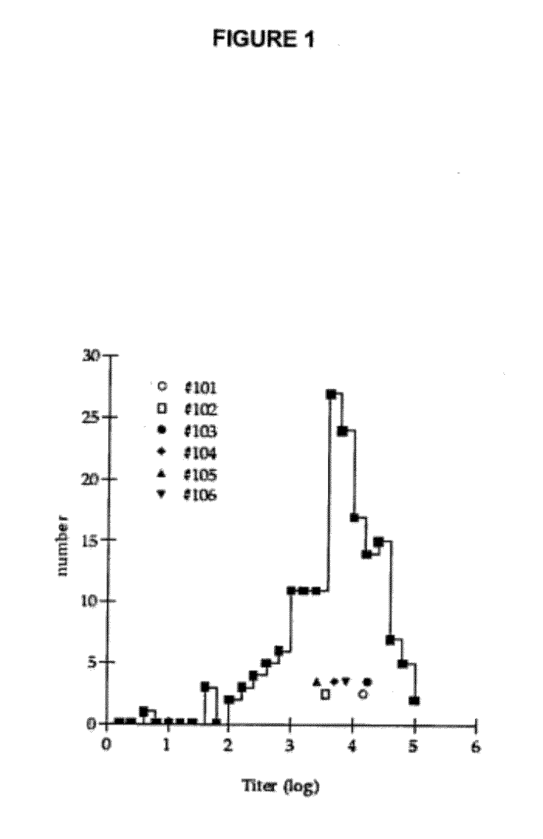
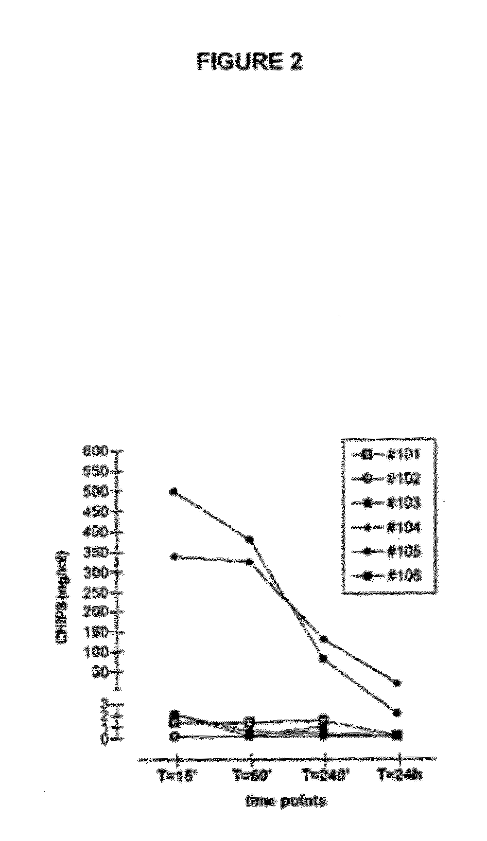
[0319]CHIPS is encountered by a majority of the human population early in life since Staphylococcus aureus is a common bacterium and the CHIPS gene is present in over 60% of S. aureus strains. Consequently, circulating anti-CHIPS antibodies that interfere with CHIPS binding to the C5aR have been detected in human serum (Wright et al., 2007, Mol Immunol 44:2507). Circulating antibodies may neutralize the biological effect of proteins. Such neutralizing antibodies may affect the efficacy of drugs, like the presence of anti-IFN-β antibodies in MS patients (Bertolotto et al., 2000, Immunopharmacology 48:95). Furthermore, circulating specific antibodies may form immune complexes (ICs) with the protein. Immune complexes are known to cause disease if deposited in blood vessels or organs, e.g. the kidney. In addition, IC mediated activation of the classical complement pathway leads to leukocyte activation and subsequent tissue damage. Therefore, the potential of CHIPS to function as an anti-inflammatory molecule is hampered by the specific antibodies.
[0320]Besides reacting with pre-existing antibodies, a recombinant protein administered to humans can potentially induce a new TH-cell dependent immune response. It can be difficult to predict whether recombinant or foreign proteins administered to humans are likely to induce a TH-cell dependent antibody response; e.g. animal models are not always applicable for human responses. However, T-cell epitope algorithms have been developed for evaluation of candidate biopharmaceutical TH-cell epitope content (De Groot et al., 2001, Vaccine 19:4385; Desmet, Spriet & Lasters, 2002, Proteins 48:31; Desmet et al., 2005, Proteins 58:53). Recent data confirm that these models can be utilized to predict human antibody responses (Koren et al., 2007, Clin Immunol 124:26).
[0321]The N-terminal amino acids of CHIPS were previously shown to be essential for FPR binding (Haas et al., 2005, J Mol Biol 353:859). As a result, N-terminal truncation will specify the CHIPS activity towards C5aR binding. To design a molecule with specific C5aR blocking activity and low interaction with human antibodies, a directed evolution approach was employed. The new improved CHIPS variant could be discovered by the use of DNA recombination, molecular modelling, site-directed mutagenesis and rounds of screening for decreased specific antibody interaction as well as preserved C5aR binding (see Example B above). In this study, we characterized the properties of the new CHIPS variant, called ADC-1004, with respect to interaction with IgG from human serum, complement activation, C5aR inhibition, immune complex formation and T-cell epitope content in different assays to evaluate the feasibility of using this protein as a pharmaceutical. Our data demonstrate that the IgG binding of a protein can be significantly decreased, while still retaining its biological function. ADC-1004 is indeed a protein with potent antagonistic activity towards the C5aR. Pre-existing human antibodies display very weak interaction with ADC-1004 and we speculate that an immediate response to neutralize ADC-1004 would most probably not occur in humans. Also, we show that ADC-1004 does not induce any of the antibody mediated effects to the same extent as full-length CHIPS1-121 when compared in different in vitro models.
Materials & Methods
Cloning, Expression and Purification of Recombinant Proteins
[0322]Wild-type CHIPS1-121 was cloned, expressed and purified as described earlier (10). CHIPS31-113 and ADC-1004 were created by truncation and mutagenesis (manuscript in preparation). These CHIPS variants were then cloned and cultivated as described above, but were purified from inclusion bodies. Briefly, 100 ml E. coli culture was pelleted and the pellets were frozen overnight at −20° C. Inclusion bodies were purified by washing in three different buffers; buffer 1: 50 mM Tris pH 8.0, 1 mM EDTA, 25% Sucrose, buffer 2: mM Tris pH 8.0, 0.2M NaCl, 0.5% Sodium Deoxycholate, 2 mM EDTA, and buffer 3: 10 mM Tris pH 8.0, 0.25% Sodium Deoxycholate, 1 mM EDTA. The pellet was dissolved, sonicated on ice and incubated with shaking at RT for 30 min in each buffer. Inclusion bodies were then pelleted by centrifugation at 12,000 rpm after each round of washing. Purified inclusion bodies were solubilized in 50 mM Tris-HCl, 0.2 M NaCl, 2 mM EDTA, 7 M GuHCl pH 8.0 and refolded by rapid dilution into PBS (final protein concentration max 100 μg/ml) and incubated with rotation at RT o/n. Then, after concentration on AmiconUltra spin filters (Millipore, Billerica, Mass.), the proteins were further purified by gel filtration on a HiLoad 16/60 Superdex 75 prep grade column (GE Healthcare, Uppsala, Sweden). Protein concentrations were determined by QuantiPro BCA assay kit (Sigma Aldrich, St Louis, Mo.).
Isolation of Human Neutrophils
[0323]Human neutrophils were prepared from buffy coats obtained from Lund University Hospital (Lund, Sweden), by Percoll (Sigma-Aldrich) density gradient centrifugation. Remaining erythrocytes were lysed with ice-cold H2O for 30 s. Cells were finally collected in RPMI-1640/0.05% BSA.
Cell Culture
[0324]U937 cells (human promonocytic cell line) transfected with the C5aR (U937/C5aR) were a generous gift from Dr. E. Prossnitz (University of New Mexico, Albuquerque, N. Mex.). Cells were grown in 75 cm2 cell culture flasks in a 5% CO2 incubator at 37° C. and were maintained in RPMI 1640 medium with L-glutamine (Cambrex, Verviers, Belgium) and 10% FBS (Cambrex, Verviers, Belgium).
Interaction with Human Serum
[0325]Human pooled serum or sera from 128, 28 and 127 healthy individuals (3H Biomedical, Uppsala, Sweden) was tested for reactivity with CHIPS1-121, CHIPS31-113, ADC-1004, respectively in ELISA. Human pooled serum was also tested for binding to Streptokinase (Aventis-Behring, King of Prussia, Pa.) and Anakinra (Amgen Inc., Thousand Oaks, Calif.). The plate was coated with equimolar amounts of the proteins or PBS. Serially diluted human serum was added and allowed to bind. IgG binding to CHIPS variants were detected with a HRP conjugated rabbit anti-human IgG (Dako A/S, Glostrup, Denmark), followed by development with Super Signal ELISA Pico Chemiluminescent Substrate (Pierce, Rockford, Ill.). Serum diluted 1/40 000 binding to coated CHIPS1-121 was used to regenerate a cut-off value. The luminescence was measured and plotted against the dilution factor and analysed in a non linear curve fitting model to calculate the dilution at which cut-off was reached, which was reported as the titer.
Complement Deposition
[0326]Complement deposition mediated by interaction between anti-CHIPS antibodies from human serum and CHIPS variants was studied in ELISA. Plates were coated with equimolar amounts of CHIPS1-121, CHIPS31-113, ADC-1004 or PBS, and incubated with human serum. Complement deposition was quantified with a mouse anti-human C3c antibody (Quidel, San Diego, Calif.) and a HRP conjugated anti-mouse Ig (Dako A/S) followed by addition of Super Signal ELISA Pico Chemiluminescent Substrate. Luminescence was measured and plotted against the serum concentration.
Biological Activity Assays
[0327]C5a induced chemotaxis of neutrophils was measured in a ChemoTx transwell system with a filter size of 8.0 μm (Neuro Probe, Gaithersburg, Md.). 5×10⁶/ml human neutrophils were labelled with 4 μM Calcein-AM (Sigma Aldrich) for 20 minutes at RT under gentle agitation, washed once in HBSS with 1% HSA and the cells were resuspended to 2.5×10⁶/ml in HBSS with 1% HSA. Cells were further incubated for 15 minutes at RT with a titration of CHIPS variants. C5a (Sigma Aldrich) as added to the lower compartment of the wells to a final concentration of 1 nM. The upper wells were assembled and labelled cells were added to the upper compartments. Labelled cells were added to the lower compartment in control wells to represent total count. The plate was incubated for 30 minutes at 37° C. with 5% CO2. Then the filters were rinsed with PBS to remove non-migrating cells and fluorescence was measured at an excitation of 485 nm and emission of 530 nm. Results are presented as % inhibition of chemotaxis as compared to cells without addition of CHIPS.
[0328]Purified CHIPS variants were tested for their ability to inhibit the C5a induced calcium mobilization in human neutrophils. Briefly, 5×10⁶/ml neutrophils were incubated with 2 μM Fluo-3AM (Sigma Aldrich) in RPMI 1640 medium with 0.05% BSA for 30 min at room temperature, washed twice and resuspended in RPMI 1640 medium with 0.05% BSA to a concentration of 106 cells/ml. Cells were preincubated with a 3-fold dilution series of CHIPS variants at room temperature for 30 min. Basal fluorescence level was measured on each sample for ˜10 s before C5a (Sigma Aldrich) (final concentration 3 nM) was added and the sample quickly placed back in the sample holder to continue measurement in a FACScalibur flow cytometer (BD Biosciences, San José, Calif.). Samples were analyzed after gating the cell population on forward and side scatter. Results are expressed as percentage inhibition of cells without addition of CHIPS.
T-Cell Epitope Analysis
[0329]CHIPS variants were analyzed for T-cell epitope content using the Epibase® system (Desmet, Spriet & Lasters, 2002, Proteins 48:31; Desmet et al., 2005, Proteins 58:53) according to patent EP 1 226 528.
Molecular Modelling
[0330]Modelling was performed by the use of the available CHIPS31-121 NMR structure (PDB code: 1XEE) (18) and the PyMol molecular graphics program (DeLano, 2002, The PyMol Molecular Graphics System. Delano Scientific, San Carlos).
Results
ADC-1004
[0331]ADC-1004 is a new CHIPS variant, based upon the CHIPS31-113 sequence, with seven amino acid substitutions, namely; K40E, D42V, N77H, K100R, K105R, N111K and G112A. ADC-1004 was discovered by mutagenesis and screening of CHIPS libraries for decreased specific antibody interaction as well as preserved C5aR binding. The CHIPS31-121 fold has previously been described to consist of an N-terminal amphipathic α-helix and a four-stranded anti-parallel β-sheet as determined by multi-dimensional NMR (18). This model was used to show the structural distribution of the ADC-1004 mutations which contribute to the ADC-1004 characteristics that we have characterized in this study (FIG. 18).
ADC-1004 Shows Low-Level Interaction with IgG in Human Serum
[0332]In order to evaluate the usefulness of ADC-1004, its interaction with IgG in human serum was studied in ELISA. The IgG titer towards ADC-1004 was compared to the titers towards CHIPS1-121, CHIPS31-113, Streptokinase and Anakinra (a recombinant IL-1 receptor antagonist, approved for treatment of rheumatoid arthritis) using human serum from healthy volunteers. The titer for Anakinra was included as a negative control since no titer could be detected towards this molecule. The ADC-1004 antibody titer was shown to be significantly lower than the titers of CHIPS1-121 (172 times lower), CHIPS31-113 (9 times lower) and Streptokinase (15 times lower). (FIG. 19 A). Furthermore, analysis of the titer of individual sera showed that a significantly higher number of individuals are responsive towards CHIPS1-121 and CHIPS31-113 than to ADC-1004 (FIG. 19 B). The individuals giving the highest IgG response towards CHIPS1-121 are also the highest responders towards CHIPS31-113 and ADC-1004.
ADC-1004 Induces Low-Level Antibody-Dependent Complement Activation
[0333]Antibodies in the circulation can interfere with the action of recombinant proteins in many ways. Besides the neutralizing effect of the anti-CHIPS antibodies, the interaction between CHIPS and antibodies could potentially activate the complement cascade. Such an activation might lead to unwanted effects such as leukocyte activation and resulting tissue damage.
[0334]To study complement activation, CHIPS variants were coated in ELISA plates and incubated with serial dilutions of human serum and deposition of complement fragment C3c was quantified. In accordance with the results from the serum IgG interaction experiments presented above, ADC-1004 mediated the lowest C3c deposition. At 10% serum, ADC-1004 mediated C3c deposition was shown to be six times lower than that induced by CHIPS1-121 and three times lower than CHIPS31-113 induced C3c deposition (FIG. 20 A). In addition, analysis of sera from 28 individuals also showed a significantly lower C3c deposition upon incubation with ADC-1004 as compared to CHIPS1-121 and CHIPS31-113 at 10% serum (FIG. 20 B).
ADC-1004 Inhibits C5a Induced Neutrophil Activation and Migration
[0335]CHIPS inhibits C5a dependent neutrophil activation and migration by blocking the C5aR. To investigate whether ADC-1004 was functional in inhibiting C5a induced chemotaxis and activation, this CHIPS variant was compared to CHIPS1-121 and CHIPS31-113 in two assays.
[0336]First, C5aR signalling inhibition was investigated by measuring the mobilization of intracellular calcium by flow cytometry. Serial dilutions of the CHIPS variants were preincubated with Fluo-3 labeled neutrophils and the cells were stimulated with C5a. The flow cytometry data show that the concentration needed for 50% inhibition of calcium release (IC50) of ADC-1004 was 0.4 nM±0.12, which is a mean value calculated from three independent experiments. The mean value was the same as the IC50 of CHIPS31-113 and twice that of the IC50 of CHIPS1-121. A representative example is shown in FIG. 21A. Second, neutrophil migration was studied in a transwell system where serial dilutions of the CHIPS variants were preincubated with Calcein labeled neutrophils in the upper compartment of the transwell. The labeled neutrophils were then allowed to migrate towards a C5a gradient over the membrane to the lower compartment. The results show that the IC50 of ADC-1004 was the same as the IC50 of CHIPS31-113 and approximately four times higher than the IC50 of CHIPS1-121 (FIG. 21B).
ADC-1004 T-Cell Epitope Content
[0337]T-cell epitopes are peptides of a protein that are needed to ensure TH-cell activation. The T-cell epitope content of ADC-1004 was compared to that of wild-type full-length CHIPS (CHIPS1-121) in an in silico model (Epibase®; (Desmet, Spriet & Lasters, 2002, Proteins 48:31; Desmet et al, 2005, Proteins 58:53)) to estimate the probability of ADC-1004 inducing a new TH-cell dependent antibody response. Table 7 shows that the total number of predicted epitopes is lower in ADC-1004 than in CHIPS1-121. Moreover, the predicted epitopes in ADC-1004 are different and less promiscuous than those found in CHIPS1-121 (data not shown).
Discussion
[0338]Complement fragment C5a is a powerful peptide that exerts proinflammatory effects through the C5aR, a mechanism involved in many inflammatory disorders. The C5aR can thus be considered an important drug target in these diseases (Monk et al., 2007, Br J Pharmacol. 152(4):429-48; Ricklin & Lambris, 2007, Nat Biotechnol 25:1265). The C5aR antagonist CHIPS is one of the proteins produced by the human pathogen S. aureus to evade the host response by modulating the innate immune system (De Haas et al., 2004, Agent. J Exp Med 199:687; Lee at al., 2004, J Infect Dis 190:571; Rooijakkers et al., 2005, Nat Immunol 6:920; Jongerius et al., 2007, J Exp Med 204:2461). Antibodies directed against CHIPS have been found in human sera (Wright et al., 2007, Mol Immunol 44:2507); hence this protein is not likely to be tolerated by humans and the CHIPS activity in vivo is likely to be neutralized. In essence, CHIPS is a potent C5aR antagonist but with limited use in humans due to pre-existing specific antibodies.
[0339]To increase the feasibility to use this protein in vivo, we have created an improved functional unity, called ADC-1004. This CHIPS variant harbours seven amino acid substitutions (see Example B above), which together contribute to the new characteristics of the ADC-1004 protein. Interestingly, despite the high number of mutations that totally alter the interaction with human antibodies, the biological function to inhibit the C5aR is retained. Presumably some of the amino acid substitutions in fact stabilize the CHIPS fold rather than being directly involved in decreasing antibody binding to ADC-1004 (see Example B above).
[0340]We have determined the IgG titer towards ADC-1004 compared to titers towards full-length CHIPS1-121 and the truncated CHIPS31-113 variant as well as to titers towards two existing biopharmaceuticals. For comparison Streptokinase an existing pharmaceutical derived from a bacterial protein was used. Streptokinase is and has been used for treatment of myocardial infarction for more than 30 years (ISIS-2. ISIS-2 (Second International Study of Infarct Survival) Collaborative Group., 1988, Randomised trial of intravenous streptokinase, oral aspirin, both, or neither among 17,187 cases of suspected acute myocardial infarction, Lancet 2:349). ADC-1004 showed low interaction with human IgG from human serum, both pooled serum and serum from different individuals. The titer of ADC-1004 was 172 times lower than the full-length CHIPS1-121 titer and 15 times lower than for Streptokinase. Also, no individuals were strongly responsive towards ADC-1004, while several individuals showed especially high titers towards full-length CHIPS1-121.
[0341]This low interaction with human antibodies was shown to be advantageous, since ADC-1004 did not induce antibody mediated effects to the same extent as full-length CHIPS1-121 or CHIPS31-113. When studying complement activation by immobilizing the CHIPS variants in ELISA and adding serum, ADC-1004 proved to be a poor inducer of complement activation, generating six times lower C3c deposition than full-length CHIPS1-121. These data suggest that immune-mediated adverse effects are less likely to be induced upon administration of ADC-1004 than the other CHIPS variants tested.
[0342]Remarkably, the introduced mutations did not remove the biological function of ADC-1004. ADC-1004 showed an IC50 value equal to that of CHIPS31-113 and four times higher than the IC50 value of full-length CHIPS1-121 in inhibition of C5a induced neutrophil migration. There was an even smaller difference between the CHIPS variants when studying biological activity by the means of inhibition of calcium release in human neutrophils upon C5a stimulation. The mean value of ADC-1004 IC50 was the same as the IC50 of CHIPS31-113 and twice that of full-length CHIPS1-121.
[0343]Previously anti-CHIPS antibodies, which interfere with CHIPS binding to the C5aR, have been detected in human serum (Wright et al, 2007, Mol Immunol 44:2507). Of great importance for functionality in vivo, ADC-1004 was not inhibited by human serum to the same extent as full-length CHIPS1-121 as indicated in a calcium release assay (data not shown) when tested on U937 cells stably transfected with the human C5aR. Even though the interaction of ADC-1004 with IgG in human serum was undetectable when studied in ELISA, some low affinity epitopes might still be present in the protein and could be the explanation for the serum effect seen when ADC-1004 was studied in the calcium release model. Also, other Ig classes could be involved in the human response towards CHIPS. We have looked at the IgM response towards the different CHIPS variants and it is always 3-20 times lower than the IgG response.
[0344]ADC-1004 displays very weak interaction with pre-existing human IgG. However, there are other aspects of immunogenicity to take into consideration. It is also important to evaluate if it is prone to induce a TH-cell dependent immune response, like many recombinant or non-human proteins are. Foreign epitopes within a protein or the lack of glycosylation leading to the exposure of hidden epitopes as well as presence of aggregates due to suboptimal formulation are some of the causes of immunogenicity seen in biopharmaceuticals (Schellekens, 2003, Nephrol Dial Transplant 18:1257). However, T-cell epitopes within a recombinant protein are more important to take into consideration if the protein is going to be developed for treatment of chronic disorders, i.e. if it is to be administered more than once to the same patient. ADC-1004 is a small protein and would probably be most beneficial in the treatment of acute inflammations, since it is likely to have a short half-life in circulation due to its small size. To verify that the T-cell dependent immunogenicity of ADC-1004 was not increased as compared to the full-length CHIPS1-121, we estimated the T-cell epitope content of the CHIPS variants in an in silico model. ADC-1004 was shown to contain a moderate number of potential T-cell epitopes (in total 11 strong TH epitopes), lower than that of CHIPS1-121 and comparable to that of other proteins studied previously in this model (Van Walle et al., 2007, Expert opinion on biological therapy 7:405). For example, the chimeric antibody Rituximab approved for treatment of non-Hodgkin's lymphoma and RA was shown to contain in total 22 strong TH epitopes and the human antibody Ofatumumab currently in clinical trials was shown to contain in total six strong TH epitopes. It is also important to take into consideration that several epitopes differ between CHIPS1-121 and ADC-1004. Therefore, a strong T-cell memory response is less likely to occur upon administration of ADC-1004 than CHIPS1-121, since the pre-existing memory T-cells are geared towards the epitopes found in CHIPS1-121. Any T-cell responses against ADC-1004 will therefore be classical, non-memory based responses. The combined approach of avoiding pre-existing antibodies, hence B-cell epitope removal, with TH-cell memory avoidance, is consistent with ADC-1004 being significantly less immunogenic that the wild-type CHIPS protein.
[0345]In conclusion, our work with CHIPS shows that the interaction of a protein with antibodies in human serum can be significantly decreased, while still retaining the biological function of the protein.
REFERENCES
[0346] 1. Guo, R. F., and P. A. Ward. 2005. Role of C5a in inflammatory responses. Annu Rev Immuno/23:821. [0347] 2. Wetsel, R. A. 1995. Expression of the complement C5a anaphylatoxin receptor (C5aR) on non-myeloid cells. Immunol Lett 44:183. [0348] 3. Zwimer, J., A. Fayyazi, and O. Gotze. 1999. Expression of the anaphylatoxin C5a receptor in non-myeloid cells. Mol Immunol 36:877. [0349] 4. Okusawa, S., K. B. Yancey, J. W. van der Meer, S. Endres, G. Lonnemann, K. Hefter, M. M. Frank, J. F. Burke, C. A. Dinarello, and J. A. Gelfand. 1988. C5a stimulates secretion of tumor necrosis factor from human mononuclear cells in vitro. Comparison with secretion of interleukin 1 beta and interleukin 1 alpha. J Exp Med 168:443. [0350] 5. Kohl, J. 2001. Anaphylatoxins and infectious and non-infectious inflammatory diseases. Mol Immunol 38:175. [0351] 6. Barnum, S. R. 2002. Complement in central nervous system inflammation. Immunol Res 26:7. [0352] 7. Monk, P. N., A. M. Scola, P. Madala, and D. P. Fairlie. 2007. Function, structure and therapeutic potential of complement C5a receptors. Br J. Pharmacol. [0353] 8. Ricklin, D., and J. D. Lambris. 2007. Complement-targeted therapeutics. Nat Biotechnol 25:1265. [0354] 9. Sumichika, H. 2004. C5a receptor antagonists for the treatment of inflammation. Curr Opin Investig Drugs 5:505. [0355] 10. De Haas, C. J., K. E. Veldkamp, A. Peschel, F. Weerkamp, W. J. Van Wamel, E. C. Heezius, M. J. Poppelier, K. P. Van Kessel, and J. A. Van Strijp. 2004. Chemotaxis Inhibitory Protein of Staphylococcus aureus, a Bacterial Antiinflammatory Agent. J Exp Med 199:687. [0356] 11. Postma, B., M. J. Poppelier, J. C. van Galen, E. R. Prossnitz, J. A. van Strijp, C. J. de Haas, and K. P. van Kessel. 2004. Chemotaxis inhibitory protein of Staphylococcus aureus binds specifically to the C5a and formylated peptide receptor. J Immunol 172:6994. [0357] 12. Wright, A. J., A. Higginbottom, D. Philippe, A. Upadhyay, S. Bagby, R. C. Read, P. N. Monk, and L. J. Partridge. 2007. Characterisation of receptor binding by the chemotaxis inhibitory protein of Staphylococcus aureus and the effects of the host immune response. Mol Immunol 44:2507. [0358] 13. Bertolotto, A., S. Malucchi, E. Milano, A. Castello, M. Capobianco, and R. Mutani. 2000. Interferon beta neutralizing antibodies in multiple sclerosis: neutralizing activity and cross-reactivity with three different preparations. [0359] Immunopharmacology 48:95. [0360] 14. De Groot, A. S., A. Bosma, N. Chinai, J. Frost, B. M. Jesdale, M. A. Gonzalez, W. Martin, and C. Saint-Aubin. 2001. From genome to vaccine: in silico predictions, ex vivo verification. Vaccine 19:4385. [0361] 15. Desmet, J., J. Spriet, and I. Lasters. 2002. Fast and accurate side-chain topology and energy refinement (FASTER) as a new method for protein structure optimization. Proteins 48:31. [0362] 16. Desmet, J., G. Meersseman, N. Boutonnet, J. Pletinckx, K. De Clercq, M. Debulpaep, T. Braeckman, and I. Lasters. 2005. Anchor profiles of HLA-specific peptides: analysis by a novel affinity scoring method and experimental validation. Proteins 58:53. [0363] 17. Koren, E., A. S. De Groot, V. Jawa, K. D. Beck, T. Boone, D. Rivera, L. Li, D. Mytych, M. Koscec, D. Weeraratne, S. Swanson, and W. Martin. 2007. Clinical validation of the “in silico” prediction of immunogenicity of a human recombinant therapeutic protein. Clin Immunol 124:26. [0364] 18. Haas, P. J., C. J. de Haas, M. J. Poppelier, K. P. van Kessel, J. A. van Strijp, K. Dijkstra, R. M. Scheek, H. Fan, J. A. Kruijtzer, R. M. Liskamp, and J. Kemmink. 2005. The structure of the C5a receptor-blocking domain of chemotaxis inhibitory protein of Staphylococcus aureus is related to a group of immune evasive molecules. J Mol Biol 353:859. [0365] 19. DeLano, W. L. 2002. The PyMol Molecular Graphics System. Delano Scientific, San Carlos. [0366] 20. Lee, L. Y., M. Hook, D. Haviland, R. A. Wetsel, E. O. Yonter, P. Syribeys, J. Vernachio, and E. L. Brown. 2004. Inhibition of complement activation by a secreted Staphylococcus aureus protein. J Infect Dis 190:571. [0367] 21. Rooijakkers, S. H., M. Ruyken, A. Roos, M. R. Daha, J. S. Presanis, R. B. Sim, W. J. van Wamel, K. P. van Kessel, and J. A. van Strijp. 2005. Immune evasion by a staphylococcal complement inhibitor that acts on C3 convertases. Nat Immunol 6:920. [0368] 22. Jongerius, I., J. Kohl, M. K. Pandey, M. Ruyken, K. P. van Kessel, J. A. van Strijp, and S. H. Rooijakkers. 2007. Staphylococcal complement evasion by various convertase-blocking molecules. J Exp Med 204:2461. [0369] 23. 1988. Randomised trial of intravenous streptokinase, oral aspirin, both, or neither among 17,187 cases of suspected acute myocardial infarction: ISIS-2. ISIS-2 (Second International Study of Infarct Survival) Collaborative Group. Lancet 2:349. [0370] 24. Schellekens, H. 2003. Immunogenicity of therapeutic proteins. Nephrol Dial Transplant 18:1257. [0371] 25. Van Walle, I., Y. Gansemans, P. W. H. I. Parren, P. Stas, and I. Lasters. 2007. Immunogenicity screening in protein drug development. Expert opinion on biological therapy 7:405.
TABLE 7 TH epitope counts for CHIPS1-121 and ADC-1004. Peptides binding to multiple HLAs of the same group (DRB1, DRB3/4/5, DP, DQ) are counted as one DRB1 Strong Medium DRB3/4/5 DQ DP CHIPS1-121 9 23 2 1 2 ADC-1004 7 19 2 1 1

Example

EXAMPLE D
In VIVO Study of the Efficacy of Chips Variant ADC-1004 In the Treatment of Acute Myocardial Infarction and Reperfusion Injury
Introduction
[0372]A percutaneous catheter-based approach was chosen in order to induce ischemia with minimum trauma, operation-induced stress and secondary changes in circulatory physiology. Myocardial infarct size was chosen as the primary effect parameter since, in the clinical setting, the long term outcome is heavily dependant on the infarct size [1]. Reduction of myocardial infarct size is also the aim of reperfusion therapy. Reperfusion therapy that achieves a good macroscopic result after opening of the coronary occlusion may yet result in persistent ST-segment elevation attributed to microvascular injury [2]. The degree of microvascular injury is associated with the duration of myocardial ischemia and the extent of myocardial infarction but may possibly also be caused by reperfusion injury. The presence of microvascular obstruction is associated with a worse clinical outcome [3]. MO was therefore chosen as a complementary effect parameter. Ex vivo MRI allows for achievement of high resolution images of the myocardial infarction and correlates closely to histology with TTC-staining [4], and was therefore chosen as the method for IS evaluation. SPECT was used to determine area at risk during ischemia. The presence of microvascular obstruction may have several pathophysiological mechanisms [5]. One of these is a neutrophil induced inflammatory response upon reperfusion. C5aR (CD88) bearing cells in the infarct area can be determined by histology. State of neutrophil activation can be monitored by CD88 and the activation marker CD18 on histology sections. A decrease of activated neutrophils upon ADC-1004 treatment would indicate a reduction in neutrophil inflammatory response.
Materials and Methods
Experimental Preparation
[0373]8 healthy domestic male and female 40-50 kg pigs were fasted overnight with free access to water. Premedication was administered with Kataminol (ketamine, Intervet AB Danderyd Sweden) and Rompun (xylazin, Bayer AG, Leverhusen, Germany) 30 minutes prior to the procedure. After induction of anesthesia with thiopental (Pentothal, Abbott, Stockholm, Sweden) 5-25 mg/kg, the animals were orally intubated with cuffed endotracheal tubes. Thereafter, a slow infusion of 1.25 μl/ml fentanyl (Fentanyl, Pharmalink AB, Stockholm, Sweden) in buffered glucose (25 mg/ml) was started at a rate of 1.5 ml/min and adjusted as needed. During balanced anesthesia thiopental (Pentothal, Abbott, Stockholm, Sweden), was titrated against animal requirements with small bolus doses. Mechanical ventilation was established with a Siemens-Elema 900B ventilator in the volume-controlled mode, adjusted in order to obtain normocapnia. Initial settings were: respiratory rate of 15/min, tidal volume of 10 ml/kg and positive end-expiratory pressure of 5 cmH2O. The animals were ventilated with a mixture of dinitrous oxide (70%) and oxygen (30%). The pigs were continuously monitored with electrocardiogram (ECG) and intraarterial blood pressure. Heparin (200 IU/kg) was given intravenously at the start of the catheterization. A 6 F introducer sheath (Onset, Cordis Co. Miami, Fla., USA) was inserted into the surgically exposed left carotid artery upon which a 6 F JL4 Wiseguide™ (Boston Scientific Scimed, Maple Grove, Minn., USA) was inserted into the left main coronary artery. The catheter was used to place a 0.014-inch PT Choice™ guide wire (Boston Scientific Scimed, Maple Grove, Minn., USA) into the distal portion of the LAD. A 3.5×15 mm Maverich™ monovail angioplasty balloon (Boston Scientific Scimed, Maple Grove, Minn., USA) was then positioned in the mid portion of the LAD, immediately distal to the first diagonal branch. All radiological procedures were performed in an experimental catheterization laboratory (Shimadzu Corp., Kyoto, Japan).
Ischemia Protocol
[0374]Ischemia was induced by inflation of the angioplasty balloon for 40 min. An angiogram was performed after inflation of the balloon and before deflation of the balloon in order to verify total occlusion of the coronary vessel and correct balloon positioning. After deflation of the balloon a subsequent angiogram was performed to verify restoration of blood flow in the previously occluded artery.
ADC-1004 Delivery Protocol
[0375]A single dose of around 4 mg/kg of ADC-1004 in 0.9% NaCl was intravenously administered to eight animals approximately 22 minutes after ischemia induction. Eight (8) animals received saline only. Plasma concentration of ADC-1004 was monitored during the experiment and the targeted plasma concentration was achieved for the four hours and 40 minutes the experiment lasted (using induction of ischemia as t=0).
Ex-vivo Assessment of Area at Risk by SPECT
[0376]Single photon emission computed tomography (SPECT) was used to assess the AAR as percent of left ventricular myocardium. Five hundred MBq of 99 mTc-tetrofosmin was administered intravenously 18 minutes before deflation of the angioplasty balloon. Ex-vivo imaging was performed with a dual head camera (Skylight, Philips, Best, the Netherlands) at 32 projections (40 s per projection) with a 64×64 matrix yielding a digital resolution of 5×5×5 mm. Iterative reconstruction using maximum likelihood-expectation maximization (MLEM) was performed with a low-resolution Butterworth filter with a cut-off frequency set to 0.6 of Nyquist and order 5.0. No attenuation or scatter correction was applied. Finally short and long-axis images were reconstructed. The endocardial and epicardial borders of the left ventricle that were manually delineated in the MR images were copied to the co-registered SPECT images. A SPECT defect was defined as a region within the MRI-determined myocardium with counts lower than the 55% of the maximum counts in the myocardium [6].
Infarct Size and Microvascular Obstruction Assessed by Ex vivo MRI
[0377]Ex vivo imaging of the heart was undertaken using a 1.5 T Philips Intera CV MR scanner (Philips, Best, the Netherlands) according to a previous described protocol [4, 7]. In brief, a gadolinium-based contrast agent (Dotarem, gadoteric acid, Gothia Medical, Billdal, Sweden) was administered intravenously (0.4 mmol/kg) 30 minutes prior to removal of the heart. The heart was removed 4 hours after initiation of reperfusion. After removal, the heart was immediately rinsed in cold saline and the ventricles were filled with balloons containing deuterated water. Three dimensional acquisition of T1-weighted images (TR=20 ms, TE=3.2 ms, flip angle=70° and 2 averages) yielded a stack of approximately 200 images with an isometric resolution of 0.5 mm covering the entire heart. Images were then acquired using a head coil and the duration of acquisition was typically 45 minutes. The MR images were analyzed using freely available software [8, 9]. The endocardial and epicardial borders of the left ventricular myocardium were manually delineated in short-axis ex vivo images. This defined the volume of left ventricular myocardium (cm3=ml). The infarct size (IS) was first determined as the volume of infarcted myocardium (cm3). The infarct volume was calculated as the product of the slice thickness (cm) and the area of hyperenhanced pixels (cm2) with a signal intensity above the infarction threshold defined as >8 SD above the mean intensity of non-affected remote myocardium. Microvascular obstruction was defined as hypointense regions in the core of the infarction which had signal intensity less than the threshold for infarction. These regions were manually included in the infarct volume. The volume of microvascular obstruction (cm3) was calculated as the difference between the infarct volume before and after manual inclusion of regions of microvascular obstruction. Furthermore, the size of microvascular obstruction was expressed as percent of percent of area at risk. Ultimately, the infarct size was expressed as percent of left ventricular myocardium. Finally, infarct size was expressed as a percentage of the area at risk (IS/AAR) in order to adjust for any difference in area at risk between the groups [10, 11].
Histology
[0378]Heart muscle tissue from infarction area of pig #7 and #8 were used in this study. Tissues were fixed in 4% formaldehyde or snap frozen in isopentan chilled on dry ice. The formaldehyde fixed tissue was embedded in paraffin and sectioned at 5 μm, dried at. 37° C. over night and stained according the methods described below. The cryopreserved tissue was sectioned at 8 μm, dried at room temperature over night and stained according to the methods described below.
[0379]Staining with CD88, Serotec, MCA 1283T was performed on 5 μm paraffin sections. Samples were de-paraffinized and an antigen retrieval treatment in boric acid (pH8.0) with boiling for 10 minutes and 20 minute cooling at room temperature was performed. Samples were then treated with 1% H2O2 in tap water for 20 min., washed three times in PBS and incubated in 5% normal goat serum in PBS for 30 minutes. Subsequently samples were incubated for 30 min with the primary antibody, mouse anti human CD88, diluted to 1 μg/ml in PBS with 5% normal goat serum. After three times wash with PBS samples were incubated 30 min with Envision mouse (Dako, K4001) again washed twice in PBS and once in Tris-HCl. Finally samples were treated with DAB for 5-10 min and Mayers hematoxylin for 5 seconds before mounted with coverslips.
[0380]Staining with CD18, abcam, ab34117 was performed on 8 μm cryosections. Samples were fixed in acetone for 10 min and treated with 1% H2O2 in tap water for 20 min, washed three times in PBS and incubated in 5% normal goat serum in PBS for 30 minutes. Subsequently samples were incubated for 60 min with the primary antibody, mouse anti pig CD18, diluted to 1 μg/ml in PBS with 5% normal goat serum. After three times wash with PBS samples were incubated 30 min with Envision mouse (Dako, K4001) again washed twice in PBS and once in Tris-HCl. Finally samples were treated with DAB for 5-10 min and Mayers hematoxylin for 5 seconds before mounted with coverslips. The slides were microscopically analyzed and with a PC-based image analysis system (Leica Q500, Cambridge, UK) the percentage of positive stained area was calculated.
Results
[0381]ADC-1004 was tested in a porcine model for acute myocardial infarction. Ischemia was induced in sixteen animals where, eight pigs received approximately 4 mg/kg ADC-1004 in saline and eight pigs received saline (control group). The animals were treated and tested as described in materials and methods.
[0382]The results shown in FIG. 22 and FIG. 23 shows that the ADC-1004 significantly reduces infarct size in relation to the ischemic area (area at risk) measured by MR/SPECT (p<0.007, Mann-Whitney U-test). This supports a clinical therapeutic effect of ADC-1004 as the long term outcome is heavily dependant on the infarct size [1]. Mean microvascular obstruction was reduced in the group treated with ADC-1004. Microvascular obstruction is a factor which is clinically proven related to the severity of a mycardial infarction [3]. One of the pathophysiological mechanisms behind microvascular obstruction may be neutrophil induced inflammatory response upon reperfusion [5]. Results from histology investigations shows numerous distinct CD88 positive cells are seen in infarction area of both the placebo sections as well as sections from ADC-1004 treatment. The number of positive cells are about 250/mm2 in both cases. Positive cells are found both in blood vessels as well as intermingling with cardiac muscle fibers. CD18 shows a more variable reaction from weakly to strongly positive cells. Placebo samples shows markedly more positive cells (FIG. 24A) compared to ADC-1004 received samples (FIG. 24B). Moreover, more strongly stained cells are seen in placebo compared to treated samples. Measurement with image analysis equipment gives that placebo expresses 2-3 more CD18 compared to ADC-1004 treated indicating reduced activation of neutrophils upon ADC-1004 administration.
[0383]Thus, the data presented indicate that acting on the C5aR by administration of ADC-1004 in an acute myocardial infarction (and thereby inhibiting the activation and inflammation by neutrophils) decreases the severity of the infarction.
REFERENCES
[0384] 1. Fox K A, Dabbous O H, Goldberg R J et al Prediction of risk of death and myocardial infarction in the six months after presentation with acute coronary syndrome: prospective multinational observational study (GRACE). Bmj. 2006, 333:1091. [0385] 2. Claeys M J, Bosmans J, Veenstra L et al Determinants and prognostic implications of persistent ST-segment elevation after primary angioplasty for acute myocardial infarction: importance of microvascular reperfusion injury on clinical outcome. Circulation. 1999, 99:1972-1977. [0386] 3. Wu K C, Zerhouni E A, Judd R M et al Prognostic significance of microvascular obstruction by magnetic resonance imaging in patients with acute myocardial infarction. Circulation. 1998, 97:765-772. [0387] 4. Kim R J, Fieno D S, Parrish T B et al Relationship of MRI delayed contrast enhancement to irreversible injury, infarct age, and contractile function. Circulation 1999, 100:1992-2002. [0388] 5. Jaffe R, Charron T, Puley G et al Microvascular Obstruction and the No-Reflow Phenomenon After Percutaneous Coronary Intervention. Circulation 2008, 117:3152-3156 [0389] 6. Ugander M, Soneson H, Heiberg E et al. A novel method for quantifying myocardial perfusion SPECT defect size by co-registration and fusion with MRI—an experimental ex vivo imaging pig heart study. Abstract. Proceedings of the Swedish Heart Association Spring Meeting 2008 [0390] 7. Götberg M, Olivencrona K G, Engblom H et al Rapid short-duration hypothermia with cold saline and endovascular cooling before reperfusion reduces microvascular obstruction and myocardial infarction size. BMC Cardivascular Disorders 2008, 8:7. [0391] 8. Heiberg E, Engblom H, Engvall J et at Semi-automatic quantification of myocardial infarction from delayed contrast enhanced magnetic resonance imaging. Scand Cardiovasc J 2005, 39:267-275. [0392] 9. Website title [http://segment.heiberg.se/] [0393] 10. Hedström E: Acute Myocardial Infarction. The relationship between duration och ischaemia and infarct size in humans—Assessment by MRI and SPECT. In PhD thesis Lund: University of Lund; 2005. [0394] 11. Hedstrom E, Frogner F, Astrom-Olsson K et al Myocardial infarct size in relation to myocardium at risk versus duration of ischemia in humans: Comparison with different species (Abstract). J Cardiovasc Magn Reson 2007, 9:363
EXAMPLE E
In vivo Study of the Efficacy of Chips Variant ADC-1004 In the Treatment of Stroke and Reperfusion Injury
Introduction
[0395]The aim of this study was to investigate whether treatment with ADC-1004 starting 1 h and 45 min after occlusion of the middle cerebral artery and 15 min prior to reperfusion and with infusion for 24 hrs infusion, would differentially influence the cerebral infarct size in a rat model of transient middle cerebral artery occlusion (tMCAO).
Materials and Methods
[0396]Temporary Middle Cerebral Artery Occlusion (tMCAO)
[0397]All surgical procedures were approved by the Ethics Committee for Animal Research at Lund University. The male Wistar rats had free access to water and food and were housed under 12 h light/12 h dark cycle. The rats were fasted overnight and subjected to 2 h transient middle cerebral artery occlusion (MCAO) using the intraluminal filament technique. Following the 2 h occlusion, a neurological assessment of deficits was performed after 1.5 hrs of reperfusion. Rats showing rotational asymmetry and dysfunctional limb placement were included. The right middle cerebral artery (MCA) was occluded by the intraluminal filament technique. Animals were anesthetized by inhalation of 4% Forene in O2: N2O (30:70) and then spontaneously ventilating in a nose mask delivering 2% Forene in O2: N2O. For continuous monitoring of blood pressure, control of blood gases and for injection of 60 IU of heparin, a catheter was inserted in the tail artery.
[0398]A heating pad connected to a rectal temperature probe was used to guarantee that body temperature was maintained at 37° C. A laser Doppler probe was glued onto right side of the skull, hence monitoring blood flow in the MCA territory. Experimental stroke was induced by first making a skin incision in the middle of the neck to expose the right common carotid artery together with the internal and external part. The external carotid was ligated and the internal carotid was encircled by a suture. In the common carotid artery, a small incision was made close to the bifurcation and a filament was introduced into the internal carotid artery and advanced until it blocked the origin of the middle cerebral artery. The blockade was registered as a decrease in the Laser Doppler signal. After this surgical procedure the rats were allowed to recover from anesthesia during the ischemic period. Reperfusion was performed 2 h later by removal of the filament when the animal once again was anaesthetized.
[0399]Prior to reperfusion a silicon catheter was inserted into the vena cava superior and advanced 1-2 cm towards the heart. The catheter was stabilized and tunnelated under the skin of the back of the neck. The catheter was connected to a swivel system that allowed the animal to freely move in a plastic bowl while being injected with either ADC-1004 or placebo (blinded). Fifteen minutes prior to the end of the MCAO the animal was injected with a bolus dose of the solution into the vena cava, followed by a continuous infusion of the solution for 24 hrs. Blood (0.25-0.5 ml) was centrifuged and the plasma frozen for further analysis. Blood was sampled 3, 19, 24 and 48 hrs after reperfusion. In sham-operated animals, identical surgery was performed apart from the insertion of the filament. All physiological parameters of animals included in the study were within normal ranges: pO2>90 mm Hg, pCO2 30-50 mm Hg and pH 7.35-45 as were the arterial blood pressure and rectal temperature during the surgery.
Infarct Evaluation
[0400]After 2 days of reperfusion the animals were anesthetized in 4% Forane and decapitated. The brains were quickly removed from the scull and placed in cold saline for 10 minutes. In a tissue slicer, the brains were cut in twelve one millimeter thick coronal slices and the sections were stained in a saline solution containing 1.0% 2,3,5-triphenyltetrazolium chloride (TTC) at 37° C. for 20 minutes. The same procedures were performed for sham-operated animal. The infarct size was assessed by computer assisted image analysis.
Results
[0401]ADC-1004 was tested in a rat model for stroke. Rats treated with placebo (saline) had a mean infarct volume of 24.5 mm3 while animals treated with ADC-1004 had a mean infarct size of 15.8 mm3.
[0402]These results clearly show a tendency towards smaller infarct size in the ADC-1004 treated animals.

Example

EXAMPLE F
In vivo Study of the Efficacy of Chips Variant ADC-1004 in the Treatment in Lung Transplantation
Introduction
[0403]Obstacles to present day lung transplantation involve (1) lack of suitable donor organs, (2) ischemia/reperfusion (I/R) injury, (3) rejection, and (4) development of bronchiolitis obliterans. Lung I/R injury after transplantation is a common cause of respiratory failure and manifests typically during the first 72 hours post-transplantation.[1] I/R injury continues to be a universal and substantive cause of morbidity and mortality in the early postoperative period, with reported rates as great as 41%.[1] The 30-day mortality of patients with I/R injury is about 40%, compared with 7% in patients without I/R injury.[2] Patients showing I/R injury necessitate prolonged mechanical ventilation with greater hospital stays and are at an increased risk of multiorgan failure. [3]
[0404]The mechanisms of I/R injury are diverse and include generation of reactive oxygen species (ROS), leukocyte activation/recruitment, complement and platelet activation, abnormalities in pulmonary vascular tone, and increased pro-coagulant activity. The production of pro-inflammatory cytokines is increased considerably in the lung after I/R. Several studies suggest that lung I/R injury is characterized by neutrophil dependent injury.[4, 5] Various studies have shown that neutrophil-activating compounds cause lung injury, neutrophil depletion attenuates lung I/R injury, and depletion of neutrophil adhesion prevents lung I/R injury.[6, 7]
[0405]ADC-1004 was used in a porcine lung transplantation model modified from ref. 8 investigating the condition of the transplanted lung during the first 6 hours and after 20 hours post transplantation. The arterial oxygen tension was considered as a marker for lung condition.
Materials and Methods
[0406]Eight Swedish native breed pigs with a weight of about 66 kg were used (4 donors and 4 recipients). All the animals received humane care in compliance with the “Guide for the Care and Use of Laboratory Animals” published by the National Institute of Health (NIH publication 85-23, revised 1985).
[0407]The general experimental set up has previously been described in ref. 8. The procedures are described in brief below:
Donor Procedure
[0408]The animals were ventilated with a Siemens Servoventilator 300 and a volume-controlled, pressure-regulated ventilation of 10 l/min (20 breaths/min; positive end-expiratory pressure, 8 cm H2O; inspired oxygen fraction, 0.5-1.0) was used. A sternotomy was performed. After systemic heparinization (4 mg/kg) a 40-cm-long 32F cannula was inserted into the pulmonary artery (PA). The left and right pleural spaces were entered. All parts of the lungs were carefully inspected, and the positive end-expiratory pressure was increased temporarily to 10 cm H2O until the atelectatic parts were eliminated. The abdominal viscera were lifted away from the diaphragm to avoid compressing the lower lobes of the lungs. The superior and inferior venae cavae were ligated. The ascending aorta was clamped. Both the right and left atrium were opened by a cut. The PA was perfused with 1 L of a cold preservation solution, supplemented with ADC-1004 in the treated animals. The mean PA perfusion pressure was kept below 20 mm Hg by keeping the preservation solution level at a height of 20 cm above the lungs. After this procedure, the lungs were removed and stored cold 4° C. for 60 hours.
Recipient Procedure
[0409]The recipient pig was sedated and anesthetized. The animal received 1.2 g of intramuscular benzylpenicillinprocain (Ilocillin; Ciba-Geigy, Basel, Switzerland). Tracheostomy was carried out, a No. 7 tracheal tube was inserted, and the animals were ventilated in the same way as the donors. Two central venous catheters were introduced via the internal jugular vein and two catheters were placed in the aorta through the carotid artery. A Foley catheter was inserted into the urinary bladder through a suprapubic cystostomy. A left thoracotomy through the sixth intercostal space was done and a left pneumonectomy performed, care being taken to leave long ends of the pulmonary veins.
[0410]The stored left lung from the donor pig was dissected free from the right lung and was subsequently transplanted into the recipient pig. ADC-1004 was given 30 minutes before reperfusion of the transplanted lung. The dose was given both as a bolus dose of 0.4 mg/kg and as an infusion of 0.4 (first treated pig) or 0.2 mg/kg/h (the second treated pig). The pig was placed in a prone position. The infusion of ADC-1004 was continued throughout the experiment. When the blood flow had been established through the transplanted lung, the time was defined as zero. The gas exchange was followed for six hours. Between nineteen and twenty hours after reperfusion a right thoracotomy and a right pneumonectomy were done through the seventh intercostal space and the pig was totally dependent for its survival on the left transplanted lung. Blood gases were then taken before and after the pneumonectomy. The fluid supply during the experimental period was kept constant for all the animals and consisted of 720 mL of 10% glucose (ie, the anaesthetic infusion of 30 mL/h). Ringer lactate 170 mL/hour was continuously given during the experiment.
Results
[0411]The two animals receiving lung transplants treated with ADC-1004 showed an improved oxygen tension after transplantation. As shown in FIG. 25 all pigs started around the same base line before transplantation. As early as 1 hour post transplantation the ADC-1004 treated animals showed an increase in oxygen gas exchange capacity in comparison with placebo treated animals, after 3 hours treated ADC-1004 animals had reached high oxygen gas exchange capacity indicating better lung condition compared to placebo treated animals. After 6 hours one of the control animals showed an increase in gas exchange capacity although not capable of reaching the levels in ADC-1004 treated pigs. The PaO2 (FiO2=1.0) in aortic blood after right pneumonectomy making the pig 100% dependant on the transplanted left lung is shown in Table 8.
[0412]These data indicate that lungs in the ADC-1004 treated group were in better condition 20 hours after transplantation compared to the placebo group. The better the gas-exchange capacity immediately after lung transplantation, the shorter will the need for intensive care treatment be and large clinical materials then show better 30-day survival. Impaired gas exchange capacity the first 24 hours after lung transplantation indicates reperfusion injury. Thus, indicating that ADC-1004 inhibition of the C5aR reduces the reperfusion injury in lung transplantation.
TABLE 8 PaO2 (FiO2 = 1.0) in aortic blood after right pneumonectomy. Control 1 Control 2 ADC-1004_1 ADC-1004_2 30.4 46.6 67.2 50.2
REFERENCES
[0413] 1. Granton J. Update of early respiratory failure in the lung transplant recipient. Curr Opin Crit Care 2006; 12:19-24. [0414] 2. McGregor C G, Daly R C, Peters S G, Midthun D E, Scott J P, Allen M S, et al. Evolving strategies in lung transplantation for emphysema. Ann Thorac Surg 1994; 57:1513-20. [0415] 3. Fiser S M, Tribble C G, Long S M, Kaza A K, Kern J A, Jones D R, et al. Ischemia-reperfusion injury after lung transplantation increases risk of late bronchiolitis obliterans syndrome. Ann Thorac Surg 2002; 73:1041-7. [0416] 4. Fiser S M, Tribble C G, Long S M, Kaza A K, Cope J T, Laubach V E, et al. Lung transplant reperfusion injury involves pulmonary macrophages and circulating leukocytes in a biphasic response. J Thorac Cardiovasc Surg 2001; 121:1069-75. [0417] 5. Leubach V E, Kron I L Pulmonary inflammation after lung transplantation. Surgery 2009; 146:1-4 [0418] 6. Tomizawa N, Ohwada S, Ohya T, Takeyoshi I, Ogawa T, Kawashima Y, Adachi M, Morishita Y. The effects of a neutrophil elastase inhibitor (ONO-5046.Na) and neutrophil depletion using a granulotrap (G-1) column on lung reperfusion injury in dogs. J Heart Lung Transplant. 1999; 18:637-45 [0419] 7. Ross S D, Tribble C G, Gaughen J R Jr, Shockey K S, Parrino P E, Kron I L. Reduced neutrophil infiltration protects against lung reperfusion injury after transplantation. Ann Thorac Surg. 1999; 67:1428-34 [0420] 8. Steen S, Kimblad P O, Sjoberg T, Lindberg L, Ingemansson R, Massa G Safe lung preservation for twenty-four hours with Perfadex. Ann Thorac Surg 1994; 57:450-7
<160> NUMBER OF SEQ ID NOS: 12
<210> SEQ ID NO: 1
<211> LENGTH: 121
<212> TYPE: PRT
<213> ORGANISM: Staphylococcus aureus
<400> SEQENCE: 1
Phe Thr Phe Glu Pro Phe Pro Thr Asn Glu Glu Ile Glu Ser Asn Lys
1 5 10 15
Lys Met Leu Glu Lys Glu Lys Ala Tyr Lys Glu Ser Phe Lys Asn Ser
20 25 30
Gly Leu Pro Thr Thr Leu Gly Lys Leu Asp Glu Arg Leu Arg Asn Tyr
35 40 45
Leu Lys Lys Gly Thr Lys Asn Ser Ala Gln Phe Glu Lys Met Val Ile
50 55 60
Leu Thr Glu Asn Lys Gly Tyr Tyr Thr Val Tyr Leu Asn Thr Pro Leu
65 70 75 80
Ala Glu Asp Arg Lys Asn Val Glu Leu Leu Gly Lys Met Tyr Lys Thr
85 90 95
Tyr Phe Phe Lys Lys Gly Glu Ser Lys Ser Ser Tyr Val Ile Asn Gly
100 105 110
Pro Gly Lys Thr Asn Glu Tyr Ala Tyr
115 120
<210> SEQ ID NO: 2
<211> LENGTH: 83
<212> TYPE: PRT
<213> ORGANISM: Artificial Sequence
<223> OTHER INFORMATION: Synthetic Construct
<400> SEQENCE: 2
Asn Ser Gly Leu Pro Thr Thr Leu Gly Glu Leu Val Glu Arg Leu Arg
1 5 10 15
Asn Tyr Leu Lys Lys Gly Thr Lys Asn Ser Ala Gln Phe Glu Lys Met
20 25 30
Val Ile Leu Thr Glu Asn Lys Gly Tyr Tyr Thr Val Tyr Leu His Thr
35 40 45
Pro Leu Ala Glu Asp Arg Lys Asn Val Glu Leu Leu Gly Lys Met Tyr
50 55 60
Lys Thr Tyr Phe Phe Arg Lys Gly Glu Ser Arg Ser Ser Tyr Val Ile
65 70 75 80
Lys Ala Pro
<210> SEQ ID NO: 3
<211> LENGTH: 38
<212> TYPE: DNA
<213> ORGANISM: Artificial Sequence
<223> OTHER INFORMATION: Synthetic Construct
<400> SEQENCE: 3
tcgcggccca gccggccatg gcctttactt ttgaaccg 38
<210> SEQ ID NO: 4
<211> LENGTH: 35
<212> TYPE: DNA
<213> ORGANISM: Artificial Sequence
<223> OTHER INFORMATION: Synthetic Construct
<400> SEQENCE: 4
gcctgcggcc gcagatctac cattaattac ataag 35
<210> SEQ ID NO: 5
<211> LENGTH: 83
<212> TYPE: PRT
<213> ORGANISM: Artificial Sequence
<223> OTHER INFORMATION: Synthetic Construct
<400> SEQENCE: 5
Asn Ser Gly Leu Pro Thr Thr Leu Gly Xaa Leu Xaa Glu Arg Leu Arg
1 5 10 15
Asn Tyr Leu Xaa Lys Gly Thr Lys Asn Ser Ala Gln Phe Glu Lys Met
20 25 30
Val Ile Leu Thr Glu Asn Xaa Gly Tyr Tyr Thr Val Tyr Leu Xaa Thr
35 40 45
Pro Leu Ala Glu Asp Arg Lys Asn Val Glu Leu Leu Gly Xaa Met Tyr
50 55 60
Lys Thr Tyr Phe Phe Xaa Lys Gly Glu Ser Xaa Ser Ser Tyr Val Ile
65 70 75 80
Xaa Xaa Pro
<210> SEQ ID NO: 6
<211> LENGTH: 83
<212> TYPE: PRT
<213> ORGANISM: Artificial Sequence
<223> OTHER INFORMATION: Synthetic Construct
<400> SEQENCE: 6
Asn Ser Gly Leu Pro Thr Thr Leu Gly Lys Leu Asp Glu Arg Leu Arg
1 5 10 15
Asn Tyr Leu Lys Lys Gly Thr Lys Asn Ser Ala Gln Phe Glu Lys Met
20 25 30
Val Ile Leu Thr Glu Asn Lys Gly Tyr Tyr Thr Val Tyr Leu Asn Thr
35 40 45
Pro Leu Ala Glu Asp Arg Lys Asn Val Glu Leu Leu Gly Lys Met Tyr
50 55 60
Lys Thr Tyr Phe Phe Lys Lys Gly Glu Ser Lys Ser Ser Tyr Val Ile
65 70 75 80
Asn Gly Pro
<210> SEQ ID NO: 7
<211> LENGTH: 83
<212> TYPE: PRT
<213> ORGANISM: Artificial Sequence
<223> OTHER INFORMATION: Synthetic Construct
<400> SEQENCE: 7
Asn Ser Gly Leu Pro Thr Thr Leu Gly Glu Leu Val Glu Arg Leu Arg
1 5 10 15
Asn Tyr Leu Lys Lys Gly Thr Lys Asn Ser Ala Gln Phe Glu Lys Met
20 25 30
Val Ile Leu Thr Glu Asn Ala Gly Tyr Tyr Thr Val Tyr Leu Tyr Thr
35 40 45
Pro Leu Ala Glu Asp Arg Lys Asn Val Glu Leu Leu Gly Lys Met Tyr
50 55 60
Lys Thr Tyr Phe Phe Arg Lys Gly Glu Ser Arg Ser Ser Tyr Val Ile
65 70 75 80
Lys Ala Pro
<210> SEQ ID NO: 8
<211> LENGTH: 83
<212> TYPE: PRT
<213> ORGANISM: Artificial Sequence
<223> OTHER INFORMATION: Synthetic Construct
<400> SEQENCE: 8
Asn Ser Gly Leu Pro Thr Thr Leu Gly Glu Leu Val Glu Arg Leu Arg
1 5 10 15
Asn Tyr Leu Lys Lys Gly Thr Lys Asn Ser Ala Gln Phe Glu Lys Met
20 25 30
Val Ile Leu Thr Glu Asn Lys Gly Tyr Tyr Thr Val Tyr Leu Tyr Thr
35 40 45
Pro Leu Ala Glu Asp Arg Lys Asn Val Glu Leu Leu Gly Lys Met Tyr
50 55 60
Lys Thr Tyr Phe Phe Arg Lys Gly Glu Ser Arg Ser Ser Tyr Val Ile
65 70 75 80
Lys Ala Pro
<210> SEQ ID NO: 9
<211> LENGTH: 83
<212> TYPE: PRT
<213> ORGANISM: Artificial Sequence
<223> OTHER INFORMATION: Synthetic Construct
<400> SEQENCE: 9
Asn Ser Gly Leu Pro Thr Thr Leu Gly Lys Leu Val Glu Arg Leu Arg
1 5 10 15
Asn Tyr Leu Asn Lys Gly Thr Lys Asn Ser Ala Gln Phe Glu Lys Met
20 25 30
Val Ile Leu Thr Glu Asn Arg Gly Tyr Tyr Thr Val Tyr Leu Tyr Thr
35 40 45
Pro Leu Ala Glu Asp Arg Lys Asn Val Glu Leu Leu Gly Arg Met Tyr
50 55 60
Lys Thr Tyr Phe Phe Lys Lys Gly Glu Ser Lys Ser Ser Tyr Val Ile
65 70 75 80
Lys Ala Pro
<210> SEQ ID NO: 10
<211> LENGTH: 83
<212> TYPE: PRT
<213> ORGANISM: Artificial Sequence
<223> OTHER INFORMATION: Synthetic Construct
<400> SEQENCE: 10
Asn Ser Gly Leu Pro Thr Thr Leu Gly Glu Leu Val Glu Arg Leu Arg
1 5 10 15
Asn Tyr Leu Lys Lys Gly Thr Lys Asn Ser Ala Gln Phe Glu Lys Met
20 25 30
Val Ile Leu Thr Glu Asn Lys Gly Tyr Tyr Thr Val Tyr Leu Tyr Thr
35 40 45
Pro Leu Ala Glu Asp Arg Lys Asn Val Glu Leu Leu Gly Lys Met Tyr
50 55 60
Lys Thr Tyr Phe Phe Lys Lys Gly Glu Ser Lys Ser Ser Tyr Val Ile
65 70 75 80
Lys Ala Pro
<210> SEQ ID NO: 11
<211> LENGTH: 83
<212> TYPE: PRT
<213> ORGANISM: Artificial Sequence
<223> OTHER INFORMATION: Synthetic Construct
<400> SEQENCE: 11
Asn Ser Gly Leu Pro Thr Thr Leu Gly Glu Leu Val Glu Arg Leu Arg
1 5 10 15
Asn Tyr Leu Lys Lys Gly Thr Lys Asn Ser Ala Gln Phe Glu Lys Met
20 25 30
Val Ile Leu Thr Glu Asn Lys Gly Tyr Tyr Thr Val Tyr Leu Tyr Thr
35 40 45
Pro Leu Ala Glu Asp Arg Lys Asn Val Glu Leu Leu Gly Arg Met Tyr
50 55 60
Lys Thr Tyr Phe Phe Lys Lys Gly Glu Ser Lys Ser Ser Tyr Val Ile
65 70 75 80
Lys Ala Pro
<210> SEQ ID NO: 12
<211> LENGTH: 83
<212> TYPE: PRT
<213> ORGANISM: Artificial Sequence
<223> OTHER INFORMATION: Synthetic Construct
<400> SEQENCE: 12
Asn Ser Gly Leu Pro Thr Thr Leu Gly Lys Leu Asp Glu Arg Leu Arg
1 5 10 15
Asn Tyr Leu Asn Lys Gly Thr Lys Asn Ser Ala Gln Phe Glu Lys Met
20 25 30
Val Ile Leu Thr Glu Asn Lys Gly Tyr Tyr Thr Val Tyr Leu Tyr Thr
35 40 45
Pro Leu Ala Glu Asp Arg Lys Asn Val Glu Leu Leu Gly Arg Met Tyr
50 55 60
Lys Thr Tyr Phe Phe Arg Lys Gly Glu Ser Lys Ser Ser Tyr Val Ile
65 70 75 80
Ile Gly Pro

PUM

PropertyMeasurementUnit
Temperature50.0°C
Fraction0.02fraction
Molar density1.0E-9mmol / cm ** 3

Description & Claims & Application Information

We can also present the details of the Description, Claims and Application information to help users get a comprehensive understanding of the technical details of the patent, such as background art, summary of invention, brief description of drawings, description of embodiments, and other original content. On the other hand, users can also determine the specific scope of protection of the technology through the list of claims; as well as understand the changes in the life cycle of the technology with the presentation of the patent timeline. Login to view more.

Similar technology patents

Screw cap

InactiveUS20090014405A1eliminate effect
Owner:OUBOTER JAN WIM ANTON

Systems, methods and computer program product for supervision & improvement of officer performance

InactiveUS20200265364A1eliminate effectensures consistency and predictability
Owner:PARHAM JONATHAN

Classification and recommendation of technical efficacy words

Why Eureka
Try Eureka