Regeneration of tissue without cell transplantation

a tissue and cell technology, applied in the direction of prosthesis, drug composition, peptides, etc., can solve the problems of inability to repair the joint, and inability to treat the underlying disease,

Inactive Publication Date: 2012-04-26
CLEMSON UNIVERSITY
View PDF3 Cites 23 Cited by
  • Summary
  • Abstract
  • Description
  • Claims
  • Application Information

AI Technical Summary

Benefits of technology

[0013]A further aspect of the invention is a method of regenerating tissue in a subject, comprising contacting the subject with a scaffold of the present invention comprising one or more biomolecules of this invention. In some aspects of the invention, the subject does not receive a cell transplantation prior to, in conjunction with, or after contacting with the scaffold. In other aspects of the invention, the subject does not receive a cell transplantation in conjunction with the scaffold. Thus, in particular aspects of the invention, the subject does not receive exogenous cells in conjunction with the scaffold.

Problems solved by technology

Articular cartilage does not mount an effective repair response to injury, resulting in progressive joint degeneration and patient disability.
Another option is artificial joint fluid injections (e.g., hyaluronic acid injection), which also do not treat the underlying disease.
However, joint replacement is not suitable for younger patients or patients with earlier stages of degeneration.
For the CARTICELL® approach, two surgical procedures are needed, which are painful and expensive.
Although other new methods use scaffolds as carriers for cell transplantation, these methods share the same problems as the CARTICELL® method.
Because cartilage does not mount a successful repair response, healing of a defect must be engineered.
Because cartilage is avascular, platelet clotting is not available to initiate an inflammatory stage.
This approach also requires additional procedures for the patient and introduces economic as well as regulatory issues.
Patients often experience a temporary reduction in symptoms after these procedures; however, the fibrocartilage that is generated is mechanically inferior to articular cartilage and degrades rapidly9,10.
Further, simple cell delivery, with or with out in vitro expansion, is not a successful strategy for cartilage defect healing.
This approach has several drawbacks, including a second surgery, scarcity of harvest sites, potential harvest site morbidity, potential immune response to traces of antibiotics and bovine products used in cell culture, difficulty in suturing the periosteal flap, frequent flap loosening, and significant economic cost10,20-22.
The fibrocartilage generated by both of these procedures offers temporary symptomatic relief, but does not produce long-term durable hyaline cartilage.
Due to the lack of practical and long-term solutions for repairing injury or damage to cartilage, methods for regeneration of durable articular cartilage without cell transplantation are urgently needed.

Method used


Image

  • Regeneration of tissue without cell transplantation
  • Regeneration of tissue without cell transplantation
  • Regeneration of tissue without cell transplantation

Examples


example 1

Scaffold Preparation

[0112]Chemically modified photocurable chitosan was synthesized according to the method described previously23. Briefly, 1 g chitosan was dissolved into methanesulfonic acid while constantly stirring for 25 minutes, followed by dropwise addition of a mixture of 1.1 g benzoyl chloride and 1.227 g methacryloyl chloride. The solution was kept at room temperature with stirring for another 30 minutes before it was added dropwise into an aqueous solution of ammonium hydroxide (100 ml 5 n ammonium hydroxide solution+600 ml DI water). The precipitate was filtered and washed 10 times with DI water to remove the reagent and solvent residues. Finally, the product was dried in vacuum over P2O5 for 2 days. The resulting chitosan has a 0.85 degree of deacetylation, a 0.4 graft degree of benzoic groups, and a 0.93 graft degree of methacrylate groups, as determined by 1H NMR spectroscopy23.

[0113]For gelatin-chitosan (Gtn-Cht) scaffold fabrication, 2 g gelatin was dissolved into ...

example 2

Synovial MSC Isolation, Culture, and Characterization

[0130]Synovial mesenchymal cells (SMSCs) were isolated from synovial membrane in the rabbit knee. Multipotency of the expanded synovial cells was assessed using standard in vitro differentiation assays for chondrogenesis, osteogenesis, and adipogenesis54 (FIG. 10). Limiting dilution assays were used to estimate a colony forming unit efficiency range of 1:13 to 1:52 and an alkaline phosphatase expression range of 1:26 to 1:413. The cell surface antigen profile of passage 2 cultures was investigated with a preliminary panel of monoclonal antibodies to CD14 (macrophage marker), CD44 (hyaluronin receptor), and CD90 (Thy-1) (SeroTech) (FIG. 11). Cell viability was 70.1% (FacsCalibur flow cytometer, CellQuest Pro software both Becton Dickinson). The cells were positive for CD44 and CD90 and negative for CD14. These markers are part of a more extensive panel for MSC antigen expression where CD44 and CD90 are considered important positive...

example 3

Cross-Linked Chitosan-Gelatin Elastic Scaffold with Biomolecule Delivery

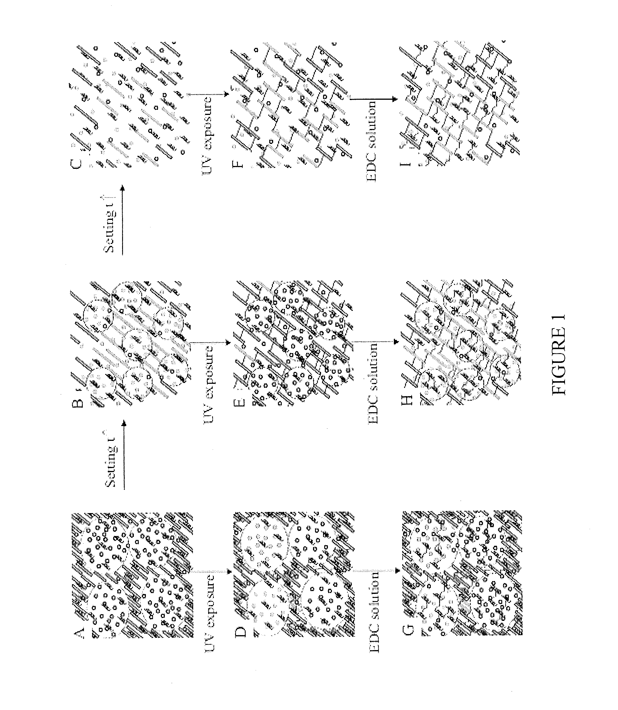
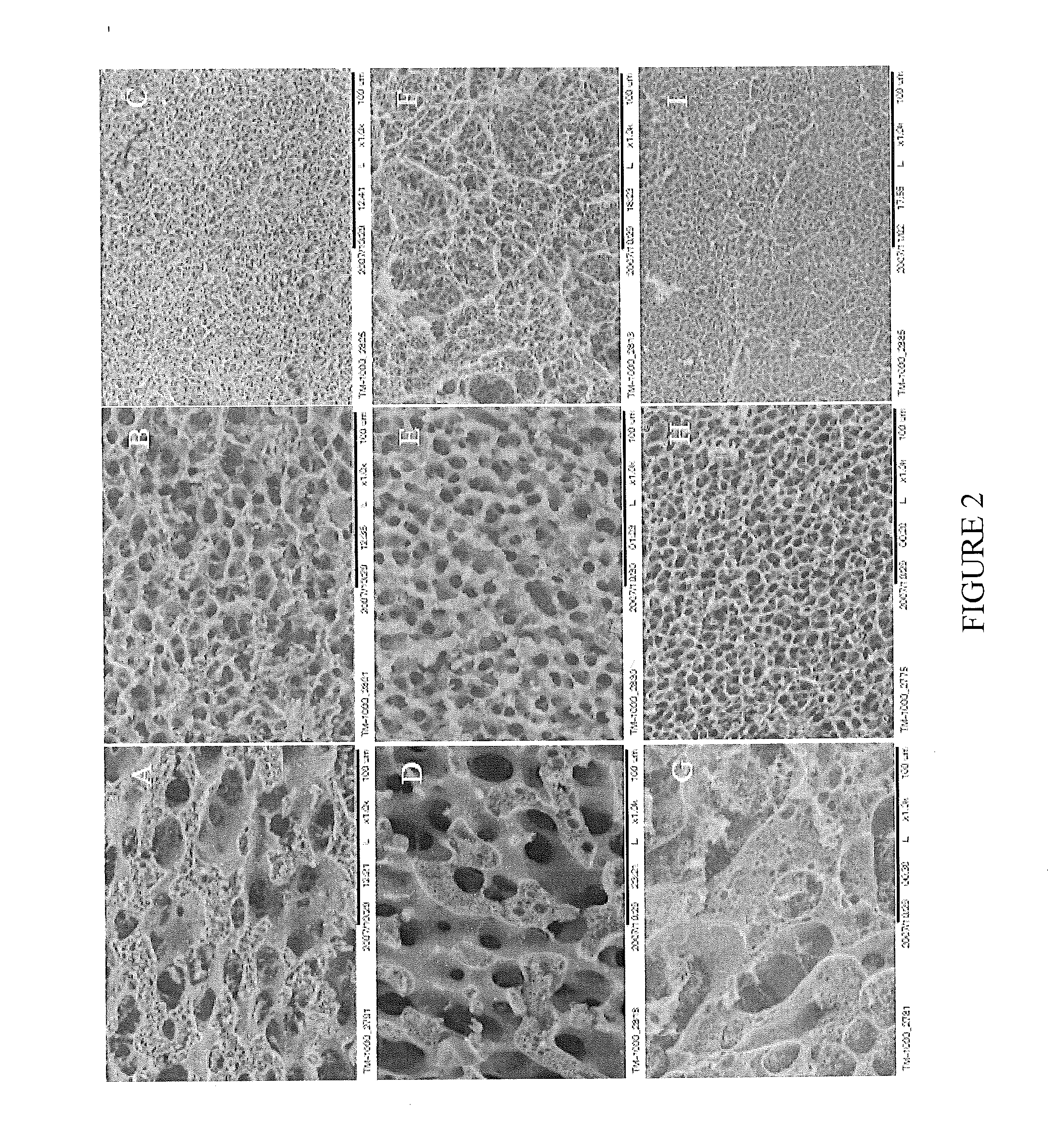
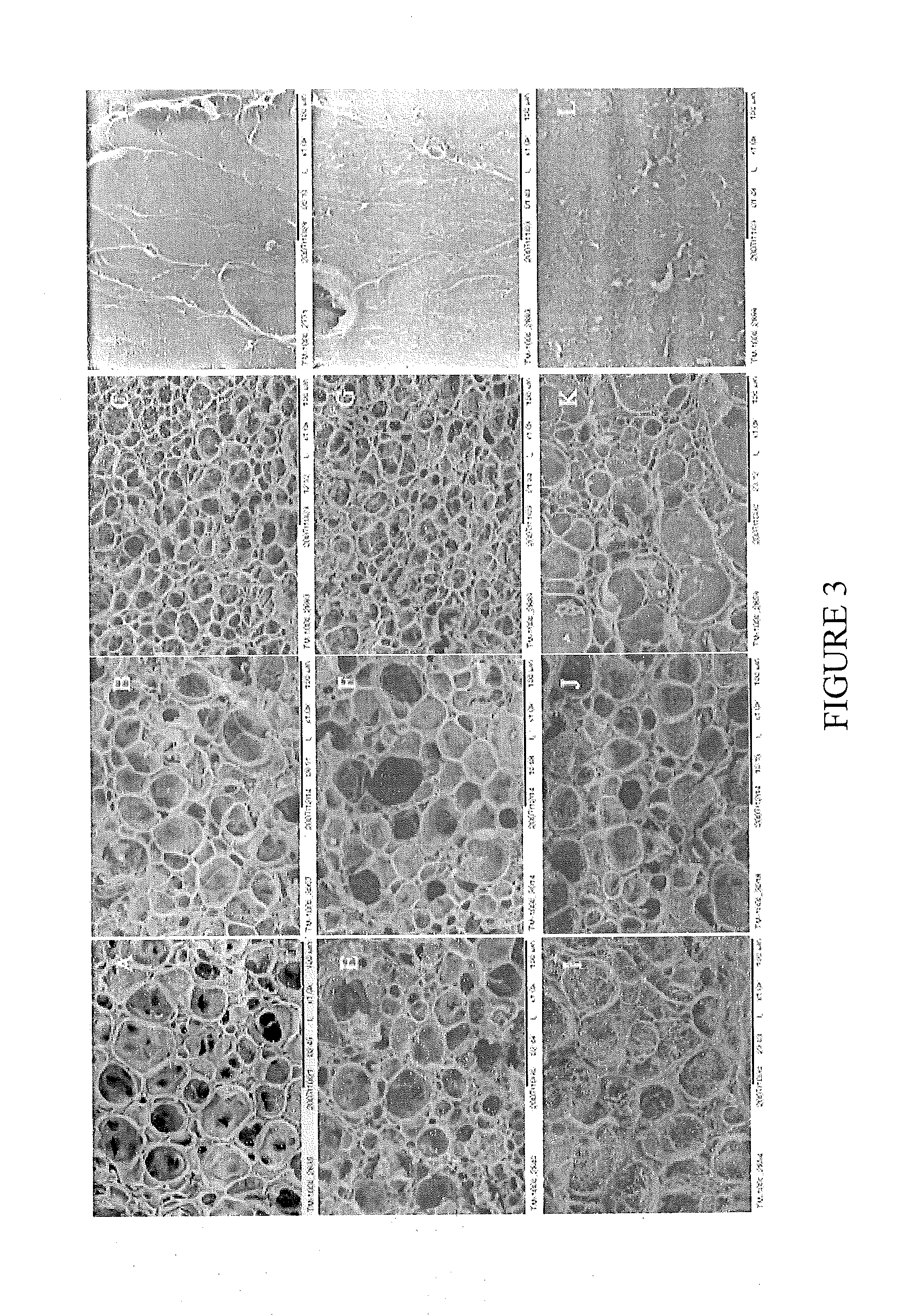
[0131]Highly elastic scaffolds from the natural polymers, chitosan and gelatin, were prepared as described above. Both the chitosan and gelatin were chemically modified to enable polymerization when exposed to light. Because chitosan is positively charged and gelatin is a polyampholyte with both negatively charged and positively charged patches, chitosan and gelatin interact electrostatically, leading to a transition from segregative phase separation to the mixing state. This transition enables controlled formation of a 3D porous structure without using porogens. By varying the chitosan-to-gelatin ratio, setting time, and gelatin crosslinking, 3D porous hybrid scaffolds can be achieved with tunable microstructures across the nano, micro, and macro length scales (FIG. 12). The scaffolds exhibit superior elasticity that has not been previously achieved in any other natural biopolymers except elastin. In vitro cult...


PUM

PropertyMeasurementUnit
temperatureaaaaaaaaaa
setting timeaaaaaaaaaa

Abstract

The present invention provides methods and compositions for tissue regeneration without cell transplantation.

Description

STATEMENT OF PRIORITY[0001]This application claims the benefit, under 35 U.S.C. §119(e), of U.S. Provisional Application No. 61 / 168,769, filed Apr. 13, 2009, the contents of which are incorporated by reference herein in their entirety.FEDERAL SUPPORT OF THE INVENTION[0002]Aspects of this invention were funded under Grant No. EPS-0447660 of the National Science Foundation USA. The U.S. Government has certain rights in this invention.FIELD OF THE INVENTION[0003]The present invention relates to compositions and methods for tissue regeneration without cell transplantation.BACKGROUND OF THE INVENTION[0004]Articular cartilage does not mount an effective repair response to injury, resulting in progressive joint degeneration and patient disability. The prevalence of clinical osteoarthritis in the United States was 27 million in 20051, with over 21 million physician visits incurred2. For patients with focal cartilage injury / damage, there are several treatment options. One option is anti-infl...

Claims


Application Information

Patent Timeline
Patent Type & Authority Applications(United States)
IPC IPC(8): A61K38/14A61K38/30A61K38/22A61K38/20C07K17/10A61P43/00C08H1/00A61K9/14A61P19/04B01J19/12A61K38/18C07K2/00B82Y5/00
CPCA61F2/30756A61F2002/30766A61L27/227A61L27/26A61L27/52A61L27/54A61L27/56A61L2300/426A61L2300/414A61L2300/252C08L5/08C08L89/06A61P19/04A61P43/00
Inventor WEN, XUEJUNQIU, YONGZHIVANDEN BERG-FOELS, WENDY S.
Owner CLEMSON UNIVERSITY
Why Eureka
Try Eureka