Methods to enhance cell migration and engraftment

a cell migration and cell technology, applied in the field of cell engraftment, can solve the problems of less than 5% improvement, too low to generate a significant impact, and insufficient functional outcomes of existing msc therapies, and achieve enhanced in vivo engraftment rate, enhanced spontaneous migration and directional migration of cells, and improved migratory activity

Active Publication Date: 2012-06-14
VERSITECH LTD
View PDF2 Cites 12 Cited by
  • Summary
  • Abstract
  • Description
  • Claims
  • Application Information

AI Technical Summary

Benefits of technology

[0009]A method to process undifferentiated cells for engraftment, such as MSCs, by functionally selecting subpopulations of undifferentiated cells with better migratory activity has been developed. The method enhances both spontaneous migration and directional migration of the cells towards specific chemo-attractants. The method also provides a means to enhance the in vivo engraftment rate of undifferentiated cells during tissue repair and regeneration.

Problems solved by technology

Despite the great potential of MSC-based therapies, the functional outcomes of existing MSC therapies have not been satisfactory, at least partly due to the extremely poor engraftment rate (usually <1-2%) of MSCs at the target tissues such as hearts in the treatment of myocardial infarction (Price et al.
Local injection of MSCs with and without carriers is able to improve engraftment in the target tissue such as heart and intervertebral disc, but the extent of improvement is less than 5%, which is too low to generate a significant impact.
Moreover, the invasiveness associated with the open surgery-based injection into inner organs such as heart and spine, and the inapplicability of local injection in diseases where multiple tissues are involved, such as osteogenic imperfecta and muscle dystrophy, prevents this approach from being widely used in regenerative medicine.
Currently, no effective method exists to improve the engraftment rate of systemically supplemented MSCs.
Nevertheless, these approaches also have significant drawbacks such as the long term uncertainty of genetically manipulated cells, the safety issue of virally transduced cells and the poor cost-effectiveness of in vivo supplementation of chemoattractants.
More importantly, none of these attempts address the heterogeneity problem of MSCs.

Method used


Image

  • Methods to enhance cell migration and engraftment
  • Methods to enhance cell migration and engraftment
  • Methods to enhance cell migration and engraftment

Examples


example 1

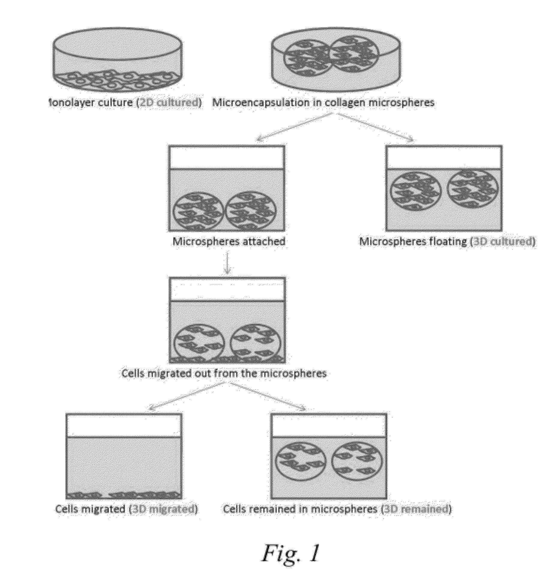
Microencapsulation of hMSCs Using Collagen Barrier

[0053]Materials and Methods

[0054]Bone marrow aspirates were collected from two healthy bone marrow donors (Subject 1 and 2) with informed consents. hMSCs were cultured in full medium consisting of Dulbecco's modified Eagle's medium-low glucose (DMEM-LG), 10% fetal bovine serum (FBS), 100 U / ml penicillin, 100 mg / ml streptomycin and 1% GlutaMax™ at 37° C. with 5% CO2. Cells at passage 4 were subcultured as traditional 2D (monolayer) cultures. The initial cell seeding density of traditional 2D culture was 6.25×10⁴ per 100 mm tissue culture dish.

[0055]After trypsinization, cells at passage 5 were labeled “2D cultured cells.” Some of these cells were used for subsequent microencapsulation. hMSCs were trypsinized using 0.05% Trypsin-EDTA (Gibco).

[0056]Cells were microencapsulated in a collagen barrier as described by Chan, et al. Biomaterials 28 (2007) 4652-4666. Rat-tail collagen type 1 solution (BD Biosciences) was neutralized by 1N NaOH...

example 2

Transwell Migratory Activities of Different hMSC Subpopulations

[0063]Materials and Methods

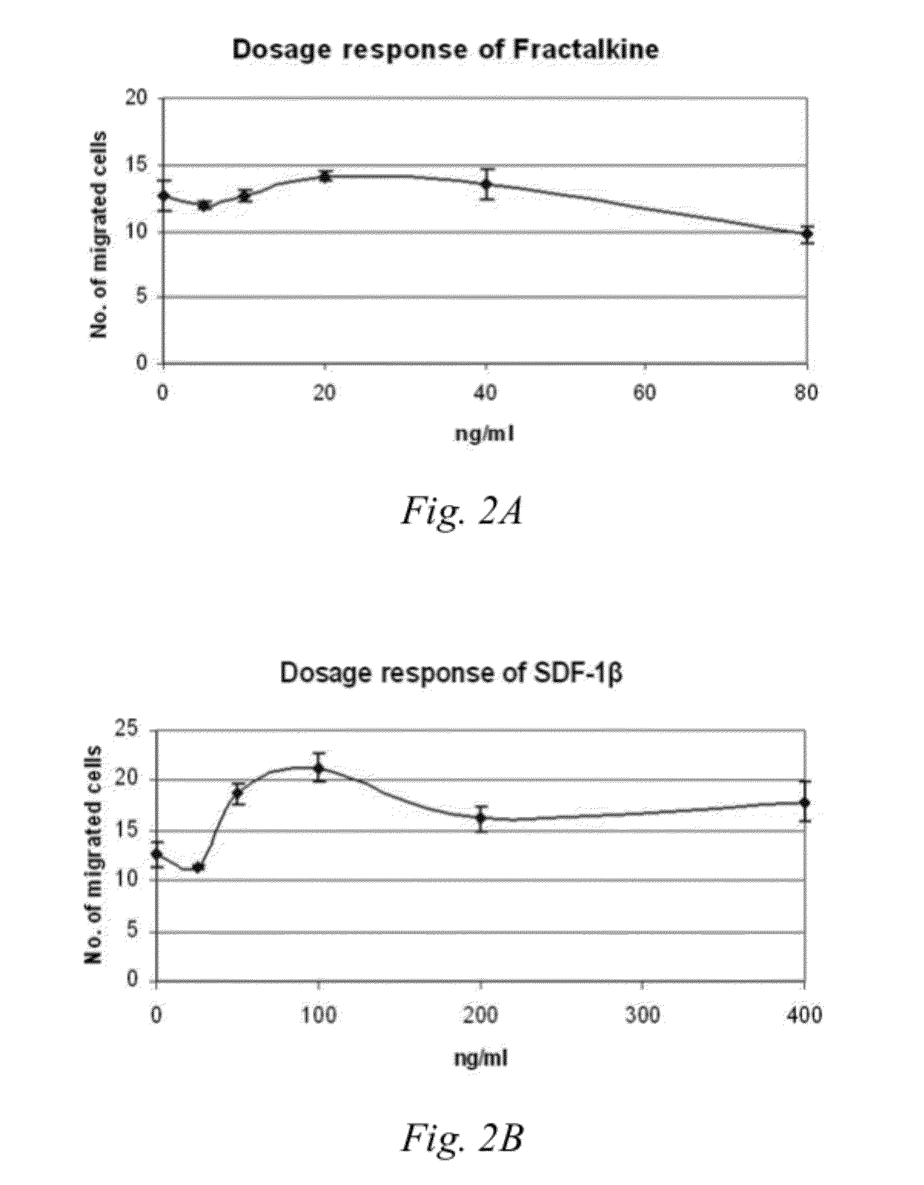
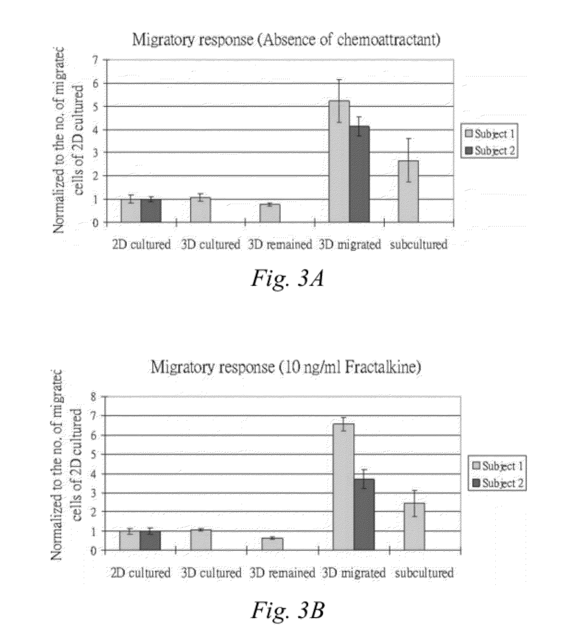
[0064]Serum free medium alone or in the presence of either 10 ng / ml Fractalkine (CX3CL1; Peprotech) or 50 ng / ml SDF-10 (CXCL12; Peprotech) in a total volume of 800 μl was added into the lower chamber of a 24-well plate transwell (BD Biosciences). A cell culture insert 8 μm pore size (BD Falcon™ Cell Culture Inserts, catalog #353097) was gently placed into the well. An aliquot of 5×10⁴ hMSCs collected from the different treatment groups of Example 1: 2D cultured cells, 3D cultured cells, 3D remained cells, 3D migrated cells or Subcultured cells, was suspended in 250 μl serum free medium and added into the insert in the upper chamber of the transwell. The cells were then incubated at 37° C. with 5% CO2 for 16 hours.

[0065]The insert was then removed from the well and the non-migrating cells from the upper side of the membrane were removed gently using a cotton bud. The lower side of the membrane w...

example 3

Engraftment of 3D Migrated Cells in Hepatectomized NOD / SCID Mice

[0072]Materials and Methods

[0073]8 week old, 25 gram NOD / SCID mice were anesthetized and then the median, left and caudate lobes of the liver, as well as the gall bladder, were removed, leaving the right lobe of liver. Two to three hours were allowed for the mice to recover after the surgery. After that, the mice were anesthetized again. Cell injection was done via the tail vein. Two million migrated cells suspended in 100 μl 1×PBS were used for cell injection.

[0074]At 48 hours 1 week and 1 month, mice were sacrificed and the liver collected for human cell marker analysis using flow cytometry and immunohistochemistry. For flow cytometry, cells from the harvested liver were isolated by incubating with collagenase for 20 minutes followed by filtering through the cellular sieve (BD Biosciences). The cell suspension was centrifuged and the supernatant was removed. Blood cells in the cell suspension were lysed by incubating ...


PUM

PropertyMeasurementUnit
concentrationsaaaaaaaaaa

Abstract

A method is provided to functionally select cells with enhanced characteristics relevant to cell engraftment, including both spontaneous migration and directional migration towards specific chemo-attractants. The cells are preferably undifferentiated cells, such as mesenchymal stem cells. The method involves entrapping or encapsulating the cells in a biomaterial barrier, optionally inducing cell migration, and selecting cells that migrated through the barrier. The cells selected by this method have better migratory activities and enhanced in vivo engraftment to injured tissues when they are supplemented systemically.

Description

CROSS REFERENCE TO RELATED APPLICATIONS[0001]This application is a continuation-in-part of co-pending U.S. patent application Ser. No. 11 / 750,863, filed May 18, 2007, which claims benefit of and priority to U.S. Provisional Patent Application No. 60 / 801,975 filed May 19, 2006. This application claims benefit of U.S. Provisional Patent Application No. 61 / 354,871, filed Jun. 15, 2010. All of the above are hereby incorporated by reference in their entirety.FIELD OF THE INVENTION[0002]The invention is generally related to the field of cell engraftment, more particularly to methods and compositions for enhancing both spontaneous migration and directional migration of transplanted cell populations.BACKGROUND OF THE INVENTION[0003]Cell-based therapy, particularly using undifferentiated cells, e.g., multipotent or pluripotent cells stem cells, presents a promising approach for regenerative medicine and tissue engineering (Short et al. Arch Med. Res. 2003 34(6):565-71, Barrilleaux et al. Tis...

Claims


Application Information

Patent Timeline
Patent Type & Authority Applications(United States)
IPC IPC(8): A61K35/12C12N5/02A61P43/00C12N5/00A61K35/28
CPCA61K9/1635A61K2035/128C12N5/0663C12N2533/54A61K35/28C12N5/0665C12N2533/90C12N5/0696C12N11/04C12N5/0606C12N2533/56C12N2533/50C12N2533/80C12N5/0666A61P43/00
Inventor CHAN, BARBARA PUIWONG, HOI LINGWONG, MEI YICHAN, GODFREY CHI-FUNGYANG, ZHEN FAN
Owner VERSITECH LTD
Why Eureka
Try Eureka