
Modular Assembly of Tissue Engineered Constructs

a tissue and construct technology, applied in the direction of vascular endothelial cells, biochemistry apparatus and processes, hepatocytes, etc., can solve the problems of limiting the protection provided by cells, difficult to get cells deposited on the outside of the scaffold to migrate to the interior, and unable to achieve the migration of cells to the interior of the scaffold, etc., to achieve sufficient structural rigidity and strength, and sufficient porosity and channel size

Inactive Publication Date: 2012-07-05
SEFTON MICHAEL V +1
View PDF1 Cites 11 Cited by
  • Summary
  • Abstract
  • Description
  • Claims
  • Application Information

AI Technical Summary

Benefits of technology

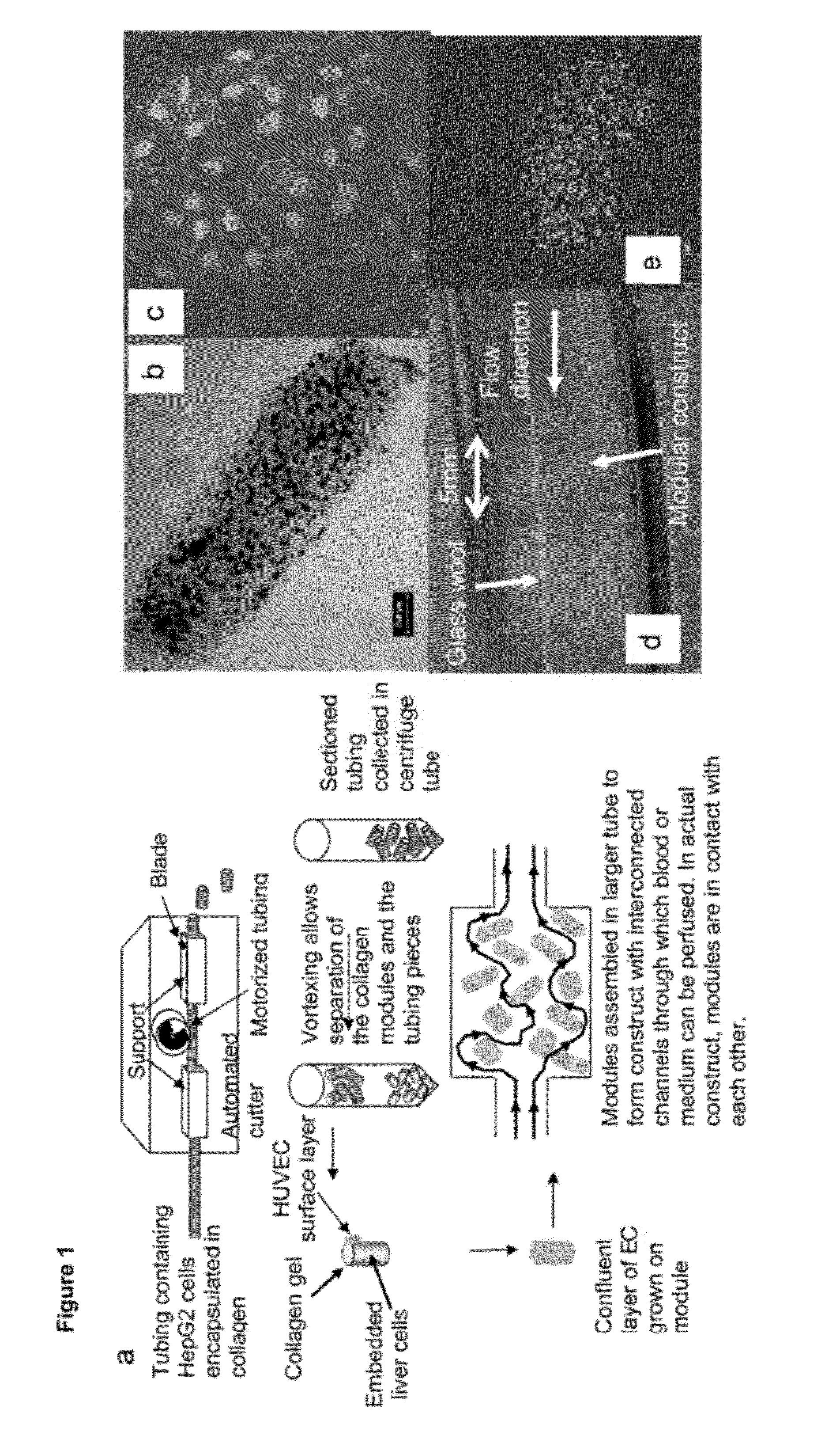
The present invention provides a new modular approach to the fabrication of tissue constructs that is scaleable, with a largely uniform cell distribution, and can accommodate multiple cell types. The resulting tissue constructs have adequate porosity to enable perfusion with a fluid and are perfuseable. The invention also includes a new method for creating a porous structure by filling an enclosure with discrete modules that have interconnected channels. The modules contain tissue-specific cells embedded within a material that provides the three-dimensional discrete and separable form to the modules. The modules can be assembled to form a larger scale functional structure (the construct) upon assembly of a multiplicity of modules. The construct then displays a set of functional characteristics that combine, build upon, and extend the functional characteristics of the discrete modules. The invention also includes a new method for producing microcapsules with a high degree of porosity.

Problems solved by technology

One fundamental difficulty in creating large three-dimensional organs is the creation of a vascularised support structure in the engineered tissue or tissue construct.
All of these can lead to tissue constructs with different characteristic features, but in all cases there are constraints on nutrient, waste and oxygen diffusion that restrict construct size to that for which the viability and function of the cellular components can be supported by the limited rate of diffusion.
(Tissue Engineering, 4(2):117-130, 1998), VEGF is but one angiogenic factor and issues associated with the functional maturity of the vessels and the need for multiple factors may limit this strategy.
Pre-seeded cells may be lost on implantation due to insufficient adhesion, as shown by Williams (Cell Trans, 4(4):401-410, 1995), and thus the protection from thrombosis provided by the cells may be limited due to the incomplete cell coverage of the support structure.
The requirement for EC attachment means that the materials used for cell encapsulation [e.g., alginate (Lim et al., 1980) or HEMA-MMAError!Bookmark not defined.] are not suitable for preparing vascularized constructs, since microcapsules are typically designed to have a surface that prevents cell attachment so as to minimize the fibrotic response on encapsulation.
In the preparation of tissue constructs by seeding cells in a scaffold, it is often difficult to get cells deposited on the outside of a scaffold to migrate to the interior; typically they populate just the periphery of the scaffold, at best an outer millimetre or so.
However, this method does not scale well for larger constructs or larger animals.
However the scalability of dynamic seeding techniques remains questionable.
However, it does not address another limitation of current tissue constructs: the difficulty of mixing two or more different cells types together without the concern that the faster growing cell type will overtake the slower one.
(Science, Jan. 24; 231(4736):397-400, 1986), circumvents this problem, but this method is not universally applicable.

Method used


Image

  • Modular Assembly of Tissue Engineered Constructs
  • Modular Assembly of Tissue Engineered Constructs
  • Modular Assembly of Tissue Engineered Constructs

Examples


embodiment 2

Gelatin Modules, Hand Cutting

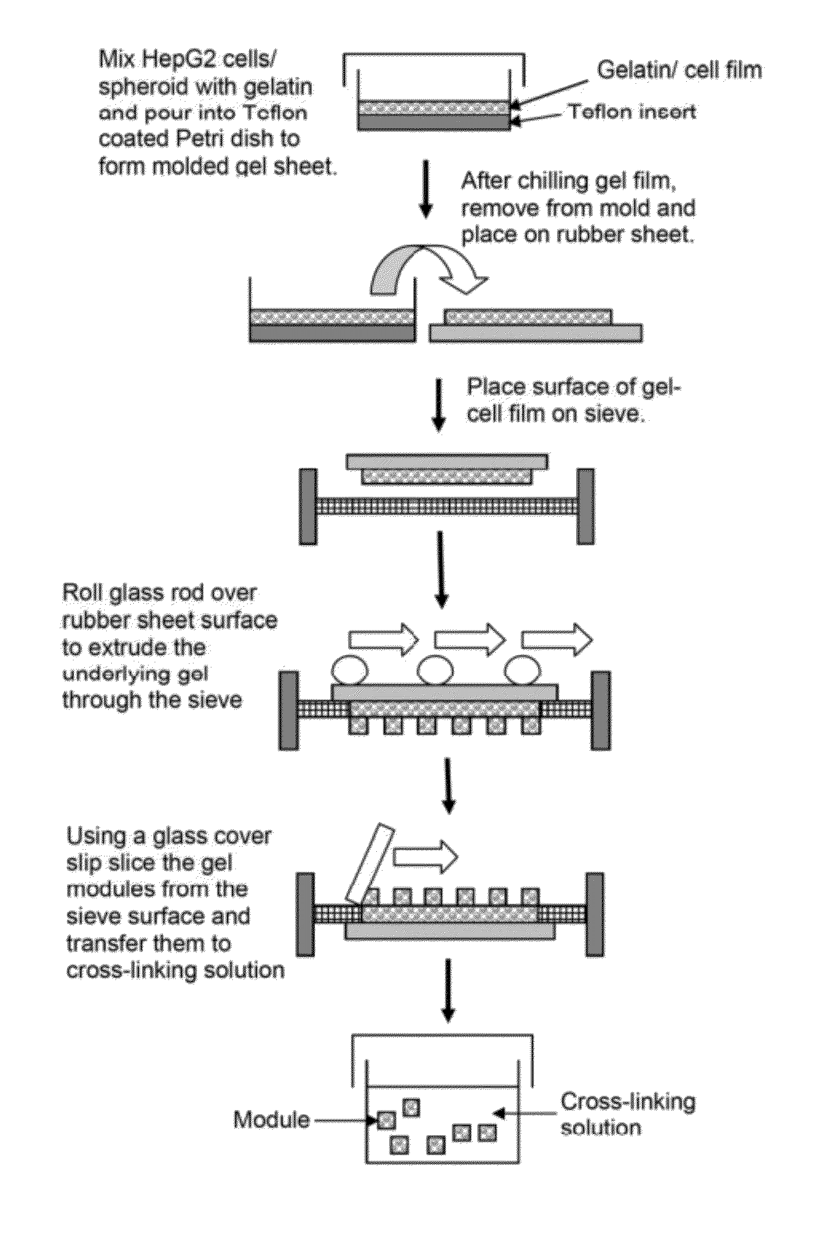
[0077]In an alternative embodiment, gelatin modules (˜120 μm diameter×1 mm long) containing HepG2 spheroids were prepared inside a glass micropipette (0.282 mm ID, Drummond microcap) prewashed with Pluronic L101. HepG2 spheroids were prepared by culture in αMEM with serum on bacteriological polystyrene culture dishes for 4 days; at this time spheroids were approximately 100 μm in diameter and contained roughly 300 cells each. Spheroids were suspended in 55 μl of 300 bloom, type A gelatin (25 wt %) liquid (˜40° C.) and a droplet of the gel-spheroid suspension was placed onto a sterilised glass slide, from which it was drawn into the glass micropipette. After 20-30 minutes refrigeration, (enough time to ensure gelation) the gel-spheroid modules were expelled from the glass capillary into a sterile solution of very dilute glutaraldehyde (0.05%) in PBS. After 20 minutes the modules were washed twice in PBS followed by a 1-2 hour wash in cell culture medium. ...

embodiment 4

Poloxamine Based Materials

[0085]In another embodiment modules were prepared using a synthetic collagen-mimetic material that was stiffer than collagen (and therefore resistant to compaction) but that like collagen allows both cell encapsulation and cell growth on the surface. This collagen-mimetic material was a poloxamine-collagen semi-interpenetrating networkError! Bookmark not defined.; poloxamine is a four-arm PEO-PPO block copolymer derivative, Tetronic™ 1107. Methacryloyl groups were added to the ends of the poloxamine (FIG. 6) and a solution of the poloxamine with collagen also in the same solution was photo-crosslinked. Cells (HepG2) were embedded easily and at high viability (Sosnik et al., Tissue Eng., 2005). The poloxamine-collagen material was much stiffer (2,000 to 7,000 Pa for polymer concentrations between 6 to 8%) than collagen alone (˜50 Pa) as was evident also in the cylindrical shape of these modules which was preserved through many weeks of culture.

[0086]A positi...

embodiment 6

In vivo Enclosure

[0089]HUVEC covered modules were implanted into an omental pouch, an enclosure to be filled with modules, in nude rats. The omental pouch is prepared by folding the omentum up towards the stomach and suturing (7‘o’ silk sutures) along the left and right edges of the omentum and along the top of the pouch but leaving an opening for the placement of the modules. Modules, suspended in PBS are placed into the omental pouch using a sterile 1000 μL micropipette tip, while preaggregated modules (e.g., prepared by incubation at high density in a small well) are placed into the pouch with tweezers. The opening is sutured closed to completely enclose the modules. FIG. 8 shows that collagen gel modules (in green) coated with HUVEC have channels (see arrow, order of 100 μm in “width”) that persist up to 21 days after implantation in the omental pouch. Without HUVEC the collagen modules remodel and do not appear to form channels. Some of these channels (FIG. 9 right; UEA-1 lecti...


PUM

PropertyMeasurementUnit
lengthaaaaaaaaaa
diameteraaaaaaaaaa
diameteraaaaaaaaaa

Abstract

Scaleable, vascularised tissue constructs that are composed of a multiplicity of cell containing, discrete and separable modules, methods of fabricating same and uses thereof. The tissue construct is a tissue substitute used in tissue transplantation or substitution or for the purpose of in vitro mimic of normal tissue.

Description

CROSS-REFERENCE TO RELATED APPLICATIONS[0001]This application is a continuation of U.S. application Ser. No. 12 / 457,507, filed Jun. 12, 2009, which application was published on Dec. 16, 2010, as U.S. Publication No. US2010 / 0316690, the contents of which is incorporated herein by reference in its entirety.FIELD OF THE INVENTION[0002]This invention relates to scalable, vascularised tissue constructs that are comprised of a multiplicity of cell containing, discrete and separable modules, methods of fabricating the same, and uses thereof.BACKGROUND OF THE INVENTION[0003]It is desirable to create an unlimited supply of vital organs, such as hearts, livers and kidneys, for example, for transplantation through tissue engineering. In the past, there have been many suggested approaches to tissue engineering. One fundamental difficulty in creating large three-dimensional organs is the creation of a vascularised support structure in the engineered tissue or tissue construct. A tissue construct...

Claims


Application Information

Patent Timeline
Patent Type & Authority Applications(United States)
IPC IPC(8): C12N5/071A61F2/00
CPCC12N5/0062C12N2533/54C12N2502/28C12N5/0671
Inventor SEFTON, MICHAEL V.MCGUIGAN, ALISON
Owner SEFTON MICHAEL V
Learn More