Solid Forms for Tissue Repair

a tissue repair and solid form technology, applied in the direction of macromolecular non-active ingredients, peptide/protein ingredients, depsipeptides, etc., can solve the problems of stress shielding to the surrounding bone, limited autologous bone tissue, fatigue failure of implants, etc., to enhance cartilage or bone formation, regeneration or enhancement

Active Publication Date: 2012-07-26
CARTIHEAL 2009
View PDF1 Cites 14 Cited by
  • Summary
  • Abstract
  • Description
  • Claims
  • Application Information

AI Technical Summary

Benefits of technology

[0131]By optimizing the specific positioning of a scaffold the porous crystalline structure of a coral scaffolds of this invention, described below, is accessible to beneficial components located within a tissue milieu. For example, the porous crystalline structure of coral allows in-growth of blood vessels to create a blood supply for the cartilage that will infiltrate the scaffold during cartilage repair. By penetrating into a bone marrow void, mesenchymal stem cells located within the bone marrow now have access to the exposed surface of the scaffold. In one embodiment, the region of the scaffold penetrating into a bone marrow void attracts mesenchymal stem cells from the bone marrow and promotes blood vessel formation to the site of cartilage repair. In one embodiment, the region of the scaffold penetrating into a bone marrow void promotes adhesion, proliferation, or differentiation or a combination thereof, of the mesenchymal stem cells attracted to the scaffold.

Problems solved by technology

Synthetic materials such as metals and bone cements have also been used for restoring and reconstructing bone for many years, but often result in stress-shielding to the surrounding bone and fatigue failure of the implant.
Another possibility is autologous bone grafting, although the supply of autologous bone tissue is limited and its collection is painful, with the risk of infection, hemorrhage, cosmetic disability, nerve damage, and loss of bone function.
In addition, significant morbidity is associated with autograft harvest sites.
However, these scaffolds often are mechanically unstable and do not readily contribute to the creation of tissue structures with a specific predefined shape for transplantation.
To obtain mechanical strength, chemical modification is required, which may lead to toxicity.
Defects and degeneration of the articular cartilage surfaces of joints causes pain and stiffness.
Damage to cartilage which protects joints can result from either physical injury as a result of trauma, sports or repetitive stresses (e.g., osteochondral fracture, secondary damage due to cruciate ligament injury) or from disease (e.g. osteoarthritis, rheumatoid arthritis, aseptic necrosis, osteochondritis dissecans).
Orthopedic prevention and repair of damaged joints is a significant burden on the medical profession both in terms of expense and time spent treating patients.
Attempts to re-grow hyaline cartilage for repair of cartilage defects remain unsuccessful.
Techniques utilizing donated tissue from autografts, allografts, or xenografts are wholly unsatisfactory as autografts add additional trauma to a subject and allografts and xenografts are limited by immunological reactivity to the host subject and possible transfer of infective agents.
Surgical attempts to utilize materials other than human or animal tissue for cartilage regeneration have been unsuccessful.

Method used


Image

  • Solid Forms for Tissue Repair
  • Solid Forms for Tissue Repair
  • Solid Forms for Tissue Repair

Examples


example 1

Applications of Coralline-Based Scaffolding of this Invention

[0311]Coralline-based scaffolding of this invention may be inserted into cartilage, bone or a combination thereof, in a subject in need thereof.

[0312]In some embodiments, such placement will include drilling in the area to expose the site in which implantation is desired, and tight fitting of the scaffold within the defect / site.

[0313]For implantation for cartilage repair, regeneration, etc., scaffolds are implanted in the desired cartilage site and within proximally located bone, so that, in this way, the coral scaffold is grafted through two types of tissue, cartilage and bone. FIG. 1 schematically depicts orientation of a cartoon of a scaffold of this invention within a site of cartilage / bone repair.

[0314]Scaffolds may be prepared according to any embodiment as described herein, as will be appreciated by the skilled artisan.

[0315]The scaffolds are envisioned for use in veterinary applications, as well as in the treatment...

example 2

Restoration of an Osteochondral Defect

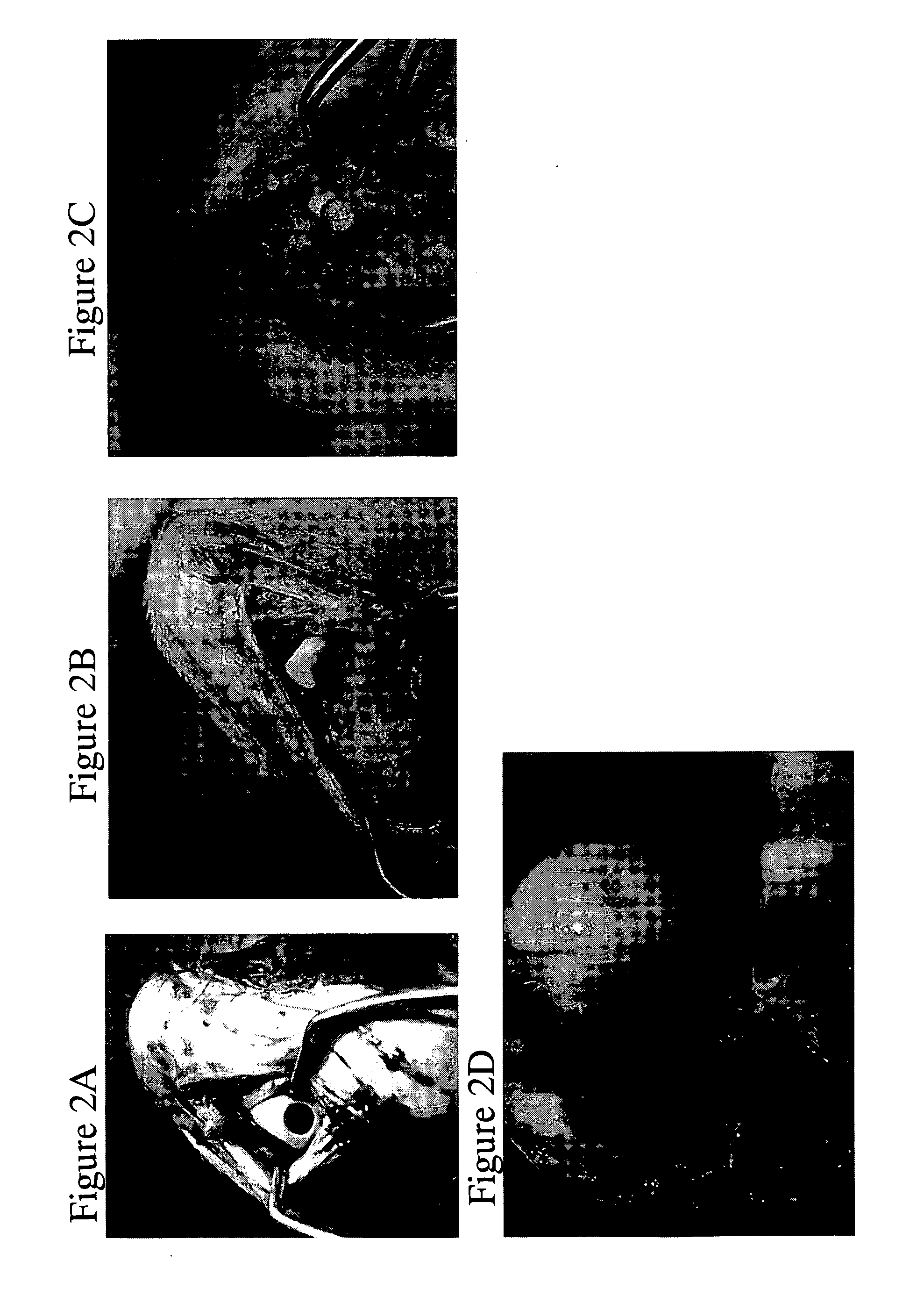
[0317]Restoration of an osteochondral defect was performed in mature goats using rounded implants which were 6 mm in diameter and 8 mm in length. A 5.5×8 mm core of cartilage and bone tissue was drilled out of, the medial femoral condyle of each goat (FIG. 2A) and the implant pressed fit into the site of cartilage and bone repair (FIGS. 2B and 2C).

[0318]Some animals harvested at 2.5 weeks post surgery exhibited signs that the implant was well incorporated into the native tissue and cartilage tissue was developed proximal to implant, moreover signs for: vascularisation can be seen (FIG. 2C).

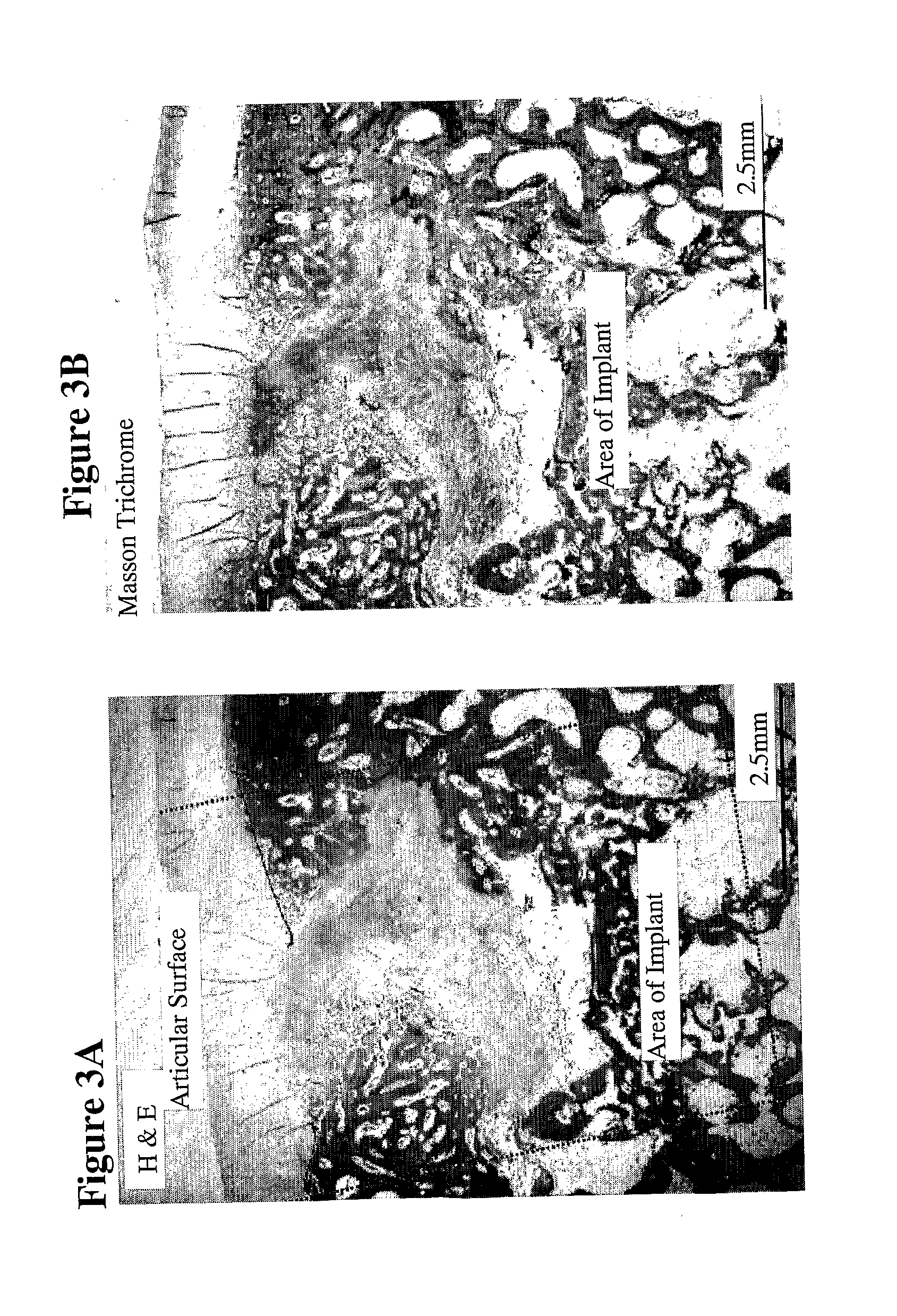
[0319]A group of animals were sacrificed and tissue was harvested from the implant site 9 weeks post surgery. H&E and Masson Trichrome histological evaluation of the tissue (FIGS. 3A and 3B, respectively) showed that area of the implant was replaced by newly formed cartilage and woven bone and the cartilage was smooth and almost completely regenerated. Safrani...

example 3

Preparation of a Multiphasic Solid Aragonite Scaffold

[0320]To create a multi-phasic scaffold varying in terms of the pore volume (porosity) of each phase, and / or varying in terms of the diameter of the voids present in each phase, plugs of 5.2 mm in diameter and 7.5 mm in length were positioned within a silicon holder whereby only the top 1 mm of the plug was exposed, and the holder with the plug was placed in an inverted position, and immersed into the reaction mixture, such that only the top 1 mm of the plug was in direct contact with the mixture.

[0321]The plug was first immersed in a 5% disodium salt solution for two hours at room temperature, followed by addition of a 99% formic acid solution to yield a final concentration of 0.5%, where the plugs were immersed again in the solution for an additional 20 minutes. The mixture was discharged and the plugs were washed in distilled water overnight, under conditions of approximately 0.2-0.00001 Bar pressure via the application of a v...


PUM

PropertyMeasurementUnit
heightaaaaaaaaaa
diameteraaaaaaaaaa
thicknessaaaaaaaaaa

Abstract

This invention provides aragonite- and calcite-based scaffolds for the repair, regeneration, enhancement of formation or a combination thereof of cartilage and / or bone, which scaffolds comprise at least two phases, wherein each phase differs in terms of its chemical content, or structure, kits comprising the same, processes for producing solid aragonite or calcite scaffolds and methods of use thereof.

Description

BACKGROUND OF THE INVENTION[0001]Surgical intervention and grafting are sometimes necessary to restore mechanical function and reconstruct the morphology of bone and cartilage, resulting from trauma, tumors, or abnormal bone developments.[0002]Synthetic materials such as metals and bone cements have also been used for restoring and reconstructing bone for many years, but often result in stress-shielding to the surrounding bone and fatigue failure of the implant. Another possibility is autologous bone grafting, although the supply of autologous bone tissue is limited and its collection is painful, with the risk of infection, hemorrhage, cosmetic disability, nerve damage, and loss of bone function. In addition, significant morbidity is associated with autograft harvest sites. These problems may be overcome by engineering tissue using scaffolds made of synthetic or natural biomaterials that promote the adhesion, migration, proliferation, and differentiation of bone marrow stem cells, a...

Claims


Application Information

Patent Timeline
Patent Type & Authority Applications(United States)
IPC IPC(8): A61K38/18A61K47/36A61K47/42A61P29/00A61F2/08A61K35/12A61K9/00A61P19/04A61P19/02A61K38/30A61K47/04A61P31/00
CPCA61F2/28A61L27/54A61F2/3094A61F2002/30011A61F2002/30224A61F2002/30807A61F2002/3092A61F2230/0069A61F2250/0023A61F2310/00341A61L27/3604A61L27/427A61L27/56A61L2430/02A61L2430/06A61L27/34A61L27/3834A61F2/30756A61L27/025C08L5/08A61P19/00A61P19/02A61P19/04A61P29/00A61P31/00A61L27/18A61L27/20A61L27/3847A61L27/3852A61L27/52A61L2300/414A61L2300/606
Inventor ALTSCHULER, NIRVAGO, RAZI
Owner CARTIHEAL 2009
Why Eureka
Try Eureka