Rapid, semi-automated method to detect respiratory virus infected cells in direct specimens

Inactive Publication Date: 2012-08-09
VERIDEX LCC +1
View PDF3 Cites 1 Cited by
  • Summary
  • Abstract
  • Description
  • Claims
  • Application Information

AI Technical Summary

Benefits of technology

[0012]Further advantages provided by this invention are the functional simplicity in design, ruggedness, compactness, AC or DC power options, and substantially lower purchase and operating costs relative to conventional commercial devices with comparable performance characteristics. The features and improvements of the devices of this invention, exemplified as compact clinical cell cytometer, make them particularly useful for operation in small, non-complex and in unsophisticated laboratories or field conditions prevalent in resource-poor countries.
[0013]A further improvement is in the individual illumination/light capture components of the cytometer. The LEDs are positioned to illuminate along the long axis of the car

Problems solved by technology

Viral Culture, DFA, Flow cytometry and similar complex analytical systems remain largely inaccessible for routine clinical use in resource-poor countries due to high instrument and reagents costs, lack of technical support.
They generally require sophisticated techniques, which involve the use of instruments that are expensive both in terms of initial cost and maintenance as well as requiring highly trained personnel.
This makes the conventional systems unsuitable for use in mid size and small hospitals as well as laboratories of resource-poor countries.
However, the sensitivity of these tests is widely reported to be

Method used


Image

  • Rapid, semi-automated method to detect respiratory virus infected cells in direct specimens
  • Rapid, semi-automated method to detect respiratory virus infected cells in direct specimens
  • Rapid, semi-automated method to detect respiratory virus infected cells in direct specimens

Examples


example 1

Detection of Virus-Infected Cells Using Rare Cell Analysis

[0041]Objective; We studied an adapted cell detection technology, originally developed to detect ultra-rare tumor cells in circulation, to capture and detect virus-infected cells present in various cell culture systems and patient NP samples.

Relevance: Clinical diagnosis of viral infection often involves direct specimen analysis using Direct Fluorescent Antibody (DFA) followed by virus culture in susceptible cell lines. DFA is faster but is labor intensive, provides a subjective result and is not as sensitive as culture or molecular methods that may require 6 to 48 hrs for a result.

Methodology: Virus culture was done using standard methods using A549, Mink Lung, SKBr-3, and RMix (DHI) as susceptible host cell lines. Ferrofluids (FF) were prepared as colloidal suspensions of 200 nM magnetic particles coated with antibodies to specific antigen targets on cells. Suspensions of cells were fixed with cell fixative solution, washed...

example 2

Feasibility Study of a Rapid, Semi-Automated Method to Detect Respiratory Virus Infected Cells in Direct Specimens

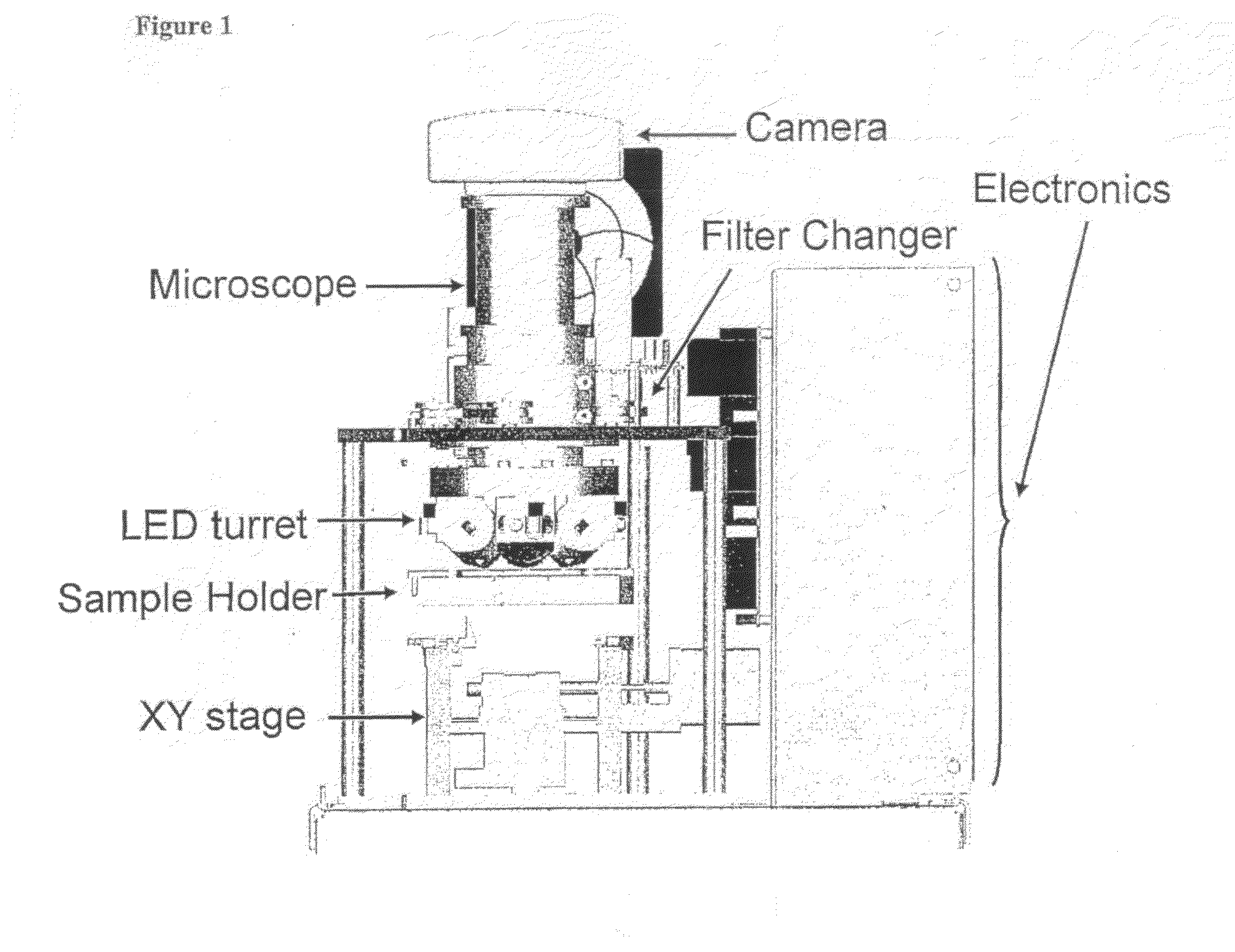
[0042]Objective: The objective of this study was to demonstrate that a rapid, liquid specimen processing procedure could readily differentiate positive and negative cell culture in <2 min using EasyCount™, an automated fluorescent microscope with cell counting capability. EasyCount™ uses a CCD camera to visualize counter stained cells for a total cell count and interrogates those same cells to identify those that are infected with virus by detecting fluorescein.

Methods: We chose R-Mix and Influenza A (Flu) as a prototype cell culture / virus system to simulate a direct specimen scenario for automated detection purposes. Flu virus was inoculated in duplicate onto 24-well R-Mix plates, centrifuged at 700×g for 60 min, and incubated at 37° C. for 20 hr. One plate was fixed, stained and counted to confirm the viral input. The cells from the replicate plate were released and ce...


PUM


Abstract

A novel cytometer system, methods and algorithms which provide a rugged, affordable and easy-to-use device in remote locations. All cells in a biological sample are fluorescently labeled, with target cells also magnetically labeled. Non-magnetically labeled cells are imaged for viability. Labeled sample is placed between two wedge-shaped magnets to selectively move the magnetically labeled cells for observation. A LED illuminates the cells and a CCO camera captures the images of the fluorescent light emitted by the target cells. Image analysis performed with a novel algorithm provides a cell count that can be related to the target cell concentration of the original sample.

Description

CROSS REFERENCE TO RELATED APPLICATIONS[0001]This application is a non-provisional application, which is incorporated by reference herein and claims priority, in part, of U.S. Provisional Application No. 60 / 932,698, filed 16 Apr. 2007, now abandoned.FIELD OF THE INVENTION[0002]This invention relates generally to simple and low cost electronic optical devices, methods and algorithms for enumeration of microscopic particles distributed in a two-dimensional plane. More specifically, the present invention relates to the adaptation of automated cell enumeration platforms for the direct detection of virus-infected cells in a fluid sample such as nasopharyngeal samples.BACKGROUND OF THE INVENTION[0003]The enumeration of absolute levels of cells and their subsets in body fluids is of primary importance in determining the state of health of human beings and mammals in general. The primary analytical platform for performing such analyses is viral culture, direct immunofluorescence (DFA), or f...

Claims


Application Information

Patent Timeline
IPC IPC(8): G01N21/17
CPCG01N33/5091G01N2333/11G01N33/56983G01N33/54326
Inventor CONNELLY, MARK CARLECOUMANS, FRANK A.W.PAGE, JIMMYRAO, GALLA CHANDRAJOLLICK, JR., JOSEPH
Owner VERIDEX LCC
Why Eureka
Try Eureka